Article

Safety and Local Efficacy of Laser Ablation for the Extrahepatic Metastasis of Hepatocellular Carcinoma: An Available Treatment Strategy

1
Department of Medical Ultrasound, Division of Interventional Ultrasound, the First Affiliated Hospital, Sun Yat-sen University, No. 58 Zhongshan 2nd Road, Guangzhou 510080, China
2
Department of Medical Oncology, the First Affiliated Hospital, Sun Yat-sen University, No. 58 Zhongshan 2nd Road, Guangzhou 510080, China
*
Author to whom correspondence should be addressed.
Coatings 2020, 10(10), 951; https://doi.org/10.3390/coatings10100951
Submission received: 28 August 2020 / Revised: 28 September 2020 / Accepted: 28 September 2020 / Published: 1 October 2020
(This article belongs to the Special Issue Thin Film Laser Damage, Ablation, Deposition and Structuring)

Abstract

:
Thermal ablation plays an important role in the treatment of extrahepatic metastasis of hepatocellular carcinoma (HCC). Yet laser ablation (LA), as a safe thermal ablative modality, is less investigated in this field. In this study, the safety and local effectiveness of LA in the treatment for the extrahepatic metastasis of HCC were evaluated. From May 2012 to May 2019, 17 patients (13 males and 4 females; mean age, 54.1 ± 14.6 years; age range, 34–80 years), who underwent LA for treatment of extrahepatic metastasis of HCC at the First Affiliated Hospital of Sun Yat-sen University, were retrospectively enrolled in this study. Local effectiveness, complications, local tumor progression (LTP), and overall survival (OS) were evaluated. Finally, a total of 28 LA treated extrahepatic metastatic lesions of HCC were reviewed. Neither LA-related mortality nor major complication occurred. Complete ablation (CA) was achieved in 20 out of 28 lesions (71.4%). During the follow-up (mean, 19.5 ± 12.8 months; range, 5–42.7 months), LTP developed in 4 out of 20 lesions with CA (20%). Four patients died of tumor progression or multiple organ dysfunction syndrome. The accumulative one- and three-year OS rates were 79.0% and 65.8%, respectively. In conclusion, LA is a safe and effective therapeutic option in the treatment of extrahepatic metastasis of HCC. Further studies are necessary to evaluate the benefit of LA.

1. Introduction

Hepatocellular carcinoma (HCC) is one of the most common malignancies worldwide, claiming approximately 700,000 deaths per year [1,2]. The Barcelona Clinic Liver Cancer (BCLC) staging system is a recommended staging model for HCC, endorsed by major academic societies of liver disease [3]. According to the BCLC staging, liver resection remains one of the first-line treatments for patients with early-stage HCC. However, it is reported that the cumulative five-year recurrence rate after liver resection is over 60%, owing to its nature of recurrence [4]. Recurrent HCC presences more severe aggressiveness, including extrahepatic metastasis or vascular invasion, and causes the majority of deaths [5]. For patients with advanced-stage HCC, systemic therapy is recommended and Sorafenib has acted as the first-line systemic agent in the past decade [6]. Other systemic agents such as Lenvatinib have shown noninferior performance in overall survival compared with Sorafenib in advanced HCC [7]. In addition, immune checkpoint inhibitors (anti-PD-1, anti-PD-L1, and anti-CTLA-4 antibodies) have exhibited potential therapeutic effects in advanced HCC [8,9,10].
Previous studies have demonstrated that Sorafenib combined with locoregional tumor control was superior to Sorafenib alone in improving long-term outcomes in advanced HCC [5,11,12]. Radiofrequency ablation (RFA), the most popular and widely used thermal ablation modality, is playing an important role not only in the treatment of early-stage HCC with curative intention, but also in the management of advanced-stage HCC as an adjuvant. The application of RFA in extrahepatic metastasis of HCC to the lung, bone, adrenal glands, and other locations has been reported in previous studies [13,14,15,16,17]. Laser ablation (LA), however, is less investigated and hardly used than that of RFA for now. LA is a minimally invasive method that kills tumors in solid organs by directing low-power laser energy inside a tumor tissue through one or more optic fibers [18]. With energy absorption and conversion, temperature within the tumor reaches up to 140 °C, resulting in coagulative necrosis [19]. Compared with RFA, LA is considered a safer option for HCC patients with “critical lesions” which are at high-risk locations where RFA is considered to be aggressive [20,21,22,23]. For extrahepatic metastases in the hepatic portal or retroperitoneal space, LA has advantages than RFA in the following aspects. First, the delivered energy is concentrated, resulting in a relatively small ablative zone and less damage to the surrounding tissue [24]. Second, the laser beam is propagated through optic fibers encapsulated in a 21-gauge needle that causes minimal puncture injury and minimizes the risk of severe complications such as fatal hemorrhage [25]. However, data on the management of extrahepatic metastasis of HCC by LA are scarce [25,26,27].
The purpose of our study was to evaluate the safety and local effectiveness of US-guided percutaneous LA in the treatment of extrahepatic metastatic HCC at various locations.

2. Materials and Methods

The protocol of this single-center retrospective study was approved by the Institutional Review Board of the First Affiliated Hospital of Sun Yat-sen University and the requirement for informed consent was obtained from all patients after the nature of the procedures had been fully explained.
A retrospective study was carried out to review patients who underwent US-guided percutaneous LA for extrahepatic metastasis after the initial treatment of intrahepatic HCC lesions in the First Affiliated Hospital of Sun Yat-sen University from May 2015 to May 2019. Eventually, 17 consecutive patients were enrolled in the study. The diagnosis and evaluation of extrahepatic metastatic lesions were based on imaging data including contrast-enhanced computed tomography (CECT), contrast-enhanced magnetic resonance imaging (CEMRI), contrast-enhanced ultrasound (CEUS) or positron emission tomography/computed tomography (PET/CT). All patients were examined and evaluated by an interdisciplinary tumor board including surgeons, radiologists, hepatologists, and oncologists. All patients had a history of curative treatment for HCC including surgical resection or thermal ablation. None of the patients was suitable for surgical resection of the extrahepatic metastatic lesions. The patients were neither suitable for RFA nor other thermal ablations, due to the location of the lesions being adjacent to important organs. The inclusion criteria were as follow: (1) aged 18–80 years; (2) previous initial treatment for primary HCC with curative intention, and confirmed extrahepatic metastases to organs or cavities visible by US/CEUS; (3) extrahepatic metastases at high-risk locations for RFA (less than 1cm from a major blood vessel, gastrointestinal tract or important organs such as kidney or spleen); (4) the largest extrahepatic metastases ≤5 cm in diameter; (5) preserved liver function at Child–Pugh Grade A or B; (6) an Eastern Cooperative Oncology Group (ECOG) performance status 0 or 1; (7) no severe coagulopathy (e.g., platelets <50,000/mL, prothrombin time ratio ≥50%); (8) absence of other major comorbidities (severe cardiopulmonary diseases, cerebrovascular disease, infection, etc.).
For each patient, age, gender, etiology, liver function test (albumin, total bilirubin), alpha-fetoprotein level (AFP), platelet account (PLT), size, and number of lesions were collected within one week before the LA. The Child–Pugh score and albumin–bilirubin (ALBI) score were calculated according to each formula [28].

2.1. LA Procedure

The LA equipment and procedure were described in a previous study [18]. Laser ablation was performed by a diode laser beam fiber ablation system (Echolaser X4, Elesta s.r.l., Florence, Italy) in a continuous mode, at 1064 nm of the wavelength of which the penetration of light in the infrared spectrum is optimal [25]. The beam was propagated through 300 µm quartz-core bare fibers (Bare Fiber PLA L, Asclepion Laser Technologies, Jena, Germany) with a flat tip inserted through 21-gauge needles. LA was guided and monitored by real-time ultrasound using Acuson Sequoia 512 (Siemens Medical Solutions, Mountain View, CA, USA) equipped with a 4V1 vector transducer (frequency range, 1.0–4.0 MHz), or Aplio 500 (Toshiba Medical Systems, Tokyo, Japan) equipped with a PVT-375 BT convex transducer (frequency range, 1.9–6.0 MHz). Pre-LA CEUS examination was performed with the same ultrasound equipment.
All procedures of LA were performed by a single experienced operator (X.Y.X. with experience in tumor ablation more than 20 years) with the real-time ultrasound guidance. LA was performed with conscious analgesic sedation (intravenous administration of 0.1 mg of fentanyl, 5 mg of droperidol, and 0.1 mg of tramadol hydrochloride) and local anesthesia (5 mL of 1% lidocaine). Vital signs were continuously monitored during the procedure. Prior to laser ablation, a CEUS examination had been performed to determine the border, location, and size of the extrahepatic metastatic lesions. The contrast agent, 2.4 mL SonoVue (Bracco, Milan, Italy), was injected intravenously by bolus injection followed by a 10 mL saline flush. The contrast agent began to accumulate in the target lesions right after the injection of the contrast agent and the duration lasted for 10–30 s, followed by a distinguished washout.
Both the number and arrangement of fibers in ablation were determined based on the size, shape, and location of the lesions. For lesions up to 1 cm, a single fiber was used. For lesions ranging between 1 and 2 cm, two fibers were employed at 1.5 cm apart between the fibers. For larger lesions, up to four fibers were placed in a square configuration. From a single laser source, laser light was applied for 4–6 min delivering 1200–1800 J per fiber. The pullback technique and multiple laser irradiations were adopted to achieve complete ablation as appropriate. If necessary, LA combined with percutaneous ethanol injection was applied with the intention of minimizing thermal injury to the surroundings or creating an adequate volume of necrosis. The decision to combine with ethanol therapy was made by the radiologists. When the whole lesion consisted of a hyperechoic zone, ablation was complete and the fibers were retracted. All patients underwent a CEUS examination immediately after LA to determine the technical success of the procedure and to assess ablation-related complications.

2.2. Follow-Up

One month after ablation, CEUS and contrast-enhanced computed tomography (CECT) were taken to evaluate the technical effectiveness. Thereafter, patients underwent CEUS, CECT or CEMRI with protocols pertaining to the ablation zone, and laboratory tests including serum AFP and liver function quarterly during the first 2 years, and 4–6 monthly afterwards. Chest X-ray, CT of the chest, PET-CT or bone scan were performed when clinically indicated. Incomplete ablation (ICA) is defined as the presence of arterial contrast enhancement and portal-venous washout on CEUS/CECT/CEMRI or the uptake of isotope on PET/CT imaging within the LA site one month after LA [29]. For tumors with complete ablation (CA) after LA, local tumor progression (LTP) was evaluated. LTP is defined as a new or growing tissue adjacent to the ablation site. Overall survival (OS) is defined as the time between the date of LA and death, or between the date of LA and patient’s last visit otherwise the end of follow-up. ICA and LTP were assessed on a tumor-by-tumor basis and OS was assessed on a patient-by-patient basis.
Complications were reported using the Dino–Clavien classifications [30]. A major complication is defined as clinical events leading to additional therapeutic interventions or prolonged hospitalization [31].

2.3. Statistical Analysis

All statistical analyses were performed using SPSS (SPSS Statistics, Version 19.0; IBM, Armonk, NY, USA). Clinical characteristics of patients and tumors were presented as descriptive statistics. Continuous variables were compared using the independent sample t-test or the Mann–Whitney U test. Categorical variables were compared using the Pearson χ2 test or Fisher exact tests. The cumulative OS rates were estimated using the Kaplan–Meier method. A two-tailed p-value of less than 0.05 was considered the indication of a significant difference.

3. Results

3.1. Baseline Clinical Characteristics

The baseline clinical characteristics of the patients are summarized in Table 1. A total of 17 patients (13 males and 4 females) with 28 extrahepatic metastatic lesions of HCC were enrolled, with a mean age of 54.1 ± 14.6 years (range 34–80 years). The majority (n = 12) had hepatitis B virus infection. The mean size of the extrahepatic metastatic tumors was 2.2 ± 1.1 cm (n = 28, range: 0.7–4.9 cm). Multiple metastatic lesions were identified in seven patients. Seven patients received Sorafenib concomitant with LA. All of the patients were classified as CTP Grade A. The majority (n = 11) were classified as ABLI grade 1 and the others (n = 6) were classified as ALBI grade 2.

3.2. LA Response and Complications

LA strategies are summarized in Table 2. A combination of ethanol injection was conducted in five lesions including three lesions in retroperitoneum and two lesions in the hepatic hilum (mean diameter 3.8 ± 1.0 cm, range 2.7–4.9 cm), with a mean volume of 8.8 ± 3.1 mL. Finally, ICA was identified in eight lesions while the other 20 lesions were classified as CA (Figure 1). Additional LA was conducted in two ICA lesions. Both failed to achieve CA after additional treatment. Therefore, the CA rate after LA for extrahepatic metastases was calculated at 71.4% (20/28).
No major complications such as bleeding, hematoma, biliary leakage, cholangitis, and intestinal perforation occurred in patients during and after the LA procedures. Three patients had low-temperature fever lasting for three to five days after ablation. One patient complained about a mild abdominal pain after ablation. The symptoms are relieved after symptomatic treatment.

3.3. Factors Associated with ICA
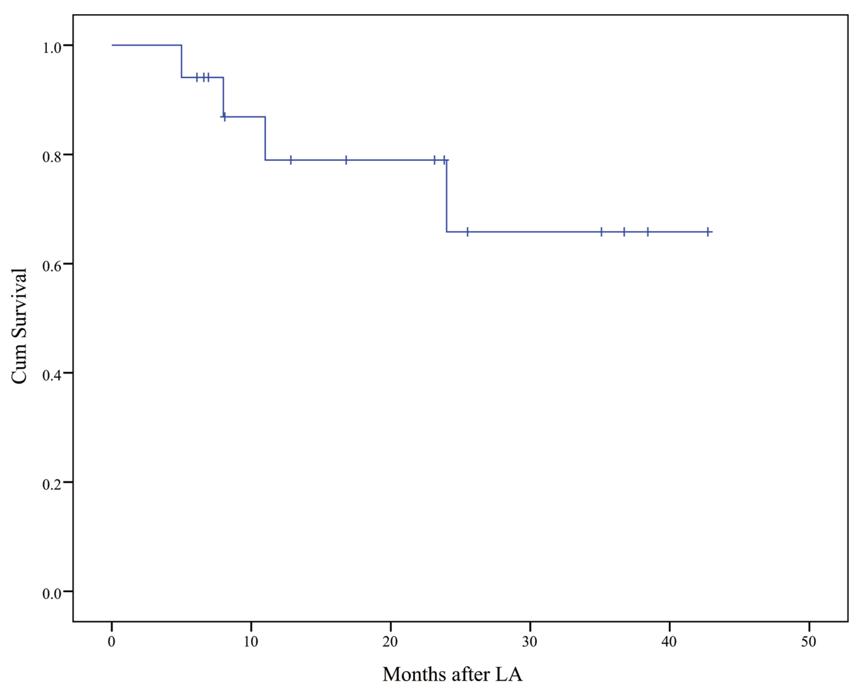
Lesion-related and procedure-related factors are compared between CA group (n = 20) and ICA group (n = 8; Table 3). The diameter and volume of the lesion are associated with ICA (p < 0.05).

3.4. Follow-Up

The mean follow-up period was 19.5 ± 12.8 months (range, 5–42.7 months). During the follow-up, LTP was detected at 3, 4, 8, and 11 months from four tumors with CA after LA. Locations of the four tumors were abdominal wall (n = 2), retroperitoneal space adjacent to the celiac trunk (n = 1) and intraabdominal seeding (n = 1). The LTP rate was 20% (4 of 20). One patient with LTP lesion on the abdominal wall was treated with percutaneous ethanol injection and subsequent surgical resection. The other three patients continued with systemic therapy with Sorafenib. Four patients died of tumor progression or multiple organ dysfunction syndrome, and 13 were alive at their last visits. The accumulative one- and three-year OS rates were 79.0% and 65.8%, respectively, after LA for extrahepatic metastases of HCC (Figure 2).

4. Discussion

This study summarizes the percutaneous US-guided LA in the treatment of extrahepatic metastasis of HCC and the local efficacy and safety of such therapeutic methods are evaluated. Previous studies reported that LA in the treatment of metastatic malignancy was applied to the sites of retroperitoneum, portal vein, and lymph nodes. However, most of them were in the form of a case report [25,26,27,32]. To our best knowledge, this is the first study to describe the application of LA in the treatment of extrahepatic metastasis of HCC at various sites, as well as evaluated the local efficacy and safety of LA.
In the present study, 20 out of 28 (71.4%) extrahepatic metastatic lesions of HCC achieved complete ablation by LA and none of the patients experienced major complications after the LA procedure. These results demonstrated that LA could provide a safe therapeutic option with a relatively high success rate in the treatment of critical lesions at high-risk locations where other thermal ablation techniques are considered to be aggressive. For intrahepatic HCC, LA could deliver a complete ablation rate at 82.0–97.5% [33,34,35]. It was reported that the application of RFA in the treatment of metastatic malignancy at various sites had a local tumor control rate at 78.9–95.8% [16,17,36,37]. However, due to a small sample size of LA, it is not appropriate to compare the results of LA in this study with other studies of thermal ablations because there are great differences in the approaches of treatment, number of patients, origin of tumors, sites of metastasis, tumor size, and progression of disease, etc.
This study has discovered that the size and volume of the target lesion are the main factors associated with the local efficacy of LA. Although treatment strategies such as arrangement and layout of multiple optic fibers, pullback techniques, and a combination of ethanol injection are currently in place, however, the relatively small ablative zone created by a single optic fiber acts the main limitation of LA in treating larger lesions [38]. A previous study demonstrated that tumor size affected the complete tumor ablation of LA [39]. In fact, tumor size is also a significant factor affecting the local efficacy of ablation in other thermal ablation techniques including RFA and MWA [40,41,42]. Combined RFA and ethanol injection could increase the range of coagulative necrosis and enlarge the ablation volume [43]. Compared with RFA alone, the procedure of RFA combined with ethanol has been proven to facilitate better local tumor control and long-term survival in the treatment of HCC [44,45,46]. A previous animal study has demonstrated that LA combined with ethanol can coagulate significantly larger volumes of tumors and reduce residual tumors [47]. In the current study, five lesions were treated by LA combined with ethanol. The mean diameter of the lesions was 3.8 ± 1.0 cm and the mean volume of injected ethanol was 8.8 ± 3.1 mL. Two lesions were at the hepatic hilum and close to the extrahepatic bile duct and the other three in the retroperitoneum adjacent to the aorta. Potential thermal injury to the bile duct and incomplete ablation resulting from the heat sink effect were the major concerns. Although ethanol was introduced, ICA was identified in three lesions after LA, including one lymph node in the hepatic hilum and two retroperitoneal lymph nodes. The low success rate of LA combined with ethanol could associate with difficult locations. The arrangement of the optic fiber and the angle of ethanol injection was also limited by the complicated surroundings around the target lesions, thus resulting in a low success rate.
The LTP rate in this study was 20%, which was comparable with the previous studies in thermal ablation for both primary HCC and extrahepatic metastasis of HCC [17,37,48,49]. The presence of LTP is related to several factors, including the margin of ablation, and size, location, and gross type of tumors [50,51,52,53]. This study identified LTP in four lesions, located at the abdominal wall, retroperitoneal space adjacent to the celiac trunk, and intraabdominal cavity. None of the lesions was treated with a combined ethanol injection. Although CA was achieved in these lesions, the inadequate safety margin of ablation caused by the difficult locations would contribute to the LTP.
Eleven patients experienced recurrences beyond the ablative sites and four patients died during follow-up. These findings were not presented in detail because the evidence was not strong by a small and heterogenous dataset. The main goal of this study was to prove the concept that LA is safe and effective as a treatment option for extrahepatic metastasis. Further studies are necessary to evaluate the long-term benefit of LA.
This study has a number of limitations. Firstly, it was a retrospective study with a small sample size, with different primary treatments for HCC in all patients. Some patients suffered intrahepatic recurrence simultaneously when extrahepatic metastasis was identified. All above might lead to bias in clinical outcomes. Secondly, the pathological examination was not obtained before LA in this study, but the preoperative clinical diagnosis was based on typical imaging for all the patients. Finally, there was no matched control group of patients treated by surgery or other thermal ablation techniques for comparison, of which the clinical value of LA as a treatment option for extrahepatic metastasis of HCC should be further investigated.

5. Conclusions

In conclusion, LA as an alternative option is safe and effective in the treatment of extrahepatic metastasis of HCC. Further studies are necessary to evaluate the benefit of LA.

Author Contributions

Conceptualization, B.L. and H.L.; formal analysis, H.L. and B.Z.; methodology, H.L., B.Z., G.H., and M.L.; project administration, B.L.; software, H.L., B.Z., J.L., and X.L.; supervision, X.X. and B.L.; validation, H.L. and B.Z.; writing—original draft, H.L., B.Z., G.H., X.L., M.L., J.L., and B.L.; writing—review and editing, B.Z, G.H., X.L., M.L., J.L., X.X., and B.L. All authors have read and agreed to the published version of the manuscript.

Funding

This research was funded by the Training Project for Young Teacher of Sun Yat-sen University (Grant No. 18ykpy10).

Conflicts of Interest

The authors declare no conflict of interest.

References

  1. Ferlay, J.; Soerjomataram, I.; Dikshit, R.; Eser, S.; Mathers, C.; Rebelo, M.; Parkin, D.M.; Forman, D.; Bray, F. Cancer incidence and mortality worldwide: Sources, methods and major patterns in GLOBOCAN 2012. Int. J. Cancer 2015, 136, E359–E386. [Google Scholar] [CrossRef] [PubMed]
  2. Erratum: Global cancer statistics 2018: GLOBOCAN estimates of incidence and mortality worldwide for 36 cancers in 185 countries. CA Cancer J. Clin. 2020, 70, 313. [CrossRef] [PubMed] [Green Version]
  3. Forner, A.; Reig, M.; Bruix, J. Hepatocellular carcinoma. Lancet 2018, 391, 1301–1314. [Google Scholar] [CrossRef]
  4. Hasegawa, K.; Kokudo, N.; Makuuchi, M.; Izumi, N.; Ichida, T.; Kudo, M.; Ku, Y.; Sakamoto, M.; Nakashima, O.; Matsui, O.; et al. Comparison of resection and ablation for hepatocellular carcinoma: A cohort study based on a Japanese nationwide survey. J. Hepatol. 2013, 58, 724–729. [Google Scholar] [CrossRef]
  5. Peng, Z.; Chen, S.; Wei, M.; Lin, M.; Jiang, C.; Mei, J.; Li, B.; Wang, Y.; Li, J.; Xie, X.; et al. Advanced Recurrent Hepatocellular Carcinoma: Treatment with Sorafenib Alone or in Combination with Transarterial Chemoembolization and Radiofrequency Ablation. Radiology 2018, 287, 705–714. [Google Scholar] [CrossRef] [Green Version]
  6. Faivre, S.; Rimassa, L.; Finn, R.S. Molecular therapies for HCC: Looking outside the box. J. Hepatol. 2020, 72, 342–352. [Google Scholar] [CrossRef] [Green Version]
  7. Kudo, M.; Finn, R.S.; Qin, S.; Han, K.H.; Ikeda, K.; Piscaglia, F.; Baron, A.; Park, J.W.; Han, G.; Jassem, J.; et al. Lenvatinib versus sorafenib in first-line treatment of patients with unresectable hepatocellular carcinoma: A randomised phase 3 non-inferiority trial. Lancet 2018, 391, 1163–1173. [Google Scholar] [CrossRef] [Green Version]
  8. Cheng, A.L.; Hsu, C.; Chan, S.L.; Choo, S.P.; Kudo, M. Challenges of combination therapy with immune checkpoint inhibitors for hepatocellular carcinoma. J. Hepatol. 2020, 72, 307–319. [Google Scholar] [CrossRef] [Green Version]
  9. Zongyi, Y.; Xiaowu, L. Immunotherapy for hepatocellular carcinoma. Cancer Lett. 2020, 470, 8–17. [Google Scholar] [CrossRef]
  10. Bouattour, M.; Mehta, N.; He, A.R.; Cohen, E.I.; Nault, J.C. Systemic treatment for advanced hepatocellular carcinoma. Liver Cancer 2019, 8, 341–358. [Google Scholar] [CrossRef]
  11. Choi, G.H.; Shim, J.H.; Kim, M.J.; Ryu, M.H.; Ryoo, B.Y.; Kang, Y.K.; Shin, Y.M.; Kim, K.M.; Lim, Y.S.; Lee, H.C. Sorafenib alone versus sorafenib combined with transarterial chemoembolization for advanced-stage hepatocellular carcinoma: Results of propensity score analyses. Radiology 2013, 269, 603–611. [Google Scholar] [CrossRef] [PubMed]
  12. Yuan, H.; Lan, Y.; Li, X.; Tang, J.; Liu, F. Large hepatocellular carcinoma with local remnants after transarterial chemoembolization: Treatment by sorafenib combined with radiofrequency ablation or sorafenib alone. Am. J. Cancer Res. 2019, 9, 791–799. [Google Scholar] [PubMed]
  13. Carrafiello, G.; Laganà, D.; Ianniello, A.; Nicotera, P.; Fontana, F.; Dizonno, M.; Cuffari, S.; Fugazzola, C. Radiofrequency thermal ablation for pain control in patients with single painful bone metastasis from hepatocellular carcinoma. Eur. J. Radiol. 2009, 71, 363–368. [Google Scholar] [CrossRef]
  14. Kashima, M.; Yamakado, K.; Takaki, H.; Kaminou, T.; Tanigawa, N.; Nakatsuka, A.; Takeda, K. Radiofrequency ablation for the treatment of bone metastases from hepatocellular carcinoma. AJR Am. J. Roentgenol. 2010, 194, 536–541. [Google Scholar] [CrossRef]
  15. Doreille, A.; N’Kontchou, G.; Halimi, A.; Bouhafs, F.; Coderc, E.; Sellier, N.; Seror, O. Percutaneous treatment of extrahepatic recurrence of hepatocellular carcinoma. Diagn. Interv. Imaging 2016, 97, 1117–1123. [Google Scholar] [CrossRef] [PubMed]
  16. Mu, L.; Sun, L.; Pan, T.; Lyu, N.; Li, S.; Li, X.; Wang, J.; Xie, Q.; Deng, H.; Zheng, L.; et al. Percutaneous CT-guided radiofrequency ablation for patients with extrahepatic oligometastases of hepatocellular carcinoma: Long-term results. Int. J. Hyperth. 2018, 34, 59–67. [Google Scholar] [CrossRef] [PubMed]
  17. Huang, J.; Xie, X.; Lin, J.; Wang, W.; Zhang, X.; Liu, M.; Li, X.; Huang, G.; Liu, B.; Xie, X. Percutaneous radiofrequency ablation of adrenal metastases from hepatocellular carcinoma: A single-center experience. Cancer Imaging 2019, 19, 44. [Google Scholar] [CrossRef] [Green Version]
  18. Pacella, C.M.; Bizzarri, G.; Magnolfi, F.; Cecconi, P.; Caspani, B.; Anelli, V.; Bianchini, A.; Valle, D.; Pacella, S.; Manenti, G.; et al. Laser thermal ablation in the treatment of small hepatocellular carcinoma: Results in 74 patients. Radiology 2001, 221, 712–720. [Google Scholar] [CrossRef] [Green Version]
  19. Ritz, J.P.; Lehmann, K.S.; Mols, A.; Frericks, B.; Knappe, V.; Buhr, H.J.; Holmer, C. Laser-induced thermotherapy for lung tissue—Evaluation of two different internally cooled application systems for clinical use. Lasers Med. Sci. 2008, 23, 195–202. [Google Scholar] [CrossRef]
  20. Caspani, B.; Ierardi, A.M.; Motta, F.; Cecconi, P.; Fesce, E.; Belli, L. Small nodular hepatocellular carcinoma treated by laser thermal ablation in high risk locations: Preliminary results. Eur. Radiol. 2010, 20, 2286–2292. [Google Scholar] [CrossRef]
  21. Orlacchio, A.; Bolacchi, F.; Chegai, F.; Bergamini, A.; Costanzo, E.; Del Giudice, C.; Angelico, M.; Simonetti, G. Comparative evaluation of percutaneous laser and radiofrequency ablation in patients with HCC smaller than 4 cm. Radiol. Med. 2014, 119, 298–308. [Google Scholar] [CrossRef] [PubMed]
  22. Teratani, T.; Yoshida, H.; Shiina, S.; Obi, S.; Sato, S.; Tateishi, R.; Mine, N.; Kondo, Y.; Kawabe, T.; Omata, M. Radiofrequency ablation for hepatocellular carcinoma in so-called high-risk locations. Hepatology 2006, 43, 1101–1108. [Google Scholar] [CrossRef] [PubMed]
  23. Francica, G.; Petrolati, A.; Di Stasio, E.; Pacella, S.; Stasi, R.; Pacella, C.M. Effectiveness, safety, and local progression after percutaneous laser ablation for hepatocellular carcinoma nodules up to 4 cm are not affected by tumor location. AJR Am. J. Roentgenol. 2012, 199, 1393–1401. [Google Scholar] [CrossRef]
  24. Di Costanzo, G.G.; D’Adamo, G.; Tortora, R.; Zanfardino, F.; Mattera, S.; Francica, G.; Pacella, C.M. A novel needle guide system to perform percutaneous laser ablation of liver tumors using the multifiber technique. Acta Radiol. 2013, 54, 876–881. [Google Scholar] [CrossRef] [PubMed]
  25. Mou, Y.; Zhao, Q.; Zhong, L.; Chen, F.; Jiang, T. Preliminary results of ultrasound-guided laser ablation for unresectable metastases to retroperitoneal and hepatic portal lymph nodes. World J. Surg. Oncol. 2016, 14, 165. [Google Scholar] [CrossRef] [PubMed] [Green Version]
  26. Jiang, T.A.; Deng, Z.; Tian, G.; Chen, F.; Bao, H.; Li, J.; Wang, W. Percutaneous laser ablation: A new contribution to unresectable high-risk metastatic retroperitoneal lesions? Oncotarget 2017, 8, 2413–2422. [Google Scholar] [CrossRef]
  27. Lu, Z.-H.; Shen, F.; Yan, Z.-L.; Li, J.; Yang, J.-H.; Zong, M.; Shi, L.-H.; Wu, M.-C. Treatment of portal vein tumor thrombus of hepatocellular carcinoma with percutaneous laser ablation. J. Cancer Res. Clin. Oncol. 2009, 135, 783–789. [Google Scholar] [CrossRef]
  28. Johnson, P.J.; Berhane, S.; Kagebayashi, C.; Satomura, S.; Teng, M.; Reeves, H.L.; O’Beirne, J.; Fox, R.; Skowronska, A.; Palmer, D.; et al. Assessment of liver function in patients with hepatocellular carcinoma: A new evidence-based approach-the ALBI grade. J. Clin. Oncol. 2015, 33, 550–558. [Google Scholar] [CrossRef]
  29. Lam, V.W.-T.; Ng, K.K.; Chok, K.S.-H.; Cheung, T.-T.; Yuen, J.; Tung, H.; Tso, W.-K.; Fan, S.-T.; Poon, R.T.P. Incomplete ablation after radiofrequency ablation of hepatocellular carcinoma: Analysis of risk factors and prognostic factors. Ann. Surg. Oncol. 2008, 15, 782–790. [Google Scholar] [CrossRef]
  30. Dindo, D.; Demartines, N.; Clavien, P.-A. Classification of surgical complications: A new proposal with evaluation in a cohort of 6336 patients and results of a survey. Ann. Surg. 2004, 240, 205–213. [Google Scholar] [CrossRef]
  31. Omary, R.A.; Bettmann, M.A.; Cardella, J.F.; Bakal, C.W.; Schwartzberg, M.S.; Sacks, D.; Rholl, K.S.; Meranze, S.G.; Lewis, C.A. Quality improvement guidelines for the reporting and archiving of interventional radiology procedures. J. Vasc. Interv. Radiol. 2002, 13, 879–881. [Google Scholar] [CrossRef]
  32. Tian, G.; Jiang, T.A. US-guided percutaneous laser ablation of refractory metastatic retroperitoneal lesions: A care-compliant case report. Medicine (Baltimore) 2017, 96, e6597. [Google Scholar] [CrossRef] [PubMed]
  33. Giorgio, A.; Tarantino, L.; de Stefano, G.; Farella, N.; Catalano, O.; Cusati, B.; Del Viscovo, L.; Alaia, A.; Caturelli, E. Interstitial laser photocoagulation under ultrasound guidance of liver tumors: Results in 104 treated patients. Eur. J. Ultrasound 2000, 11, 181–188. [Google Scholar] [CrossRef]
  34. Eichler, K.; Mack, M.G.; Straub, R.; Engelmann, K.; Zangos, S.; Woitaschek, D.; Vogl, T.J. Oligonodular hepatocellular carcinoma (HCC): MR-controlled laser-induced thermotherapy. Radiologe 2001, 41, 915–922. [Google Scholar] [CrossRef]
  35. Pacella, C.M.; Bizzarri, G.; Francica, G.; Bianchini, A.; De Nuntis, S.; Pacella, S.; Crescenzi, A.; Taccogna, S.; Forlini, G.; Rossi, Z.; et al. Percutaneous laser ablation in the treatment of hepatocellular carcinoma with small tumors: Analysis of factors affecting the achievement of tumor necrosis. J. Vasc. Interv. Radiol. 2005, 16, 1447–1457. [Google Scholar] [CrossRef]
  36. Zhou, K.; Pan, J.; Yang, N.; Shi, H.-F.; Cao, J.; Li, Y.-M.; Zhang, H.-Z.; Wang, K.-F.; Chen, S.-H. Effectiveness and safety of CT-guided percutaneous radiofrequency ablation of adrenal metastases. Br. J. Radiol. 2018, 91, 20170607. [Google Scholar] [CrossRef]
  37. Hasegawa, T.; Yamakado, K.; Nakatsuka, A.; Uraki, J.; Yamanaka, T.; Fujimori, M.; Miki, M.; Sasaki, T.; Sakuma, H.; Sugimura, Y. Unresectable Adrenal Metastases: Clinical Outcomes of Radiofrequency Ablation. Radiology 2015, 277, 584–593. [Google Scholar] [CrossRef]
  38. Zhang, Y.-R.; Fang, L.-Y.; Yu, C.; Sun, Z.-X.; Huang, Y.; Chen, J.; Guo, T.; Xiang, F.-X.; Wang, J.; Lu, C.-F.; et al. Laser-induced interstitial thermotherapy via a single-needle delivery system: Optimal conditions of ablation, pathological and ultrasonic changes. J. Huazhong Univ. Sci. Technol. Med. Sci. 2015, 35, 579–584. [Google Scholar] [CrossRef]
  39. Head, H.W.; Dodd, G.D. Thermal ablation for hepatocellular carcinoma. Gastroenterology 2004, 127, S167–S178. [Google Scholar] [CrossRef]
  40. Wang, T.; Lu, X.J.; Chi, J.C.; Ding, M.; Zhang, Y.; Tang, X.Y.; Li, P.; Zhang, L.; Zhang, X.Y.; Zhai, B. Microwave ablation of hepatocellular carcinoma as first-line treatment: Long term outcomes and prognostic factors in 221 patients. Sci. Rep. 2016, 6, 32728. [Google Scholar] [CrossRef]
  41. Liang, H.Y.; Guo, Q.Y.; Sun, W.; Mao, X.N.; Wen, F.; Shan, M.; Zhao, G.; Wang, X.H.; Lu, Z.M. Sequential Use of Transhepatic Arterial Chemoembolization and Bipolar Radiofrequency Ablation in the Clinical Therapy of Hepatocellular Carcinoma. Cancer Biother. Radiopharm. 2015, 30, 427–432. [Google Scholar] [CrossRef]
  42. Ayav, A.; Germain, A.; Marchal, F.; Tierris, I.; Laurent, V.; Bazin, C.; Yuan, Y.; Robert, L.; Brunaud, L.; Bresler, L. Radiofrequency ablation of unresectable liver tumors: Factors associated with incomplete ablation or local recurrence. Am. J. Surg. 2010, 200, 435–439. [Google Scholar] [CrossRef]
  43. Lee, J.M.; Lee, Y.H.; Kim, Y.K.; Kim, S.W.; Kim, C.S. Combined therapy of radiofrequency ablation and ethanol injection of rabbit liver: An in vivo feasibility study. Cardiovasc. Intervent. Radiol. 2004, 27, 151–157. [Google Scholar] [CrossRef] [PubMed]
  44. Zhang, Y.-J.; Liang, H.-H.; Chen, M.-S.; Guo, R.-P.; Li, J.-Q.; Zheng, Y.; Zhang, Y.-Q.; Lau, W.Y. Hepatocellular carcinoma treated with radiofrequency ablation with or without ethanol injection: A prospective randomized trial. Radiology 2007, 244, 599–607. [Google Scholar] [CrossRef] [PubMed]
  45. Zhu, Z.X.; Liao, M.H.; Wang, X.X.; Huang, J.W. Radiofrequency ablation with or without ethanol injection for hepatocellular carcinoma: A systematic review and meta-analysis. Minerva Med. 2016, 107, 381–391. [Google Scholar] [PubMed]
  46. Li, Z.; Zhang, K.; Lin, S.-M.; Mi, D.-H.; Cao, N.; Wen, Z.-Z.; Li, Z.-X. Radiofrequency ablation combined with percutaneous ethanol injection for hepatocellular carcinoma: A systematic review and meta-analysis. Int. J. Hyperth. 2017, 33, 237–246. [Google Scholar] [CrossRef]
  47. Zou, X.; Liu, Q.; Zhou, X.; He, G.; Yu, M.; Han, Z.; Meng, X.; Su, H. Ultrasound-guided percutaneous laser and ethanol ablation of rabbit VX2 liver tumors. Acta Radiol. 2013, 54, 181–187. [Google Scholar] [CrossRef]
  48. Hao, Y.; Numata, K.; Ishii, T.; Fukuda, H.; Maeda, S.; Nakano, M.; Tanaka, K. Rate of local tumor progression following radiofrequency ablation of pathologically early hepatocellular carcinoma. World J. Gastroenterol. 2017, 23, 3111–3121. [Google Scholar] [CrossRef]
  49. Lee, M.W.; Kang, D.; Lim, H.K.; Cho, J.; Sinn, D.H.; Kang, T.W.; Song, K.D.; Rhim, H.; Cha, D.I.; Lu, D.S.K. Updated 10-year outcomes of percutaneous radiofrequency ablation as first-line therapy for single hepatocellular carcinoma < 3 cm: Emphasis on association of local tumor progression and overall survival. Eur. Radiol. 2020, 30, 2391–2400. [Google Scholar] [CrossRef]
  50. Kim, Y.-S.; Lee, W.J.; Rhim, H.; Lim, H.K.; Choi, D.; Lee, J.Y. The minimal ablative margin of radiofrequency ablation of hepatocellular carcinoma (> 2 and < 5 cm) needed to prevent local tumor progression: 3D quantitative assessment using CT image fusion. AJR Am. J. Roentgenol. 2010, 195, 758–765. [Google Scholar] [CrossRef]
  51. Yoon, J.H.; Lee, J.M.; Klotz, E.; Woo, H.; Yu, M.H.; Joo, I.; Lee, E.S.; Han, J.K. Prediction of Local Tumor Progression after Radiofrequency Ablation (RFA) of Hepatocellular Carcinoma by Assessment of Ablative Margin Using Pre-RFA MRI and Post-RFA CT Registration. Korean J. Radiol. 2018, 19, 1053–1065. [Google Scholar] [CrossRef] [PubMed]
  52. Kono, M.; Inoue, T.; Kudo, M.; Chishina, H.; Arizumi, T.; Takita, M.; Kitai, S.; Yada, N.; Hagiwara, S.; Minami, Y.; et al. Radiofrequency ablation for hepatocellular carcinoma measuring 2 cm or smaller: Results and risk factors for local recurrence. Dig. Dis. 2014, 32, 670–677. [Google Scholar] [CrossRef] [PubMed]
  53. Lee, D.H.; Lee, J.M.; Lee, J.Y.; Kim, S.H.; Yoon, J.H.; Kim, Y.J.; Han, J.K.; Choi, B.I. Radiofrequency ablation of hepatocellular carcinoma as first-line treatment: Long-term results and prognostic factors in 162 patients with cirrhosis. Radiology 2014, 270, 900–909. [Google Scholar] [CrossRef] [PubMed] [Green Version]
Figure 1. A 65-year old male had hepatocellular carcinoma (HCC) and was diagnosed with extrahepatic metastases to abdominal lymph nodes. US-guided laser ablation (LA) was performed. (a,b) A hypoechoic lymph node inferior to the right diaphragm (a) and right paracolon (b) were detected by the grayscale ultrasound. (ce) On the contrast-enhanced ultrasound (CEUS) images, the targeted lymph node displayed heterogeneous hyperenhancement in the early phase (c) and washout in the late phase (d,e). (f,g) With the guidance of real-time US, optic fibers were inserted into the target lesions precisely and the tips of the fibers clearly seen. (h) Hyperechoic zone around the fiber tip appeared and the range extended as the procedure proceeded. (i,j) One month later, the target lymph nodes inferior to the right diaphragm (i) and right paracolon (j) showed no enhancement on the CEUS images, indicating a complete ablation. (k,l) One month later, target lymph node inferior to the right diaphragm exhibited no enhancement in the arterial phase (k) and in the portal-venous phase (l) on the contrast-enhanced computed tomography (CECT).
Coatings 10 00951 g001aCoatings 10 00951 g001b
Figure 2. Overall survival after laser ablation for extrahepatic metastases. LA = laser ablation.
Coatings 10 00951 g002
Table 1. Baseline characteristics of the patients.
Characteristicsn = 17
Age (year)54.1 ± 14.6
  Gender
Male13
Female4
Etiology
HBV 112
HCV 22
others3
Initial treatment of liver tumor
Surgical resection13
Ablation4
Intent of LA3
Curative12
Palliative5
Child–Pugh classification
A17
B0
Child–Pugh score
516
61
ALBI grade4
111
26
Biochemical tests
AFP (μg/L) 54.5 (1.7–360.1)
ALB (g/L) 640.3 ± 5.5
TBIL (μmol/L) 713.0 ± 4.7
PLT account (×109/L) 8180 ± 56
Tumor number
Solitary10
Multiple7
Size (cm)2.2 ± 1.1
Volume (cm3)7.2 (5.6–19.5)
1 HBV = hepatitis B virus; 2 HCV = hepatitis C virus; 3 LA = laser ablation; 4 ALBI grade = albumin–bilirubin grade; 5 AFP = alpha-fetoprotein; 6 ALB = albumin; 7 TBIL = total bilirubin; 8 PLT account = platelet account. Continuous variables are expressed in mean ± standard deviation, or in median (P25–P75).
Table 2. Laser ablation strategies and outcome.
Characteristicsn = 28
Tumor size (cm)2.2 ± 1.1
Tumor volume (cm3)7.2 (5.6–19.5)
Location
Abdominal wall3
Abdominal lymph nodes4
Intraabdominal seeding1
Hepatic surface4
Adrenal gland1
Diaphragm1
Hilar lymph nodes3
Retroperitoneal lymph nodes7
Subcutaneous chest wall4
Number of fibers3 (1–4)
Power (J)3600 (640–5813)
Combination with ethanol injection
Yes5
No23
Efficacy
CA 120
ICA 28
1 CA = complete ablation; 2 ICA = incomplete complete Aablation. Continuous variables are expressed in mean ± standard deviation, or in median (P25–P75).
Table 3. Factors associated with ICA.
CharacteristicsCA 1 Lesion (n = 20)ICA 2 Lesion (n = 8)p-Value
Diameter of lesion (cm)1.84 ± 0.853.04 ± 1.100.005
Volume of lesion (cm3)6.6 (4.5–12.3)23.8 (9.5–71.8)0.008
Combination with ethanol injection
Yes230.123
No185
Number of fibers2 (1–3)4 (2.5–4)0.119
Energy (J)3600 (2760–4750)5475 (2950–6500)0.281
1 CA = complete ablation; 2 ICA = incomplete complete ablation. Continuous variables were expressed in mean ± standard deviation, or in median (P25–P75).

