 Eric Capo1,2*
 Andrea G. Bravo3
 Anne L. Soerensen4
 Stefan Bertilsson2
 Jarone Pinhassi5
 Caiyan Feng1
 Anders F. Andersson6
 Moritz Buck2†
 Erik Björn1†
- 1Department of Chemistry, Umeå University, Umeå, Sweden
- 2Department of Aquatic Sciences and Assessment, Swedish University of Agricultural Sciences, Uppsala, Sweden
- 3Institut de Ciències del Mar (ICM-CSIC), Barcelona, Spain
- 4Department of Environmental Research and Monitoring, Swedish Museum of Natural History, Stockholm, Sweden
- 5Centre for Ecology and Evolution in Microbial Model Systems – EEMiS, Linnaeus University, Kalmar, Sweden
- 6Department of Gene Technology, Science for Life Laboratory, KTH Royal Institute of Technology, Solna, Sweden
Methylmercury (MeHg), a neurotoxic compound biomagnifying in aquatic food webs, can be a threat to human health via fish consumption. However, the composition and distribution of the microbial communities mediating the methylation of mercury (Hg) to MeHg in marine systems remain largely unknown. In order to fill this knowledge gap, we used the Baltic Sea Reference Metagenome (BARM) dataset to study the abundance and distribution of the genes involved in Hg methylation (the hgcAB gene cluster). We determined the relative abundance of the hgcAB genes and their taxonomic identity in 81 brackish metagenomes that cover spatial, seasonal and redox variability in the Baltic Sea water column. The hgcAB genes were predominantly detected in anoxic water, but some hgcAB genes were also detected in hypoxic and normoxic waters. Phylogenetic analysis identified putative Hg methylators within Deltaproteobacteria, in oxygen-deficient water layers, but also Spirochaetes-like and Kiritimatiellaeota-like bacteria. Higher relative quantities of hgcAB genes were found in metagenomes from marine particles compared to free-living communities in anoxic water, suggesting that such particles are hotspot habitats for Hg methylators in oxygen-depleted seawater. Altogether, our work unveils the diversity of the microorganisms with the potential to mediate MeHg production in the Baltic Sea and pinpoint the important ecological niches for these microorganisms within the marine water column.
Introduction
Methylmercury (MeHg) is a neurotoxic compound that accumulates in aquatic food webs and may be a threat to human health related to fish consumption (Mason et al., 2012). Methylation of inorganic mercury (Hg) to MeHg is predominantly a biological process driven by anaerobic bacteria and archaea carrying the hgcA and hgcB genes (Parks et al., 2013) and takes place in various oxygen-deficient environments (e.g., sediment, water, soil). Hg methylation appears to be controlled by the activity of Hg-methylating microbes, the composition and activity of microbial communities (that indirectly modulate Hg methylation), and Hg bioavailability (Bravo and Cosio, 2019). It is broadly established that the capacity for Hg-methylation is limited to specific microbial lineages, with the most commonly reported groups found in the Deltaproteobacteria and Methanomicrobiales (Gilmour et al., 2013; Podar et al., 2015; Bravo et al., 2018; Yu et al., 2018). However, recent work has expanded this view and unraveled a higher phylogenetic diversity of microbes carrying the hgcAB genes than previously expected (McDaniel et al., 2020) calling for further analyses of microbial Hg-methylation in aquatic environments.
Recent advances in metagenomics have yielded new insights into the microbial taxonomic and functional diversity in various aquatic ecosystems (e.g., Mehrshad et al., 2016; Haro-Moreno et al., 2018; Nowinski et al., 2019). The approach has for example been applied to broadly assess the presence and diversity of genes central to biological Hg cycling in marine systems (Podar et al., 2015; Gionfriddo et al., 2016, 2020; Bowman et al., 2019; Lin et al., 2020; Tada et al., 2020; Villar et al., 2020). Podar et al. (2015) only detected hgcAB genes in a few metagenomes from marine pelagic waters (seven out of 138 metagenomes) but highlighted that limited sequencing depths of these metagenomes could have hampered detection. While a more recent study did not detect hgcA genes in waters from the Arctic and equatorial Pacific Oceans (Bowman et al., 2019), the presence of hgcAB-like genes was reported in normoxic water from the open ocean in several ocean basins (Villar et al., 2020), the mesopelagic zone of the East China Sea (Tada et al., 2020) and Antarctica sea ice (Gionfriddo et al., 2016), with a fraction of those genes being associated to a microaerophilic nitrite oxidizing bacteria. Further, Blum et al. (2013) demonstrated that between 20 and 40 % of MeHg measured in surface mixed layer of the North Pacific Ocean originated from internal production in the surface water. Finally, recent work from Gorokhova et al. (2020) reported the presence of hgcA genes from Deltaproteobacteria and Firmicutes in the gut microbiome from copepods collected in normoxic Baltic Sea water, suggesting that endogenous Hg methylation in zooplankton may be one path of Hg transfer from lower to higher trophic levels in this aquatic system.
Oceans and coastal areas, such as the West Coast of South America, the Arabian Sea, The Gulf of Mexico and the Baltic Sea, have experienced increased decline in oxygen since at least the middle of the 20th century (Breitburg et al., 2018). This phenomenon can be caused by (i) warming that decreases the solubility of oxygen in the ocean and (ii) nutrient enrichment in coastal water causing an increase of algal biomass and subsequent decomposition of sinking organic matter by microbes consuming the oxygen (Breitburg et al., 2018). Such oxygen deficient waters potentially offer ecological niches suitable for Hg-methylating microorganisms. Overall there are still important knowledge gaps concerning the process of Hg methylation in aquatic systems, in particular regarding the influence of variable redox conditions. In addition, marine particles (organic-rich particulate matter and aggregates) is hypothesized to provide both substrates for heterotrophic microbes (Azam and Long, 2001; Azam and Malfatti, 2007) and various anaerobic microenvironments (Alldredge and Silver, 1988; Bianchi et al., 2018) that could potentially favor Hg methylation, via e.g., microbial sulfate-reduction. Based on this, several studies proposed (Monperrus et al., 2007; Cossa et al., 2009; Sunderland et al., 2009; Lehnherr et al., 2011; Schartup et al., 2015) or demonstrated (Ortiz et al., 2015; Gascón Díez et al., 2016). Hg methylation in marine particles. For the Baltic Sea it has been proposed that Hg methylation in normoxic water can be associated with phytoplankton blooms via production of increased levels of phytoplankton derived OM that is also manifested as particulate organic particles sinking through the water column (Soerensen et al., 2016) that may providing suitable anoxic niches for Hg methylators. However, as far as we know there are no studies on microbial communities in relation to this phenomenon in the Baltic Sea or elsewhere.
The Baltic Sea is an ecosystem that has experienced large increases in nutrient loads and oxygen consumptions over the last century, resulting in extensive coastal and offshore zones with permanent hypoxic and anoxic water below the oxygenated surface water (Conley et al., 2011; Carstensen et al., 2014). As such, the Baltic Sea represents a model for the expansion of coastal ecosystems influenced by anoxia. Elevated MeHg concentrations in the Baltic Sea have been observed in anoxic water (>1000 fM) compared to hypoxic and normoxic water (Kuss et al., 2018; Soerensen et al., 2018). Soerensen et al. (2018) demonstrated that this was caused by increased rates of Hg methylation in the oxygen deficient water zones. They hypothesized that this process is predominantly driven by microbial sulfate-reduction because of the relatively high concentrations in dissolved sulfide in the anoxic water (up to ∼60 μM S–II). Although the concentrations of MeHg in normoxic water were generally low (13–80 fM), concentrations were higher than what could be explained by MeHg input from external sources only, and the authors could infer in situ formation of MeHg at a low rates also in normoxic water zones in the Baltic Sea (Soerensen et al., 2016, 2018). The presence and distribution of microorganisms carrying the hgcAB genes, including taxa known to reduce sulfate, could unequivocally confirm the potential for in situ MeHg formation in the Baltic Sea.
In this study, we assessed the spatial and seasonal variability of hgcAB genes in the Baltic Sea, including water column profiles, allowing us to investigate for the first time the presence and variation of Hg-methylating microbes across redoxclines in the Baltic Sea. We show the presence and relative abundance of Hg methylators in both hypoxic and anoxic water masses of the Baltic Sea, while hgcAB genes were present in very low abundance or not at all detected in normoxic waters. In addition, we found a higher proportion of hgcAB reads in metagenomes obtained from 3 μm filters compared to 0.2 μm filters, suggesting particles as an important habitat for Hg methylators. Our work provides new information on Hg-methylating microorganisms in coastal seas impacted by oxygen deficiency.
Materials and Methods
The Baltic Sea Reference Metagenome (BARM) data (Alneberg et al., 2018) used in our study is composed of 81 metagenomes combined from three datasets spanning 13 locations (Figure 1) and are selected to cover natural variation in geography, depth and seasons of Baltic Sea waters. A summary on sampling and filtration of water samples for each of the datasets is provided in Table 1. We classified the water samples into three categories based on the measured O2 concentrations: (i) normoxic water with O2 concentrations exceeding or equal to 2 mL O2.L–1 (ii) hypoxic water with detectable O2 concentrations lower than 2 mL O2.L–1 and (iii) anoxic water with no detectable O2.
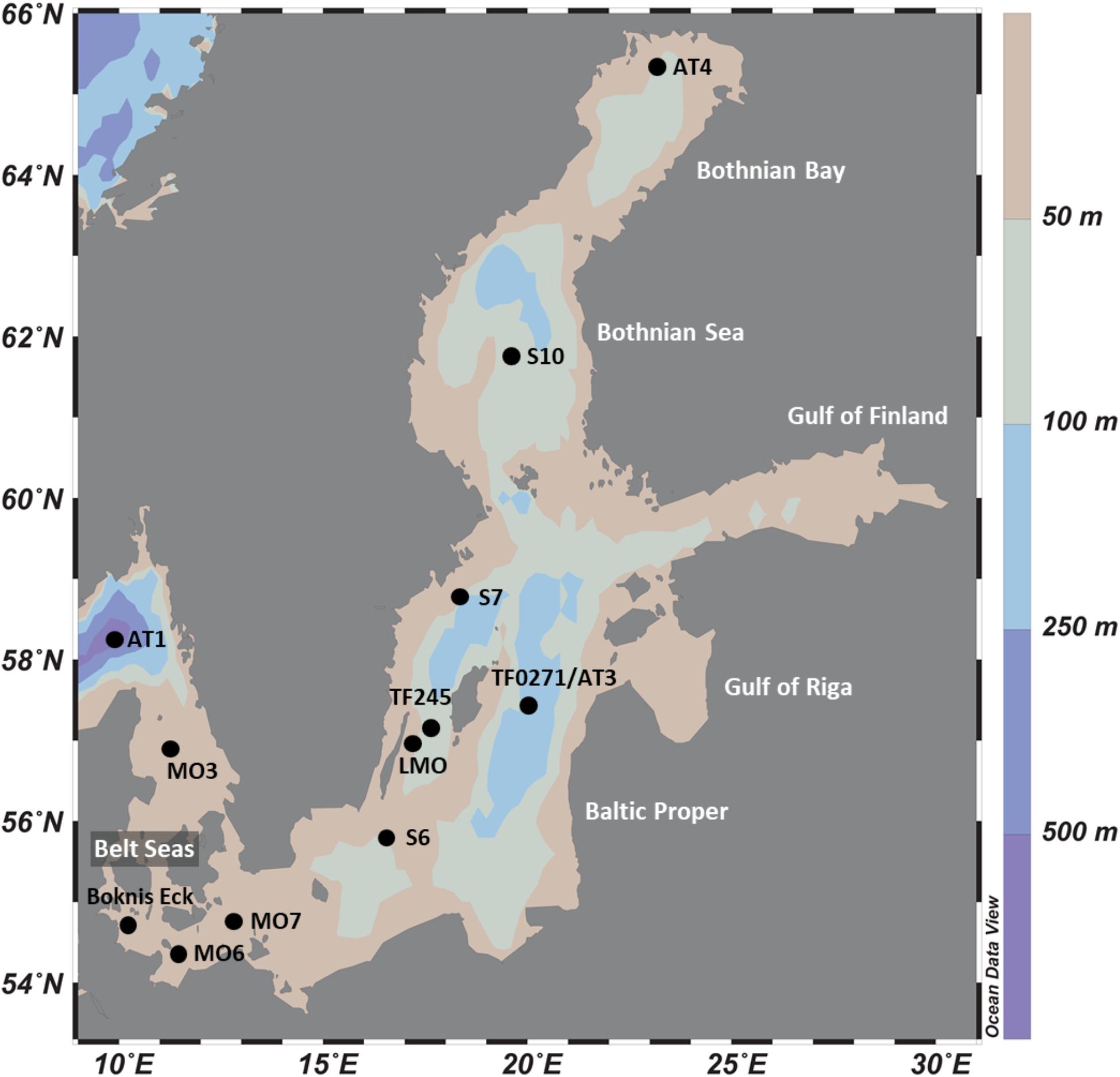
Figure 1. Location of study sites in the Baltic Sea.
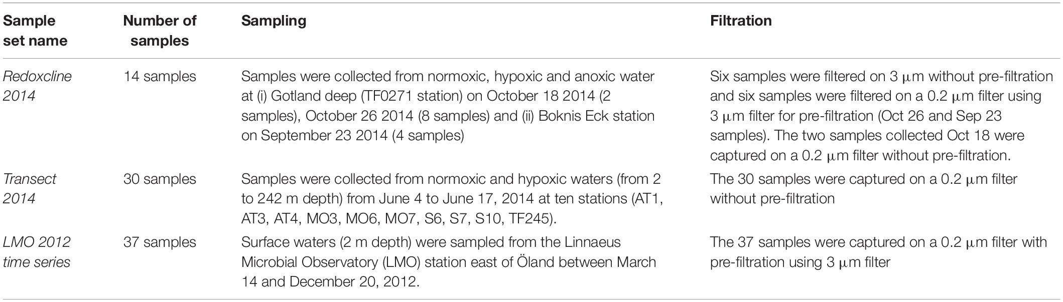
Table 1. Description of the number of metagenomes obtained with their respective sampling and filtration strategies for each of the three datasets.
Methods for DNA extractions, library preparations and sequencing as well as the initial processing of metagenomics data is described in greater details in Alneberg et al. (2018). Briefly, preprocessed reads were co-assembled using Megahit (version 1.0.2, Li et al., 2015). Functional and taxonomic annotation was performed for the 6.8 million genes found on the 2.4 million contigs > 1 kilobase. To determine the overall composition of bacterial and archaeal communities, we used the metagenomics data analyzed in Alneberg et al. (2018) and compiled in BalticMicrobeDB1.
In order to detect hgcAB homologs genes, we first analyzed the 6.8 million predicted genes with the hmmsearch program from the hmmer (3.2.1 version, Finn et al., 2011) with the HMM profiles of concatenated hgcAB amino acid sequences from Podar et al. (2015) as a reference database (Supplementary Datasheet S1). We considered genes with E-values ≤ 10–3 as significant hits resulting in a total of 3,215 genes. Only a fraction of these 3,215 genes correspond to hgcA and hgcB genes and we therefore performed a manual check of their amino acid sequences using the knowledge from the seminal paper of Parks et al. (2013) that described unique motifs in hgcA (NVWCA(A/G/S)GK) and hgcB amino acid sequences (C(M/I)EC(G/S)(A/G)C). To ensure the reliability of the outputs obtained using the HMM profiles of hgcAB amino acid sequences from Podar et al. (2015) to capture all hgcAB-like genes from the metagenomic dataset, we applied the same procedure using the HMM profiles provided by the ORNL hgcAB library (Gionfriddo et al., 2020). The two procedures resulted in the recovery of the same hgcA genes and hgcAB gene clusters from the BARM dataset.
For phylogenetic analysis, we used the amino acid sequences of the 650 hgcAB gene clusters identified as putative Hg methylators by McDaniel et al. (2020). These 650 hgcAB gene clusters were obtained from the analysis of publicly available isolate genomes and metagenome-assembled genomes (Supplementary Datasheet S2, McDaniel et al., 2020). For each phylogenetic analysis, amino acid sequences were aligned using MUSCLE (cluster method UPGMA) in the software MEGAX (Kumar et al., 2018) and approximate maximum likelihood (ML) trees were constructed using FastTree (Price et al., 2009) with JTT+CAT model of sequence evolution and 1,000 ultrafast bootstrap replicates. The trees were visualized using iTOL (Letunic and Bork, 2019) and clades were collapsed by the dominant, monophyletic phyla when possible for visualization ease.
Results
Detection of hgcAB Genes Across the Baltic Sea Metagenomes
The total DNA sequence counts (number of reads) in the 81 Baltic Sea metagenomes ranged from 1.6 to and 37 million reads (mean 12.9, sd: 9.7). Among the total of 6.8 million protein-coding genes predicted from the co-assembly, 22 hgcA-like and 12 hgcB-like genes were detected. In some cases, hgcA and hgcB genes were found side-by-side on the same contig. Overall, we detected: (i) nine hgcAB-like gene clusters, (ii) thirteen hgcA-like genes, and (iii) three hgcB-like genes (Figure 2, Supplementary Datasheet S3). The resulting 25 gene clusters or single genes were named as displayed in Figure 2. The thirteen hgcA-like genes found without a coupled hgcB gene were always found at an extremity of the respective contig, possibly explaining why no hgcB genes were detected alongside. In contrast, the three hgcB-like genes found alone on their respective contig were consistently found in the central portions of the contigs, with no downstream or upstream amino acid motifs from hgcA genes. Most of the hgcA and hgcB genes had the most common amino acid motifs, NVWCAAGK and CMECGAC, respectively, as described by Parks et al. (2013). Two gene clusters contained both the “NVWCASGK” and “CIECGAC” motifs (BARM-01 & -09) while BARM-07 was the only gene cluster with the NVWCAAGK and CIECGAC combination (Figure 2).
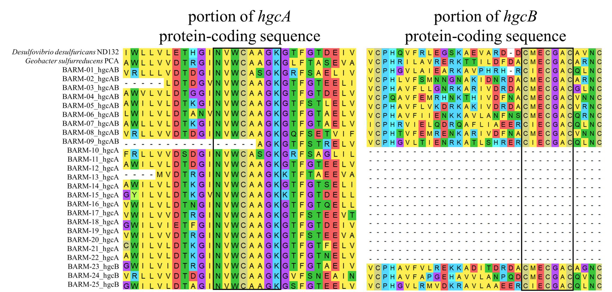
Figure 2. Alignment of conserved regions for amino acid sequences of the nine hgcAB-like gene clusters, (BARM-01 to 09) 13 hgcA-like genes (BARM-10 to 22), three hgcB-like genes (BARM-23 to 25) detected in the BARM dataset. The corresponding hgcAB amino acid sequences from Desulfovibrio desulfuricans ND132 and Geobacter sulfurreducens PCA were added in the alignment as references.
Taxonomic Identifications of hgcAB-Like Genes Found in the Baltic Sea
In order to taxonomically identify each Hg methylation gene detected in the BARM dataset, we constructed phylogenetic trees using the 650 hgcAB gene clusters generated by McDaniel et al. (2020). For the nine hgcAB-like gene clusters concatenated, we performed a phylogenetic analysis with the 650 hgcAB gene clusters (Figure 3, Supplementary Figure S1). For the 13 hgcA detected alone in their respective contig, an additional phylogenetic trees were performed using the 650 hgcA and 482 hgcB genes, respectively (Supplementary Figures S2, S3). The phylogenetic analysis revealed the presence of several hgc genes (referring hereafter either to hgcAB gene clusters, hgcA genes or hgcB genes) affiliated with the order Desulfobacterales (Deltaproteobacteria, class Desulfobacterota in GTDB classification): a member of the family Desulfobulbaceae (BARM-15 & -17), a Desulfobacula sp. (BARM-04 & -08), and a Desulforhopalus sp. (BARM-11) (Figure 3). In addition, the hgc genes BARM-02, -06 & -10 were associated with members of the orders Desulfatiglanales (naphS2 family) and Desulfarculales (Desulfarculaceae family) and Syntrophales. The hgc genes BARM-01, -07, -09 & -21 clustered together and were related – even if with very long branch length – to genes detected in various microbial phyla, with the closest related hgc genes detected in the genomes of two Spirochaetes from the family Treponemataceae and were referred thereafter as hgcAB-carrying Spirochaetes-like bacteria. Three hgc genes (BARM-05, -19 & -22) were associated with members of the recently discovered phylum Kiritimatiellaeota (Spring et al., 2016, defined as a class of Verrucomicrobia in GTDB classification), which is part of the widespread PVC superphylum (i.e., including Planctomycetes, Verrucomicrobia, Chlamydiae and Lentisphaerae). In addition, some BARM hgc genes were closely related to Firmicutes (Clostridia, BARM-14), a group of hgc genes from Euryarchaea and Chloroflexi clustered together (BARM-18, -24) and to member of the Spirochaetales order (BARM-25). Finally, seven BARM hgc genes were associated with clades including various microbial lineages and are thus classified here as unidentified (BARM-03, -12, -13, -16, -20, -23).
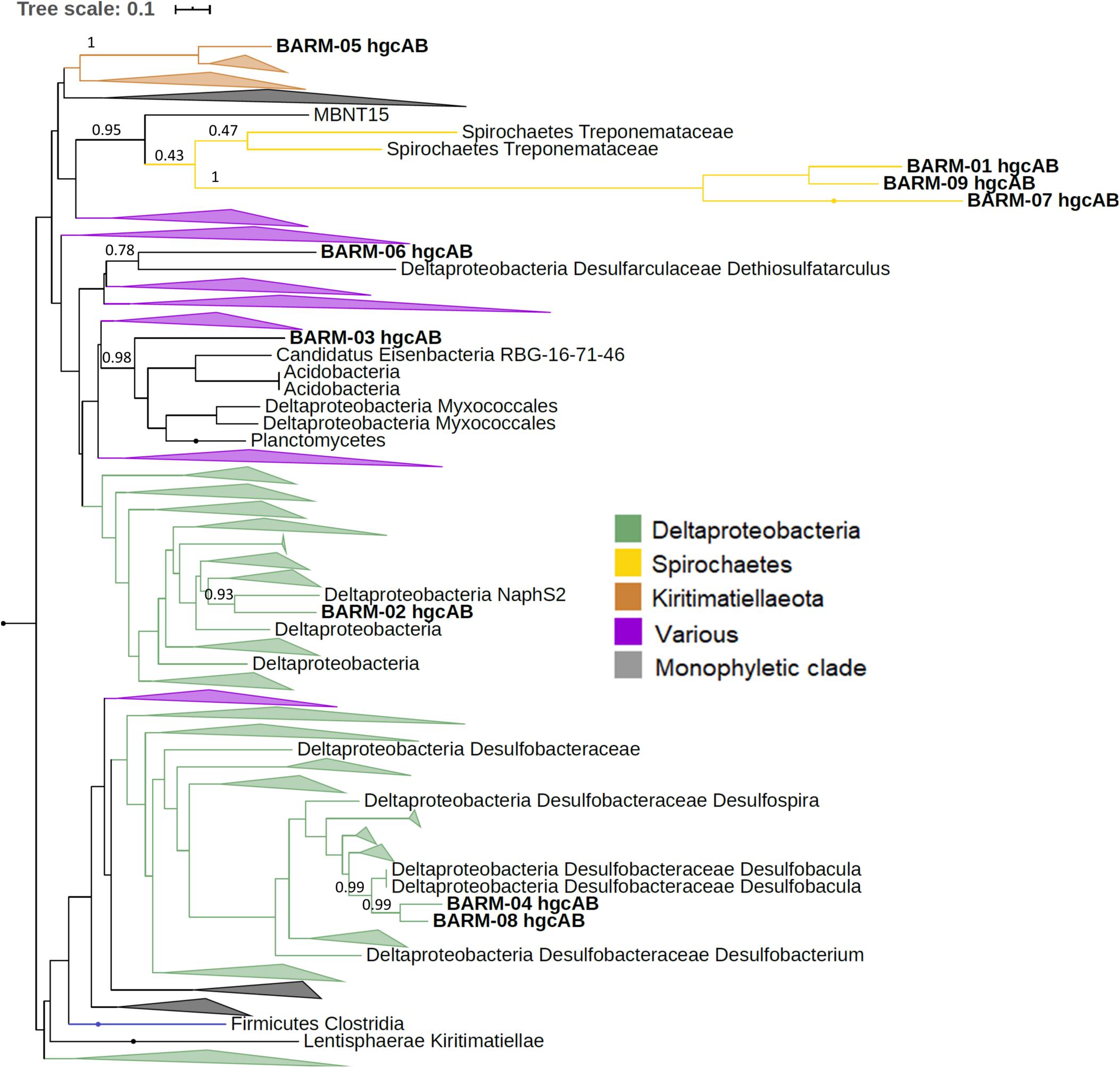
Figure 3. Maximum-likelihood phylogenetic tree of concatenated hgcAB amino acid sequences with collapsed clades. Collapsed clades are discriminated with colors based on their taxonomy. Gene clusters detected in the Baltic Sea water column (BARM) are displayed in bold. Bootstrap values obtained for nodes leading to BARM hgcAB amino acid sequences.
Relative Abundance of hgcAB Genes in the Baltic Sea Water Column
The summed relative abundance of hgcA and hgcB-like genes in the Baltic Sea water column (i.e., the number of reads for the hgcAB-like genes per total annotated reads per sample, expressed as %) ranged from undetected to 6.7 × 10⁻³ % (mean: 0.3 × 10⁻³; sd: 1.2 × 10⁻³) (Table 2, Figure 4). The highest relative abundance of hgcAB-like genes was found in the hypoxic water from 76.5 m depth at station S7 with 6.7 × 10⁻³ % (Figure 4). Elevated abundance of hgcAB-like genes was also found in hypoxic and anoxic water from the TF0271/AT3 station with the highest values at a water depth of 200 m with 6.5 × 10⁻³ %. The proportion of hgcAB genes detected in the other 10 locations was relatively low with a maximum value of 0.2 × 10⁻³ % in the hypoxic layer (87.5 m) at station TF245 (Figure 4). At the LMO station, for which only normoxic water was sampled, the highest proportion of hgcAB genes found in the 37 samples was less than 0.01 × 10⁻³ % (sample LMO 120806, Supplementary Datasheet S4). We found that the most abundant hgcAB genes in Baltic Sea anoxic water belonged to members of Deltaprotebacteria, more specifically members of Desulfobulbaceae, Desulfarculaceae, Desulfobacula sp. and Syntrophales, and to microbes closely related to Spirochaetes from the Treponemataceae family (Figure 4, Supplementary Datasheet S5). In terms of overall community composition, the metagenomes obtained from anoxic and hypoxic layers were dominated by Deltaproteobacteria, Epsilonproteobacteria, Chloroflexi and Thaumarchaeota. In contrast, metagenomes from normoxic water samples exhibited higher proportion of DNA sequences identified as Actinobacteria, Alpha- and Gammaproteobacteria, Bacteroidetes and Cyanobacteria (Figure 4).
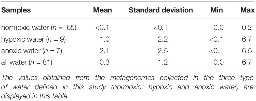
Table 2. Proportion (units in 1 × 10⁻³; normalized to total DNA sequences count of each metagenome) of hgcA- and hgcB-like genes in the 81 metagenomes.
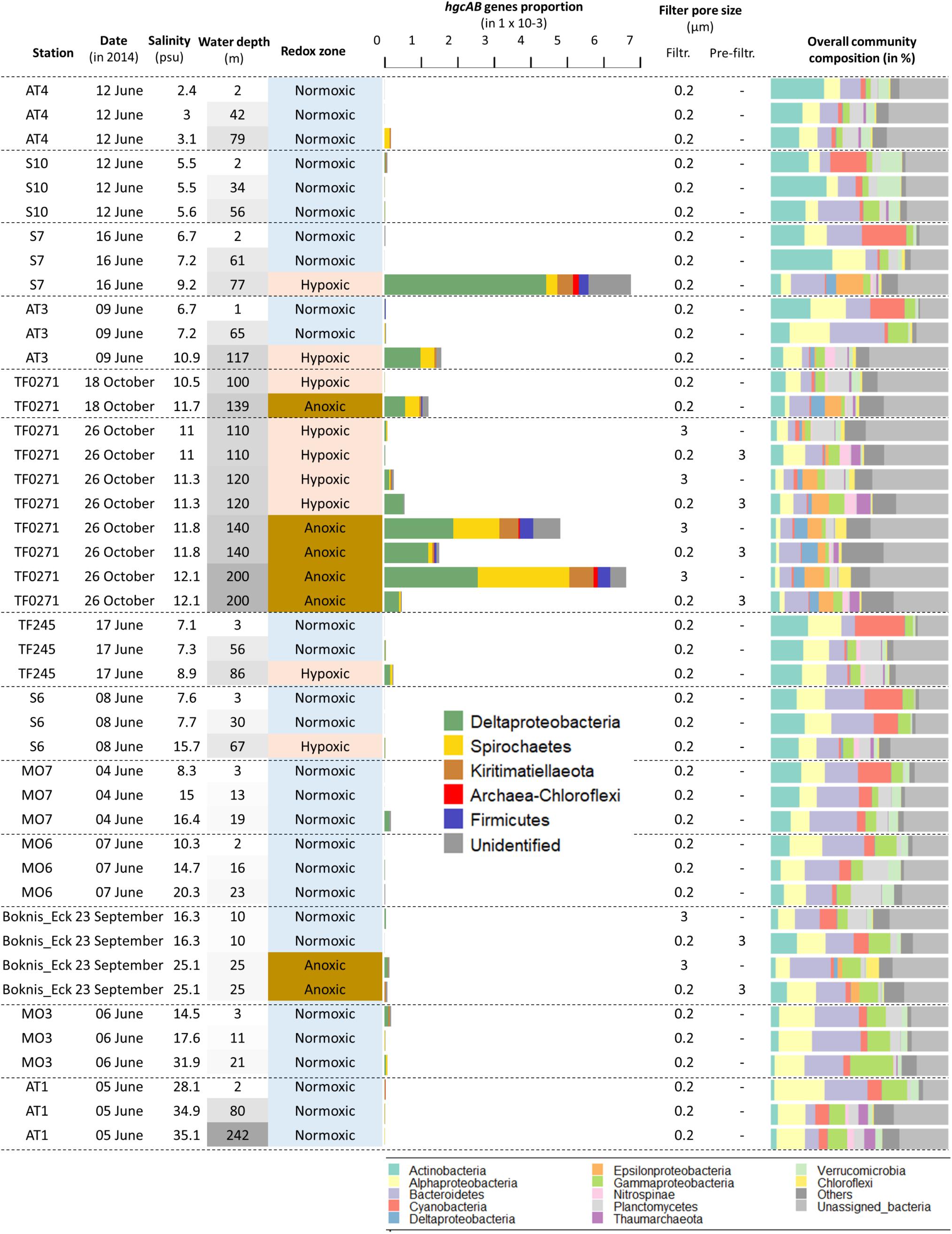
Figure 4. Relative abundance of hgcA- and hgcB-like genes in samples from the dataset “Redoxcline 2014” and “Transect 2014”. The dataset “LMO 2012” is not included because only few genes were detected and at low abundances (data shown in Supplementary Datasheet S4). The sampling date is displayed. Samples are ordered based on station locations from Bothnian Bay to Belt Seas (see Figure 1). The sampling depth (m) is color coded based on its respective gradient (darker shade of gray with increasing depth). The water redox zone is color coded based on the O2 categories defined in the text (light blue for normoxic, beige for hypoxic and brown for anoxic conditions). The abbreviations “Filtr.” and “Pre-filtr.” indicate the pore size of the filters used to obtain each metagenome and if pre-filtration were done prior to filtration, respectively. The composition of microbial communities is described from the dominant bacterial and archaeal lineages in proportion of DNA sequences related to the total number of DNA sequences identified as bacteria and archaea in the metagenomes.
Differences in hgcAB Genes Relative Abundance With Filter-Size
The quantity of hgcAB-like genes detected in metagenomes obtained from the Baltic Sea water column differed systematically between filter size fractions (Figure 4). Metagenomes obtained from the TF0271 station profile, and filtered onto 3 μm filters (hereafter referred to as the “3 μm metagenomes”), had consistently higher proportions of hgcAB-like genes (up to 6.5 × 10⁻³ %) than metagenomes obtained from 0.2 μm filters following pre-filtration with 3 μm filters (hereafter referred to as the “0.2–3 μm metagenomes”) (up to 1.5 × 10⁻³ %) (Figure 4). The relative abundance of hgcAB genes from the anoxic TF0271-Oct26 samples was three and 14 times higher in 3 μm metagenomes compared to 0.2–3 μm metagenomes for samples collected at 140 and 200 m depth, respectively (Figure 4, Supplementary Datasheet S4). At Boknis Eck, only a few samples were collected and the gene proportions were too low to properly investigate differences between filtration methods. At TF0271, hgcAB genes from Deltaproteobacteria were predominant for both 0.2–3 and 3 μm metagenomes (Figure 4). In these metagenomes, the most abundant hgcAB genes were attributed to Deltaproteobacteria from the families Desulfobulbaceae and Desulfarculales (Supplementary Datasheet S4). The 3 μm metagenomes from anoxic TF0271 samples (both 140 and 200 m) featured higher proportions of genes affiliated with Spirochaetes-like and Kiritimatiellaeota-like bacteria (Figure 4). In terms of overall community composition, the main differences between 3 μm metagenomes and 0.2–3 μm metagenomes was higher proportions of Chloroflexi in 3 μm metagenomes and of Bacteroidetes and Thaumarchaoeta in 0.2–3 μm metagenomes (Figure 4). Interestingly, the Kiritimatiellaeota proportion in hypoxic and anoxic water metagenomes from station TF0271/AT3 were more abundant in 3 μm metagenomes than in 0.2–3 μm metagenomes (Supplementary Datasheet S5). In contrast, Spirochaetes were found with same proportion in both types of metagenomes for hypoxic and anoxic water at TF0271 (Supplementary Datasheet S5).
Discussion
Our phylogenetic analysis of the Baltic Sea Reference Metagenome dataset (Alneberg et al., 2018) identified at least 18 different hgcAB gene clusters that belong to several microbial lineages (Figure 3, Supplementary Figures S2, S3). The total number of DNA sequences obtained for each metagenome was relatively low (12.9 ± 9.7 million DNA sequences) compared to the numbers typically obtained in nowadays metagenomic studies (e.g., >20 million DNA sequences per metagenone) and therefore a higher sequencing depth may have provided a more refined picture of the diversity of hgcAB-carrying microbes in the Baltic Sea water as shown by the hgcAB-like genes rarefaction curves obtained from the BARM dataset (Supplementary Figure S4). The majority of hgcAB genes detected were affiliated with Deltaproteobacteria (or Desulfobacterota with GTDB classification) notably in genomes from possible sulfate reducing bacteria (e.g., Compeau and Bartha, 1984, 1985; Gilmour et al., 2013). Some of the identified Deltaproteobacteria/Desulfobacterota belong to groups of organisms previously known or predicted to perform Hg methylation, such as a species of Desulfobacula (Gionfriddo et al., 2016), members of the Desulfobulbaceae family (Benoit et al., 2001; Colin et al., 2018), and a member of the naphS2 group (Parks et al., 2013). It is noteworthy that none of the hgcAB genes detected in the water of the Baltic Sea were closely related to known iron reducing bacteria such as the Geobacteraceae family (Fleming et al., 2006; Kerin et al., 2006; Bravo et al., 2018). Relatively high Fe(II) concentrations have previously been reported in anoxic water from the Baltic Sea (>25 nM; Pohl and Fernández-Otero, 2012) although there is seasonal and inter-annual variability in concentrations. Our finding that none of the hgcAB was closely related to Geobacteraceae suggests that other iron reducing bacteria without the capability to methylate Hg, such as Shewenella sp. (incl S. baltica), are responsible for the formation of Fe(II) in the Baltic sea water column. Indeed, Shewanellaceae are in general one to two orders of magnitude more abundant than Geobacteraceae in the metagenomes compiled in the BalticMicrobeDB (Supplementary Datasheet S5; see text footnote 1). Our results thus suggest that Hg methylation is not linked to iron-reduction in the Baltic Sea. Recent studies also identified hgcAB-carrying Nitrospirea/Nitrospinae from several environments including marine systems (Gionfriddo et al., 2016; Tada et al., 2020; Villar et al., 2020). Despite members of this genus having been detected in open ocean and seas, we did not find any hgcAB-like genes related to Nitrospina sp. in any Baltic Sea water sample. In support of this, eleven metagenome-assembled genomes (MAGs) of Nitrospinae were extracted from the Baltic Sea metagenomes (Alneberg et al., 2020), and none of these contained hgcAB-like genes (data not shown).
Microbial syntrophy, defined as an obligate mutualistic metabolism, is a process known to occur mainly in environments with shortage of favorable electron acceptors, e.g., mostly in anoxic environments (McInerney et al., 2009; Morris et al., 2013). Some syntrophic microbes were recently suggested to be involved in the Hg methylation process (Gilmour et al., 2013; Yu et al., 2018; Hu et al., 2020), but we did not identify hgcAB genes from known syntrophic microorganisms in the Baltic Sea. Instead, we identified hgcAB-carrying bacteria closely related to various groups that are still poorly described (McDaniel et al., 2020) including members of Spirochaetes and Kiritimatiellaeota phyla. For instance, among the predominant hgcAB-like genes detected in Baltic Sea, the hgcAB genes BARM-01, -07 & -09 were clustered together and found close to a number of Spirochaetes (Figure 3). However, it should be noted these gene clusters, that formed very long branches in the inferred phylogenetic tree, are in a badly resolved region of the tree also containing MBNT15, Acidobacteria, Actinobacteria and Verrucomicrobia (Supplementary Figure S1 for full description), which precludes a safe identification at present. Although, Kiritimatiellaeota phylum is not well described (Spring et al., 2016), strains from this group were recently identified as anaerobic degraders of sulfated polysaccharides compounds (Van Vliet et al., 2019). Such compounds are produced by marine algae and are generally abundant in marine waters (Helbert, 2017). The availability of sulfated polysaccharides could explained the presence of Kiritimatiellaeota in oxygen-deficient water samples from TF0271 and S7 stations.
The 81 metagenomes collected from three sampling efforts (Alneberg et al., 2018) cover substantial variations in water depth, season and location across the Baltic Sea and our study investigates the relationship between putative Hg methylating populations and these factors. We found higher relative abundance of hgcAB genes in oxygen deficient water (hypoxic and anoxic layers) compared to those observed in normoxic layers also when including an extensive time series (37 time points over the year 2012) at 2 m depth. While no MeHg concentrations were measured alongside with the 81 samples used in the present study, Soerensen et al. (2018) reported MeHg concentrations measured in samples collected from several stations in the Baltic Sea in September 2014 and we compared such data with the proportion of the hcgAB-like genes detected in the metagenomes. For instance, they reported relatively high MeHg concentrations (787 fM) in an anoxic sample collected at 200 m depth at the station TF0271/AT3 (named BY15 in their work) for which high proportion of hgcAB-like genes was also found in our study (Soerensen et al., 2018; Figure 4). This finding is in agreement with the general understanding that methylation of Hg in aquatic systems is associated with anoxic conditions (Compeau and Bartha, 1984; Gilmour et al., 1992; Eckley et al., 2005; Malcolm et al., 2010; Bravo et al., 2015). We also found relatively high proportion of hgcAB-like genes in hypoxic water samples at two locations (TF0271/AT3, 117 m and S7 77 m depth) for which MeHg concentrations in Soerensen et al. (2018) were also high (i) 459 fM at 125 m depth at TF0271/AT3 station (named BY15 station in their work) and (ii) 1625 fM at 70 m and 1375 fM at 80 m depth at S7 station (named BY32 station in their work). By contrast, very low abundance of hgcAB-like genes were found at Boknis Eck station in the anoxic water layer (25 m). We hypothesized that the absence of putative mercury methylators in this anoxic water layer is caused by its strong seasonality in oxygen concentrations (Lennartz et al., 2014) inducing a strong turnover of microbial communities that does not provide suitable conditions for the establishment of stable mercury methylating communities. It is noticeable that several studies (Goñi-Urriza et al., 2015; Bravo et al., 2016; Christensen et al., 2019) have demonstrated a poor quantitative relation between Hg methylation rate and the presence of hgcA genes (mRNA and DNA), likely because of additional important factors/processes affecting Hg methylation. Therefore the very low abundance of hgcAB-like genes in Bocknis Eck anoxic layer do not necessarily indicate the absence of mercury methylation. MeHg concentrations measured in locations near our studied stations were always found to be relatively low consistent with low relative abundances of hgcAB-like genes. Overall, the comparison between the presence of MeHg and hgcAB-like gene suggests that both anoxic and hypoxic water layers from the Baltic Sea are conducive environments for Hg methylation.
In situ formation of MeHg in normoxic waters has previously been proposed to occur in anaerobic microzones such as around marine particles such as organic-rich particulate matter and aggregates (Monperrus et al., 2007; Cossa et al., 2009; Sunderland et al., 2009; Lehnherr et al., 2011; Schartup et al., 2015) and recently in zooplankton gut (Gorokhova et al., 2020). In some cases this has been experimentally demonstrated (Ortiz et al., 2015; Gascón Díez et al., 2016). Our study expands on the role of settling particles for Hg methylation in coastal seas and support the concept of settling particles as important habitats for Hg methylators also in anoxic waters. Firstly, we found higher relative abundances of hgcAB genes in the metagenome of marine particles from anoxic (3 μm filters) compared to hypoxic (3 μm filters) or normoxic (not pre-filter with 3 μm filters) water samples (Figure 4). Considering that the 3 μm filters represent the particle and aggregated organic matter fraction, our results suggest that marine particles becomes a more suitable habitat for Hg methylators (and thus potentially constitutes an environment with high Hg methylation rate) when they reach the anoxic zone. This phenomenon is likely caused by an increased prevalence of anaerobic conditions in the marine particles from anoxic water layers. Secondly, we found a higher proportion of DNA sequences of hgcAB genes in 3 μm metagenomes than in the free-living microbes, represented by the metagenomes obtained from 0.2 to 3 μm size fraction, in anoxic waters. This finding could be explained by marine particles featuring high concentrations of organic compounds that are suitable electron donors and fuel microbial activities occurring in anoxic conditions (Bianchi et al., 2018). It is noticeable that previous studies demonstrated differences between particle-associated and free-living bacterial communities in the Baltic Sea water column, but Spirochaetes, for which hgcAB-like genes closely related to previously described hgcAB-carrying Spirochaetes were found relatively abundant in the 3 μm metagenomes from BARM dataset, were not identified as being more predominant in particle-associated bacterial communities (Rieck et al., 2015, Supplementary Datasheet S5). In contrast, Kiritimatiellaeota were found more abundant in 3 μm metagenomes than 0.2–3 μm metagenomes in Baltic Sea data (Supplementary Datasheet S5), and this is consistent with their hgcAB-genes predominance in marine particles found in our study.
Our findings advance the understanding of the diversity and distribution of genes involved in Hg methylation as well the hgcAB-carrying microbial populations in marine environments. Particularly noteworthy was the finding that most of hgcAB-carrying microbes in the Baltic Sea water column were Deltaproteobacteria/Desulfobacterota predominantly found in oxygen-deficient zones (anoxic but also hypoxic zones). In addition, the differences in the relative abundance of hgcAB genes in metagenomes obtained from 0.2 compared to 3 μm pore size filters suggest that marine particles could be a preferred niche for Hg methylating microbial communities and therefore and important potential hotspot for MeHg formation in both hypoxic and anoxic sea water zones. Finally, our phylogenetic analysis highlights that a substantial part of the Hg methylators present in the Baltic Sea, and likely in other marine environments, are still poorly described and more works are needed to isolate, characterize and describe their genetic diversity.
Data Availability Statement
Publicly available datasets were analyzed in this study. This data can be found here: https://doi.org/10.6084/m9.figshare.c.3831631.v1.
Author Contributions
EC, MB, and EB designed this work. EC and MB performed the data analysis. All authors contributed to the data interpretation and writing of the manuscript.
Funding
This work was funded by the Swedish Research Council Formas (grant 2018-01031).
Conflict of Interest
The authors declare that the research was conducted in the absence of any commercial or financial relationships that could be construed as a potential conflict of interest.
Acknowledgments
The computations were performed on resources provided by SNIC through Uppsala Multidisciplinary Center for Advanced Computational Science (UPPMAX) under Project SNIC 2019/8-322. This manuscript has been released as a pre-print at the journal bioRxiv (Capo et al., 2020). AB acknowledges the Marie Curie Individual Fellowship (H2020-MSCA-IF-2016; project-749645).
Supplementary Material
The Supplementary Material for this article can be found online at: https://www.frontiersin.org/articles/10.3389/fmicb.2020.574080/full#supplementary-material
FIGURE S1 | Maximum-likelihood phylogenetic tree of concatenated hgcAB amino acid sequences of 650 hgcAB gene clusters (McDaniel et al., 2020) and BARM hgcAB genes. Gene clusters detected in the Baltic Sea water column (BARM) are displayed in bold. Bootstrap values are displayed aside each node (1,000 ultrafast bootstrap replicates).
FIGURE S2 | Maximum-likelihood phylogenetic tree of concatenated hgcA amino acid sequences of 650 hgcA gene clusters (McDaniel et al., 2020) and BARM hgcA genes. Gene clusters detected in the Baltic Sea water column (BARM) are displayed in bold. Bootstrap values are displayed aside each node (1,000 ultrafast bootstrap replicates).
FIGURE S3 | Maximum-likelihood phylogenetic tree of concatenated hgcA amino acid sequences of 482 hgcB gene clusters (McDaniel et al., 2020) and BARM hgcA genes. Gene clusters detected in the Baltic Sea water column (BARM) are displayed in bold. Bootstrap values are displayed aside each node (1,000 ultrafast bootstrap replicates).
FIGURE S4 | Rarefaction curves of hgcAB-like gene sobtained from the BARM dataset.
DATASHEET S1 | HMM profiles of hgcAB amino acid sequences modified from Podar et al. (2015).
DATASHEET S2 | Amino acid sequences of hgcAB gene clusters identified in 650 bacterial and archaeal genomes (McDaniel et al., 2020).
DATASHEET S3 | Amino acid sequences of hgcAB-like genes detected in BARM dataset.
DATASHEET S4 | Proportion of hgcAB genes (out of the total number of reads of each metagenome) in each metagenome from the three datasets used in the present study. The taxonomic identifications associated to each gene are displayed. The environmental conditions associated to the water samples for each metagenome are displayed.
DATASHEET S5 | Proportion of DNA sequences identified to bacterial and archaeal groups for each metagenome from the three datasets used in the present study. The taxonomic identifications associated to each gene are displayed. The environmental conditions associated to the water samples for each metagenome are displayed. Fine taxonomic resolutions is displayed for orders and families from Deltaproteobacteria.
Footnotes
References
Alldredge, A. L., and Silver, M. W. (1988). Characteristics, dynamics and significance of marine snow. Prog. Oceanogr. 20, 41–82. doi: 10.1016/0079-6611(88)90053-5
Alneberg, J., Bennke, C., Beier, S., Bunse, C., Quince, C., Ininbergs, K., et al. (2020). Ecosystem-wide metagenomic binning enables prediction of ecological niches from genomes. Commun. Biol. 3, 1–10. doi: 10.1038/s42003-020-0856-x
Alneberg, J., Sundh, J., Bennke, C., Beier, S., Lundin, D., Hugerth, L. W., et al. (2018). Data descriptor: BARM and balticmicrobeDB, a reference metagenome and interface to meta-omic data for the baltic sea. Sci. Data 5:180146. doi: 10.1038/sdata.2018.146
Azam, F., and Malfatti, F. (2007). Microbial structuring of marine ecosystems. Nat. Rev. Microbiol. 5, 782–791. doi: 10.1038/nrmicro1747
Benoit, J. M., Gilmour, C. C., and Mason, R. P. (2001). Aspects of bioavailability of mercury for methylation in pure cultures of Desulfobulbus propionicus (1pr3). Appl. Environ. Microbiol. 67, 51–58. doi: 10.1128/AEM.67.1.51-58.2001
Bianchi, D., Weber, T. S., Kiko, R., and Deutsch, C. (2018). Global niche of marine anaerobic metabolisms expanded by particle microenvironments. Nat. Geosci. 11, 263–268. doi: 10.1038/s41561-018-0081-0
Blum, J. D., Popp, B. N., Drazen, J. C., Anela Choy, C., and Johnson, M. W. (2013). Methylmercury production below the mixed layer in the North Pacific Ocean. Nat. Geosci. 6, 879–884. doi: 10.1038/ngeo1918
Bowman, K. L., Collins, R. E., Agather, A. M., Lamborg, C. H., Hammerschmidt, C. R., Kaul, D., et al. (2019). Distribution of mercury-cycling genes in the Arctic and equatorial Pacific Oceans and their relationship to mercury speciation. Limnol. Oceanogr. 65, S310–S320. doi: 10.1002/lno.11310
Bravo, A. G., Bouchet, S., Guédron, S., Amouroux, D., Dominik, J., and Zopfi, J. (2015). High methylmercury production under ferruginous conditions in sediments impacted by sewage treatment plant discharges. Water Res. 80, 245–255. doi: 10.1016/j.watres.2015.04.039
Bravo, A. G., and Cosio, C. (2019). Biotic formation of methylmercury: a bio–physico–chemical conundrum. Limnol. Oceanogr. 65, 1010–1027. doi: 10.1002/lno.11366
Bravo, A. G., Loizeau, J. L., Dranguet, P., Makri, S., Björn, E., Ungureanu, V. G., et al. (2016). Persistent Hg contamination and occurrence of Hg-methylating transcript (hgcA) downstream of a chlor-alkali plant in the Olt River (Romania). Environ. Sci. Pollut. Res. 23, 10529–10541. doi: 10.1007/s11356-015-5906-4
Bravo, A. G., Zopfi, J., Buck, M., Xu, J., Bertilsson, S., Schaefer, J. K., et al. (2018). Geobacteraceae are important members of mercury-methylating microbial communities of sediments impacted by waste water releases. ISME J. 12, 802–812. doi: 10.1038/s41396-017-0007-7
Breitburg, D., Levin, L. A., Oschlies, A., Grégoire, M., Chavez, F. P., Conley, D. J., et al. (2018). Declining oxygen in the global ocean and coastal waters. Science 359:eaam7240. doi: 10.1126/science.aam7240
Capo, E., Bravo, A. G., Soerensen, A. L., Bertilsson, S., Pinhassi, J., Feng, C., et al. (2020). Marine snow as a habitat for microbial mercury methylators in the Baltic Sea. bioRxiv doi: 10.1101/2020.03.04.975987
Carstensen, J., Andersen, J. H., Gustafsson, B. G., and Conley, D. J. (2014). Deoxygenation of the Baltic Sea during the last century. Proc. Natl. Acad. Sci. U.S.A. 111, 5628–5633. doi: 10.1073/pnas.1323156111
Christensen, G. A., Gionfriddo, C. M., King, A. J., Moberly, J. G., Miller, C. L., Somenahally, A. C., et al. (2019). Determining the reliability of measuring mercury cycling gene abundance with correlations with mercury and methylmercury concentrations. Environ. Sci. Technol. 53, 8649–8663. doi: 10.1021/acs.est.8b06389
Colin, Y., Gury, J., Monperrus, M., Gentes, S., Ayala Borda, P., Goni-Urriza, M., et al. (2018). Biosensor for screening bacterial mercury methylation: example within the Desulfobulbaceae. Res. Microbiol. 169, 44–51. doi: 10.1016/j.resmic.2017.09.005
Compeau, G., and Bartha, R. (1984). Methylation and demethylation of mercury under controlled redox, pH, and salinity conditions. Appl. Environ. Microbiol. 48, 1203–1207. doi: 10.1128/aem.48.6.1203-1207.1984
Compeau, G. C., and Bartha, R. (1985). Sulfate-reducing bacteria: principal methylators of mercury in anoxic estuarine sediment. Appl. Environ. Microbiol. 50, 498–502. doi: 10.1128/aem.50.2.498-502.1985
Conley, D. J., Carstensen, J., Aigars, J., Axe, P., Bonsdorff, E., Eremina, T., et al. (2011). Hypoxia is increasing in the coastal zone of the Baltic Sea. Environ. Sci. Technol. 45, 6777–6783. doi: 10.1021/es201212r
Cossa, D., Averty, B., and Pirrone, N. (2009). The origin of methylmercury in open mediterranean waters. Limnol. Oceanogr. 54, 837–844. doi: 10.4319/lo.2009.54.3.0837
Eckley, C. S., Watras, C. J., Hintelmann, H., Morrison, K., Kent, A. D., and Regnell, O. (2005). Mercury methylation in the hypolimnetic waters of lakes with and without connection to wetlands in northern Wisconsin. Can. J. Fish. Aquat. Sci. 62, 400–411. doi: 10.1139/f04-205
Finn, R. D., Clements, J., and Eddy, S. R. (2011). HMMER web server: interactive sequence similarity searching. Nucleic Acids Res. 39, W29–W37. doi: 10.1093/nar/gkr367
Fleming, E. J., Mack, E. E., Green, P. G., and Nelson, D. C. (2006). Mercury methylation from unexpected sources: molybdate-inhibited freshwater sediments and an iron-reducing bacterium. Appl. Environ. Microbiol. 72, 457–464. doi: 10.1128/AEM.72.1.457-464.2006
Gascón Díez, E., Loizeau, J. L., Cosio, C., Bouchet, S., Adatte, T., Amouroux, D., et al. (2016). Role of settling particles on mercury methylation in the oxic water column of freshwater systems. Environ. Sci. Technol. 50, 11672–11679. doi: 10.1021/acs.est.6b03260
Gilmour, C. C., Henry, E. A., and Ralph, M. (1992). Sulfate stimulation of mercury methylation in freshwater sediments. Environ. Sci. Technol. 26, 2281–2287. doi: 10.1021/es00035a029
Gilmour, C. C., Podar, M., Bullock, A. L., Graham, A. M., Brown, S. D., Somenahally, A. C., et al. (2013). Mercury methylation by novel microorganisms from new environments. Environ. Sci. Technol. 47, 11810–11820. doi: 10.1021/es403075t
Gionfriddo, C. M., Tate, M. T., Wick, R. R., Schultz, M. B., Zemla, A., Thelen, M. P., et al. (2016). Microbial mercury methylation in Antarctic sea ice. Nat. Microbiol. 1:16127. doi: 10.1038/nmicrobiol.2016.127
Gionfriddo, C. M., Wymore, A. M., Jones, D. S., Wilpiszeski, R. L., Lynes, M. M., Christensen, G. A., et al. (2020). An improved hgcAB primer set and direct high-throughput sequencing expand Hg-methylator diversity in nature. bioRxiv doi: 10.1101/2020.03.10.983866
Goñi-Urriza, M., Corsellis, Y., Lanceleur, L., Tessier, E., Gury, J., Monperrus, M., et al. (2015). Relationships between bacterial energetic metabolism, mercury methylation potential, and hgcA/hgcB gene expression in Desulfovibrio dechloroacetivorans BerOc1. Environ. Sci. Pollut. Res. 22, 13764–13771. doi: 10.1007/s11356-015-4273-5
Gorokhova, E., Soerensen, A. L., and Motwani, N. H. (2020). Mercury-methylating bacteria are associated with copepods: a proof-of-principle survey in the Baltic Sea. PLoS One 15:e0230310. doi: 10.1371/journal.pone.0230310
Haro-Moreno, J. M., López-Pérez, M., de la Torre, J. R., Picazo, A., Camacho, A., and Rodriguez-Valera, F. (2018). Fine metagenomic profile of the Mediterranean stratified and mixed water columns revealed by assembly and recruitment. Microbiome 6, 1–19. doi: 10.1186/s40168-018-0513-5
Helbert, W. (2017). Marine polysaccharide sulfatases. Front. Mar. Sci. 4:6. doi: 10.3389/fmars.2017.00006
Hu, H., Wang, B., Bravo, A. G., Björn, E., Skyllberg, U., Amouroux, D., et al. (2020). Shifts in mercury methylation across a peatland chronosequence: from sulfate reduction to methanogenesis and syntrophy. J. Hazard. Mater. 387:121967. doi: 10.1016/J.JHAZMAT.2019.121967
Kerin, E. J., Gilmour, C. C., Roden, E., Suzuki, M. T., Coates, J. D., and Mason, R. P. (2006). Mercury methylation by dissimilatory iron-reducing bacteria. Appl. Environ. Microbiol. 72, 7919–7921. doi: 10.1128/AEM.01602-06
Kumar, S., Stecher, G., Li, M., Knyaz, C., and Tamura, K. (2018). MEGA X: molecular evolutionary genetics analysis across computing platforms. Mol. Biol. Evol. 35, 1547–1549. doi: 10.1093/molbev/msy096
Kuss, J., Krüger, S., Ruickoldt, J., and Wlost, K.-P. (2018). High-resolution measurements of elemental mercury in surface water for an improved quantitative understanding of the Baltic Sea as a source of atmospheric mercury. Atmos. Chem. Phys. 18, 4361–4376. doi: 10.5194/acp-18-4361-2018
Lehnherr, I., St. Louis, V. L., Hintelmann, H., and Kirk, J. L. (2011). Methylation of inorganic mercury in polar marine waters. Nat. Geosci. 4, 298–302. doi: 10.1038/ngeo1134
Lennartz, S. T., Lehmann, A., Herrford, J., Malien, F., Hansen, H. P., Biester, H., et al. (2014). Long-term trends at the Boknis Eck time series station (Baltic Sea), 1957-2013: does climate change counteract the decline in eutrophication? Biogeosciences 11, 6323–6339. doi: 10.5194/bg-11-6323-2014
Letunic, I., and Bork, P. (2019). Interactive Tree Of Life (iTOL) v4: recent updates and new developments. Nucleic Acids Res. 47, W256–W259. doi: 10.1093/nar/gkz239
Li, D., Liu, C.-M., Luo, R., Sadakane, K., and Lam, T.-W. (2015). MEGAHIT: an ultra-fast single-node solution for large and complex metagenomics assembly via succinct de Bruijn graph. Bioinformatics 31, 1674–1676. doi: 10.1093/bioinformatics/btv033
Lin, H., Ascher, D., Myung, Y., Lamborg, C., Hallam, S., Gionfriddo, C., et al. (2020). Mercury methylation by metabolically versatile and cosmopolitan marine bacteria. bioRxiv doi: 10.1101/2020.06.03.132969
Malcolm, E. G., Schaefer, J. K., Ekstrom, E. B., Tuit, C. B., Jayakumar, A., Park, H., et al. (2010). Mercury methylation in oxygen deficient zones of the oceans: no evidence for the predominance of anaerobes. Mar. Chem. 122, 11–19. doi: 10.1016/j.marchem.2010.08.004
Mason, R., Choi, A. L., Fitzgerald, W. F., Hammerschmidt, C. R., Lamborg, C. H., and Soerensen, A. L. (2012). Mercury biogeochemical cycling in the ocean and policy implications. Environ. Res. 119, 101–117. doi: 10.1016/J.ENVRES.2012.03.013
McDaniel, E. A., Peterson, B. D., Stevens, S. L. R., Tran, P. Q., Anantharaman, K., and McMahon, K. D. (2020). Expanded phylogenetic diversity and metabolic flexibility of mercury-methylating microorganisms. mSystems 5:e00299-20. doi: 10.1128/mSystems.00299-20
McInerney, M. J., Sieber, J. R., and Gunsalus, R. P. (2009). Syntrophy in anaerobic global carbon cycles. Curr. Opin. Biotechnol. 20, 623–632. doi: 10.1016/j.copbio.2009.10.001
Mehrshad, M., Amoozegar, M. A., Ghai, R., Shahzadeh Fazeli, S. A., and Rodriguez-Valera, F. (2016). Genome reconstruction from metagenomic data sets reveals novel microbes in the brackish waters of the Caspian Sea. Appl. Environ. Microbiol. 82, 1599–1612. doi: 10.1128/AEM.03381-15
Monperrus, M., Tessier, E., Amouroux, D., Leynaert, A., Huonnic, P., and Donard, O. F. X. (2007). Mercury methylation, demethylation and reduction rates in coastal and marine surface waters of the Mediterranean Sea. Mar. Chem. 107, 49–63. doi: 10.1016/j.marchem.2007.01.018
Morris, B. E. L., Henneberger, R., Huber, H., and Moissl-Eichinger, C. (2013). Microbial syntrophy: interaction for the common good. FEMS Microbiol. Rev. 37, 384–406. doi: 10.1111/1574-6976.12019
Nowinski, B., Motard-Côté, J., Landa, M., Preston, C. M., Scholin, C. A., Birch, J. M., et al. (2019). Microdiversity and temporal dynamics of marine bacterial dimethylsulfoniopropionate genes. Environ. Microbiol. 21, 1687–1701. doi: 10.1111/1462-2920.14560
Ortiz, V. L., Mason, R. P., and Evan Ward, J. (2015). An examination of the factors influencing mercury and methylmercury particulate distributions, methylation and demethylation rates in laboratory-generated marine snow. Mar. Chem. 177, 753–762. doi: 10.1016/j.marchem.2015.07.006
Parks, J. M., Johs, A., Podar, M., Bridou, R., Hurt, R. A., Smith, S. D., et al. (2013). The genetic basis for bacterial mercury methylation. Science 339, 1332–1335. doi: 10.1126/science.1230667
Podar, M., Gilmour, C. C., Brandt, C. C., Soren, A., Brown, S. D., Crable, B. R., et al. (2015). Global prevalence and distribution of genes and microorganisms involved in mercury methylation. Sci. Adv. 1:e1500675. doi: 10.1126/sciadv.1500675
Pohl, C., and Fernández-Otero, E. (2012). Iron distribution and speciation in oxic and anoxic waters of the Baltic Sea. Mar. Chem. 145–147, 1–15. doi: 10.1016/j.marchem.2012.09.001
Price, M., Dehal, P., and Arkin, A. (2009). FastTree: computing large minimum evolution trees with profiles instead of a distance matrix. Mol. Biol. Evol. 26, 1641–1650. doi: 10.1093/molbev/msp077
Rieck, A., Herlemann, D. P. R., Jürgens, K., and Grossart, H.-P. (2015). Particle-associated differ from free-living bacteria in surface waters of the Baltic Sea. Front. Microbiol. 6:1297. doi: 10.3389/fmicb.2015.01297
Schartup, A. T., Balcom, P. H., Soerensen, A. L., Gosnell, K. J., Calder, R. S. D., Mason, R. P., et al. (2015). Freshwater discharges drive high levels of methylmercury in Arctic marine biota. Proc. Natl. Acad. Sci. U.S.A. 112, 11789–11794. doi: 10.1073/pnas.1505541112
Soerensen, A. L., Schartup, A. T., Gustafsson, E., Gustafsson, B. G., Undeman, E., and Björn, E. (2016). Eutrophication increases phytoplankton methylmercury concentrations in a coastal sea - a baltic sea case study. Environ. Sci. Technol. 50, 11787–11796. doi: 10.1021/acs.est.6b02717
Soerensen, A. L., Schartup, A. T., Skrobonja, A., Bouchet, S., Amouroux, D., Liem-Nguyen, V., et al. (2018). Deciphering the role of water column redoxclines on methylmercury cycling using speciation modeling and observations from the baltic sea. Glob. Biogeochem. Cycles 32, 1498–1513. doi: 10.1029/2018GB005942
Spring, S., Bunk, B., Spröer, C., Schumann, P., Rohde, M., Tindall, B. J., et al. (2016). Characterization of the first cultured representative of Verrucomicrobia subdivision 5 indicates the proposal of a novel phylum. Isme J. 10, 2801–2816. doi: 10.1038/ismej.2016.84
Sunderland, E. M., Krabbenhoft, D. P., Moreau, J. W., Strode, S. A., and Landing, W. M. (2009). Mercury sources, distribution, and bioavailability in the North Pacific Ocean: insights from data and models. Glob. Biogeochem. Cycles 23:GB2010. doi: 10.1029/2008GB003425
Tada, Y., Marumoto, K., and Takeuchi, A. (2020). Nitrospina-like bacteria are potential mercury methylators in the mesopelagic zone in the East China Sea. Front. Microbiol. 11:1369. doi: 10.3389/FMICB.2020.01369
Van Vliet, D. M., Ayudthaya, S. P. N., Diop, S., Villanueva, L., Stams, A. J. M., and Sánchez-Andrea, I. (2019). Anaerobic degradation of sulfated polysaccharides by two novel Kiritimatiellales strains isolated from black sea sediment. Front. Microbiol. 10:253. doi: 10.3389/fmicb.2019.00253
Villar, E., Cabrol, L., and Heimbürger-Boavida, L. (2020). Widespread microbial mercury methylation genes in the global ocean. Environ. Microbiol. Rep. 12, 277–287. doi: 10.1111/1758-2229.12829
Keywords: mercury methylation, hgcAB, Baltic Sea, deltaproteobacteria, spirochaetes-like bacteria
Citation: Capo E, Bravo AG, Soerensen AL, Bertilsson S, Pinhassi J, Feng C, Andersson AF, Buck M and Björn E (2020) Deltaproteobacteria and Spirochaetes-Like Bacteria Are Abundant Putative Mercury Methylators in Oxygen-Deficient Water and Marine Particles in the Baltic Sea. Front. Microbiol. 11:574080. doi: 10.3389/fmicb.2020.574080
Received: 18 June 2020; Accepted: 25 August 2020;
Published: 22 September 2020.
Edited by:
Alexandre J. Poulain, University of Ottawa, CanadaReviewed by:
Daniel Seth Jones, University of Minnesota Twin Cities, United StatesMircea Podar, Oak Ridge National Laboratory (DOE), United States
Copyright © 2020 Capo, Bravo, Soerensen, Bertilsson, Pinhassi, Feng, Andersson, Buck and Björn. This is an open-access article distributed under the terms of the Creative Commons Attribution License (CC BY). The use, distribution or reproduction in other forums is permitted, provided the original author(s) and the copyright owner(s) are credited and that the original publication in this journal is cited, in accordance with accepted academic practice. No use, distribution or reproduction is permitted which does not comply with these terms.
*Correspondence: Eric Capo, eric.capo@hotmail.fr
†These authors share last authorship