Hydrogel-Based Localized Nonviral Gene Delivery in Regenerative Medicine Approaches—An Overview

 , and
, and
Abstract
:1. Introduction
1.1. Requirements for Localized Gene Delivery in Tissue Regeneration
1.2. Hydrogels as Vehicles for Nonviral Gene Delivery
2. Hydrogel-Mediated Gene Delivery in Regenerative Medicine
2.1. Musculoskeletal Tissue Repair
2.1.1. Bone Tissue
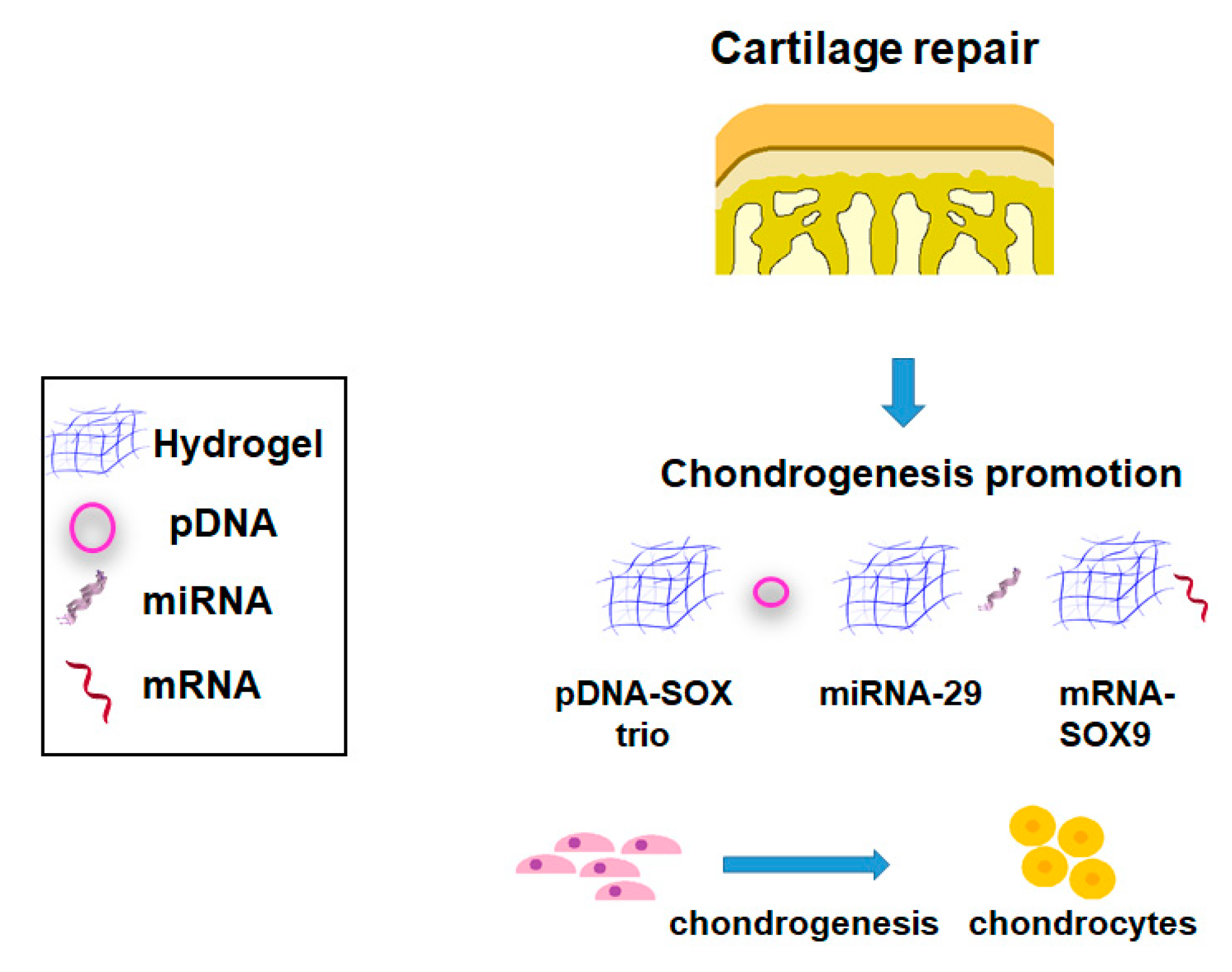
2.1.2. Cartilage
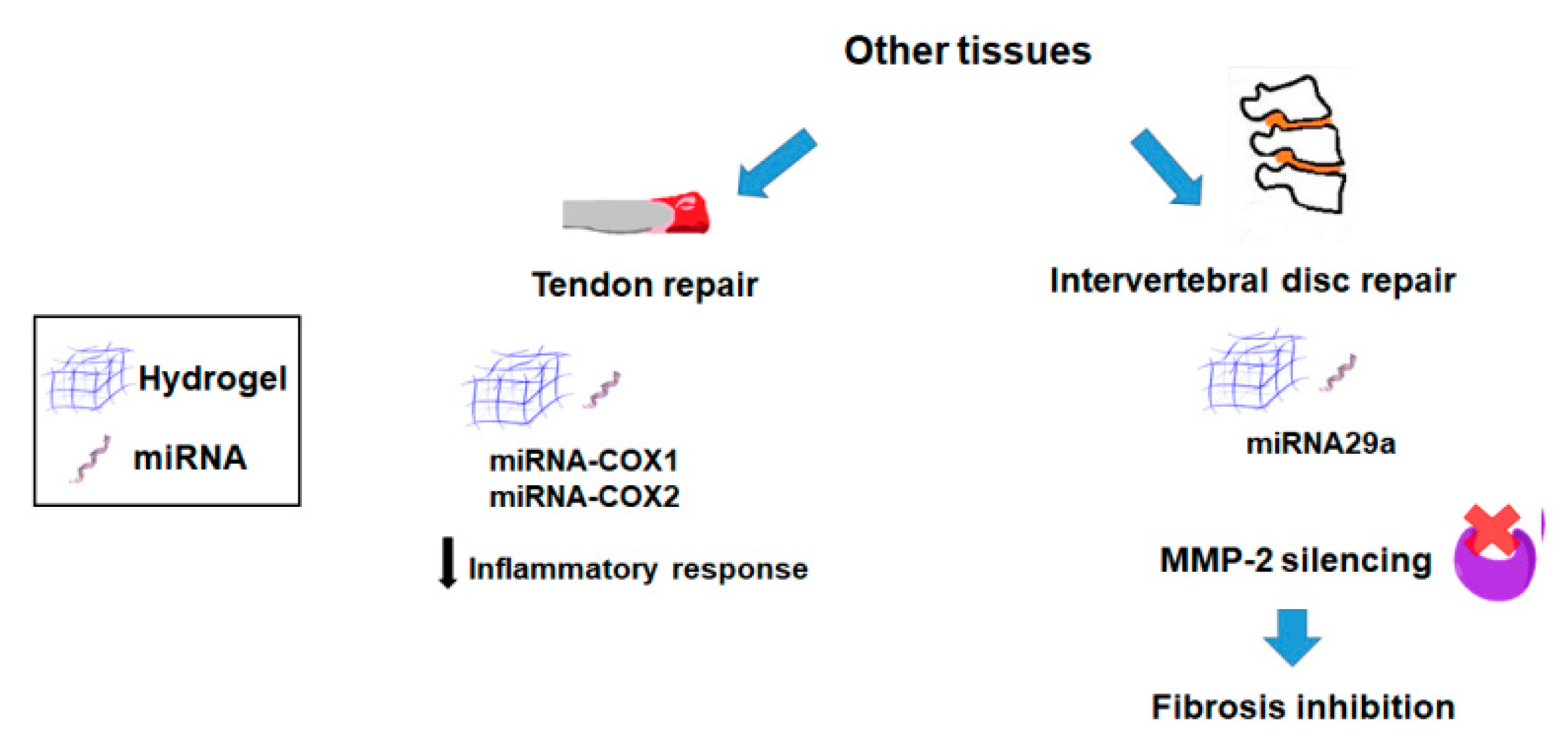
2.1.3. Other Tissues
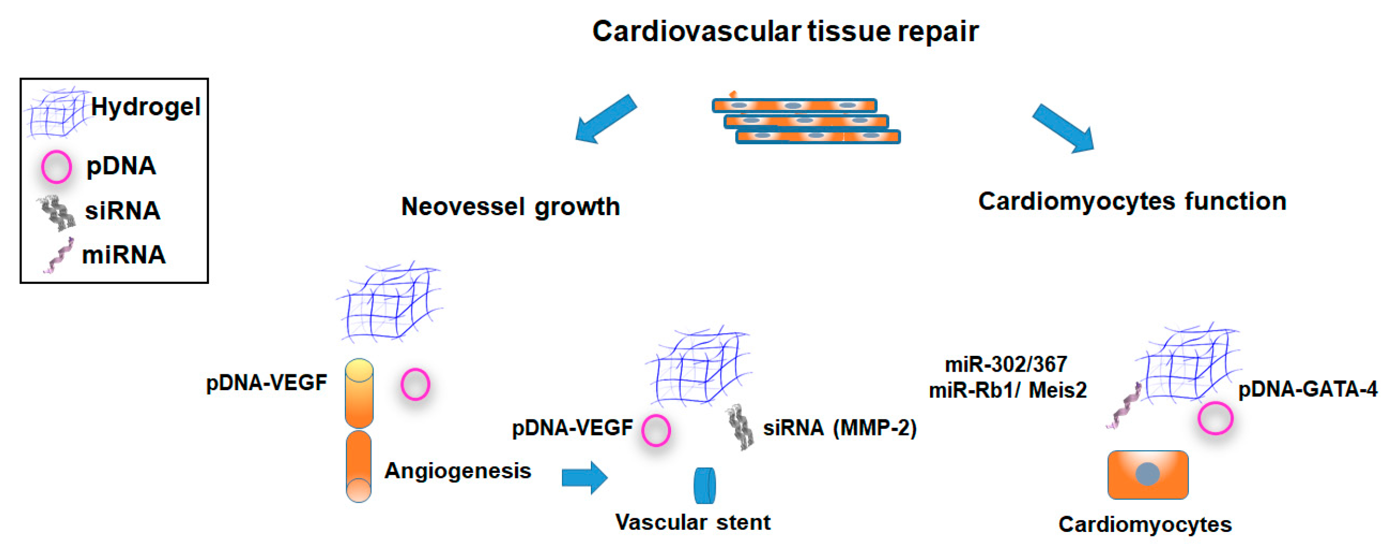
2.2. Cardiovascular Tissue Repair
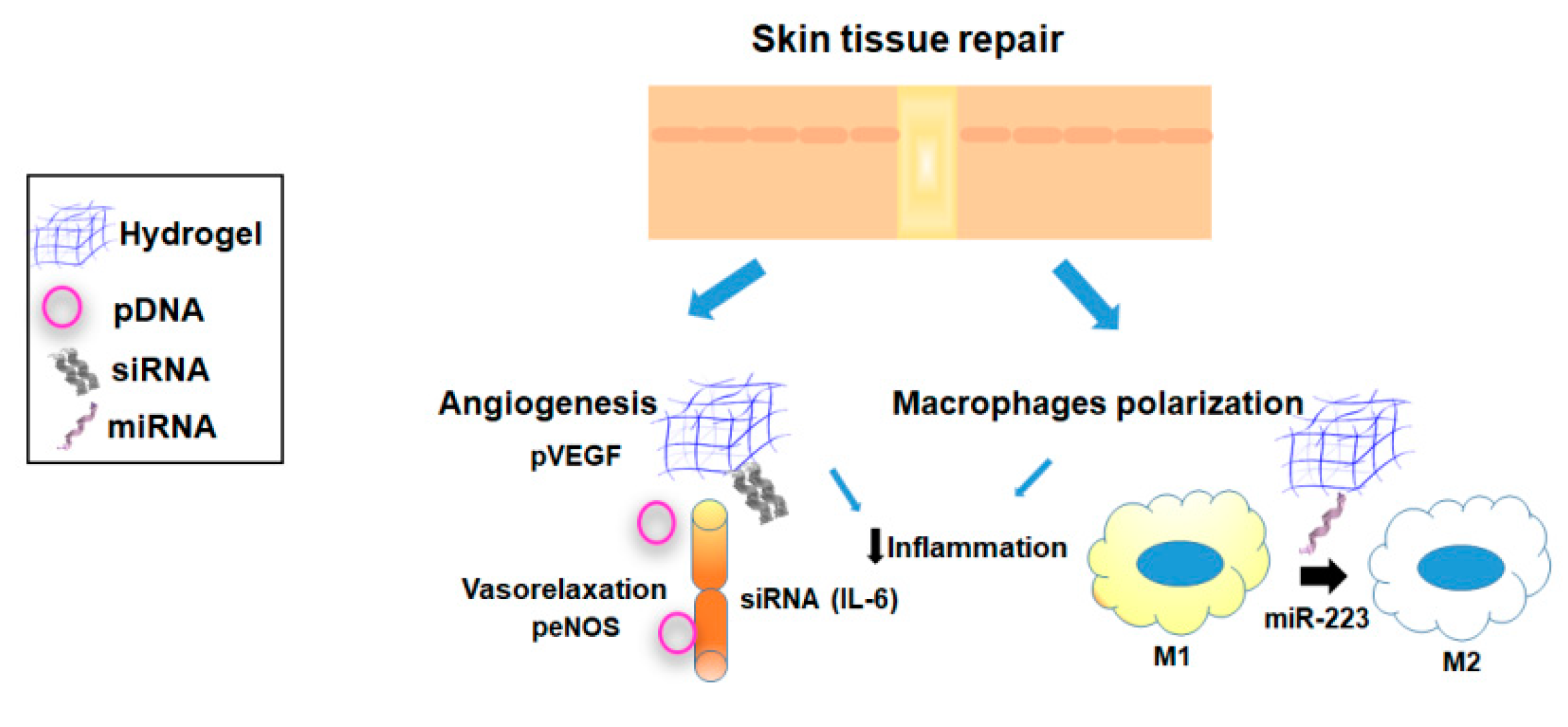
2.3. Skin Tissue Repair
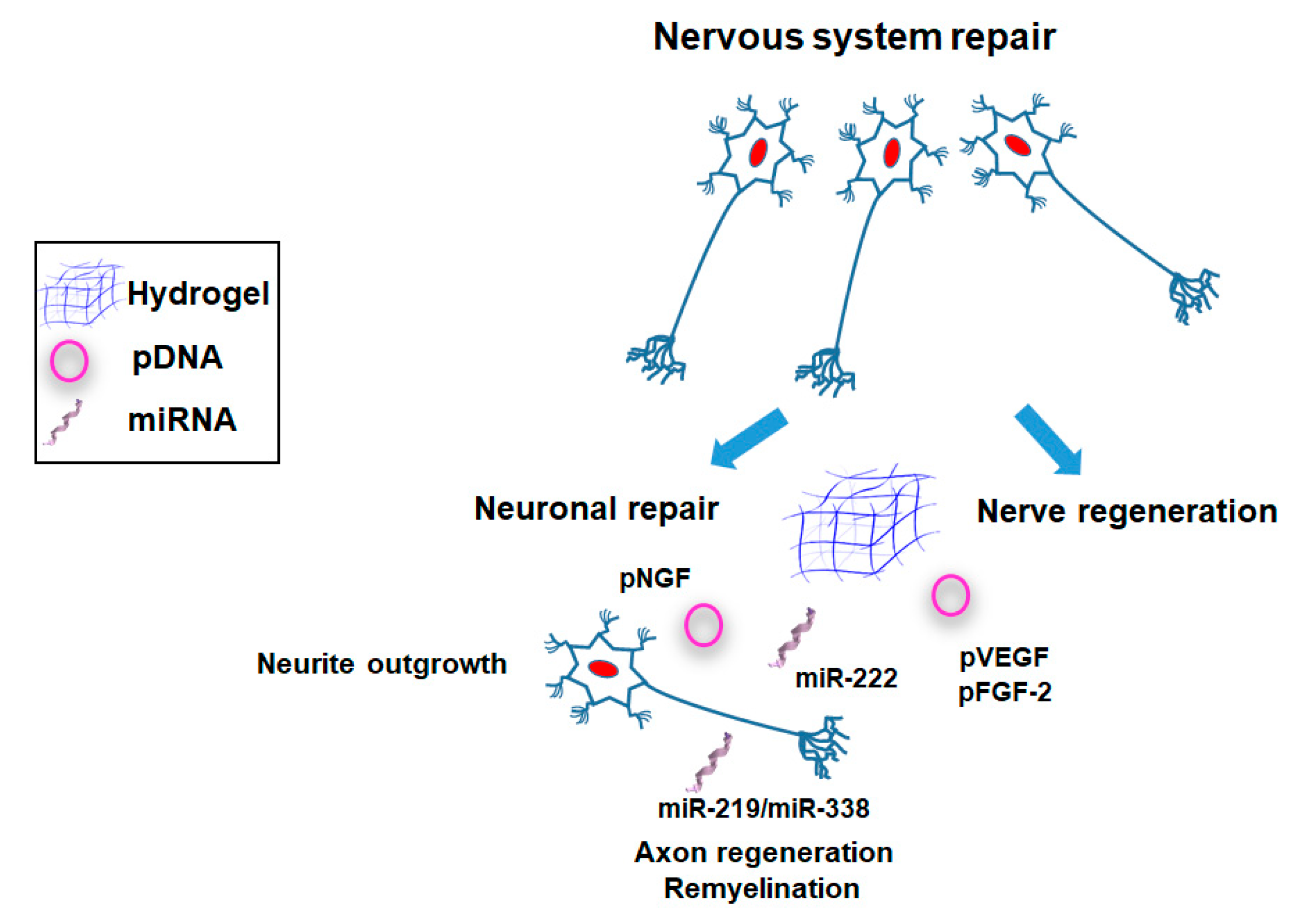
2.4. Nervous Tissue Repair
3. Discussion
Author Contributions
Funding
Acknowledgments
Conflicts of Interest
References
- Ma, C.C.; Wang, Z.L.; Xu, T.; He, Z.Y.; Wei, Y.Q. The approved gene therapy drugs worldwide: From 1998 to 2019. Biotechnol. Adv. 2020, 40, 107502. [Google Scholar] [CrossRef] [PubMed]
- Uludag, H.; Ubeda, A.; Ansari, A. At the intersection of biomaterials and gene therapy: Progress in non-viral delivery of nucleic acids. Front. Bioeng. Biotechnol. 2019, 7, 1–21. [Google Scholar] [CrossRef] [PubMed]
- Patel, S.; Athirasala, A.; Menezes, P.P.; Ashwanikumar, N.; Zou, T.; Sahay, G.; Bertassoni, L.E. Messenger RNA Delivery for Tissue Engineering and Regenerative Medicine Applications. Tissue Eng. 2019, 25, 91–112. [Google Scholar] [CrossRef] [PubMed]
- Ansari, A.S.; Santerre, P.J.; Uludaǧ, H. Biomaterials for polynucleotide delivery to anchorage-independent cells. J. Mater. Chem. B 2017, 5, 7238–7261. [Google Scholar] [CrossRef] [PubMed]
- Agrawal, N.; Dasaradhi, P.V.N.; Mohmmed, A.; Malhotra, P.; Bhatnagar, R.K.; Mukherjee, S.K. RNA Interference: Biology, Mechanism, and Applications. Microbiol. Mol. Biol. Rev. 2003, 67, 657–685. [Google Scholar] [CrossRef] [PubMed] [Green Version]
- Dong, Y.; Siegwart, D.J.; Anderson, D.G. Strategies, design, and chemistry in siRNA delivery systems. Adv. Drug Deliv. Rev. 2019, 144, 133–147. [Google Scholar] [CrossRef]
- MacFarlane, L.-A.; Murphy, P.R. MicroRNA: Biogenesis, Function and Role in Cancer. Curr. Genom. 2010, 11, 537–561. [Google Scholar] [CrossRef] [Green Version]
- Bono, N.; Ponti, F.; Mantovani, D.; Candiani, G. Non-Viral in Vitro Gene Delivery: It is Now Time to Set the Bar! Pharmaceutics 2020, 12, 183. [Google Scholar] [CrossRef] [Green Version]
- Cucchiarini, M. Human gene therapy: Novel approaches to improve the current gene delivery systems. Discov. Med. 2016, 21, 495–506. [Google Scholar]
- Sanada, F.; Taniyama, Y.; Kanbara, Y.; Otsu, R.; Ikeda-Iwabu, Y.; Carracedo, M.; Rakugi, H.; Morishita, R. Gene therapy in peripheral artery disease. Expert Opin. Biol. Ther. 2015, 15, 381–390. [Google Scholar] [CrossRef]
- Shimamura, M.; Nakagami, H.; Taniyama, Y.; Morishita, R. Gene therapy for peripheral arterial disease. Expert Opin. Biol. Ther. 2014, 14, 1175–1184. [Google Scholar] [CrossRef] [PubMed]
- Lostalé-Seijo, I.; Montenegro, J. Synthetic materials at the forefront of gene delivery. Nat. Rev. Chem. 2018, 2, 258–277. [Google Scholar] [CrossRef]
- Rey-Rico, A.; Cucchiarini, M. Controlled release strategies for rAAV-mediated gene delivery. Acta Biomater. 2016, 29, 1–10. [Google Scholar] [CrossRef] [PubMed]
- Wang, L.L.; Burdick, J.A. Engineered Hydrogels for Local and Sustained Delivery of RNA-Interference Therapies. Adv. Healthc. Mater. 2017, 6, 1–16. [Google Scholar] [CrossRef] [PubMed]
- Youngblood, R.L.; Truong, N.F.; Segura, T.; Shea, L.D. It’s All in the Delivery: Designing Hydrogels for Cell and Non-viral Gene Therapies. Mol. Ther. 2018, 26, 2087–2106. [Google Scholar] [CrossRef] [PubMed] [Green Version]
- Sahin, U.; Karikó, K.; Türeci, Ö. mRNA-based therapeutics--developing a new class of drugs. Nat. Rev. Drug Discov. 2014, 13, 759–780. [Google Scholar] [CrossRef]
- Ledo, A.M.; Senra, A.; Rilo-Alvarez, H.; Borrajo, E.; Vidal, A.; Alonso, M.J.; Garcia-Fuentes, M. mRNA-activated matrices encoding transcription factors as primers of cell differentiation in tissue engineering. Biomaterials 2020, 247, 120016. [Google Scholar] [CrossRef]
- Oh, S.; Kessler, J.A. Design, Assembly, Production, and Transfection of Synthetic Modified mRNA. Methods 2018, 133, 29–43. [Google Scholar] [CrossRef]
- Gower, R.M.; Shea, L.D. Biomaterial Scaffolds for Controlled, Localized Gene Delivery of Regenerative Factors. Adv. Wound Care 2013, 2, 100–106. [Google Scholar] [CrossRef] [Green Version]
- Rey-Rico, A.; Madry, H.; Cucchiarini, M. Hydrogel-Based Controlled Delivery Systems for Articular Cartilage Repair. Biomed Res. Int. 2016, 2016, 1215263. [Google Scholar] [CrossRef] [Green Version]
- Yan, X.; Chen, Y.R.; Song, Y.F.; Yang, M.; Ye, J.; Zhou, G.; Yu, J.K. Scaffold-based gene therapeutics for osteochondral tissue engineering. Front. Pharmacol. 2020, 10, 1–13. [Google Scholar] [CrossRef] [PubMed]
- Valle, D. Hydrogels for Biomedical Applications: Cellulose, Chitosan, and Protein/Peptide Derivatives. Gels 2017, 3, 27. [Google Scholar] [CrossRef] [PubMed] [Green Version]
- Liaw, C.-Y.; Ji, S.; Guvendiren, M. Engineering 3D Hydrogels for Personalized In Vitro Human Tissue Models. Adv. Healthc. Mater. 2018, 7, 1701165. [Google Scholar] [CrossRef] [PubMed]
- Vijayavenkataraman, S.; Yan, W.-C.; Lu, W.F.; Wang, C.-H.; Fuh, J.Y.H. 3D bioprinting of tissues and organs for regenerative medicine. Adv. Drug Deliv. Rev. 2018, 132, 296–332. [Google Scholar] [CrossRef] [PubMed]
- Singh, S.; Choudhury, D.; Yu, F.; Mironov, V.; Naing, M.W. In situ bioprinting—Bioprinting from benchside to bedside? Acta Biomater. 2020, 101, 14–25. [Google Scholar] [CrossRef] [PubMed]
- Liu, M.; Zeng, X.; Ma, C.; Yi, H.; Ali, Z.; Mou, X.; Li, S.; Deng, Y.; He, N. Injectable hydrogels for cartilage and bone tissue engineering. Bone Res. 2017, 5, 17014. [Google Scholar] [CrossRef]
- Peña, B.; Laughter, M.; Jett, S.; Rowland, T.J.; Taylor, M.R.G.; Mestroni, L.; Park, D. Injectable Hydrogels for Cardiac Tissue Engineering. Macromol. Biosci. 2018, 18, e1800079. [Google Scholar] [CrossRef]
- Sood, N.; Bhardwaj, A.; Mehta, S.; Mehta, A. Stimuli-responsive hydrogels in drug delivery and tissue engineering. Drug Deliv. 2016, 23, 748–770. [Google Scholar] [CrossRef] [Green Version]
- Zhang, Y.; Yu, J.; Ren, K.; Zuo, J.; Ding, J.; Chen, X. Thermosensitive Hydrogels as Scaffolds for Cartilage Tissue Engineering. Biomacromolecules 2019, 20, 1478–1492. [Google Scholar] [CrossRef]
- Ghobril, C.; Grinstaff, M.W. The chemistry and engineering of polymeric hydrogel adhesives for wound closure: A tutorial. Chem. Soc. Rev. 2015, 44, 1820–1835. [Google Scholar] [CrossRef]
- Chaudhari, A.A.; Vig, K.; Baganizi, D.R.; Sahu, R.; Dixit, S.; Dennis, V.; Singh, S.R.; Pillai, S.R. Future prospects for scaffolding methods and biomaterials in skin tissue engineering: A review. Int. J. Mol. Sci. 2016, 17, 1974. [Google Scholar] [CrossRef]
- McCrary, M.R.; Jesson, K.; Wei, Z.Z.; Logun, M.; Lenear, C.; Tan, S.; Gu, X.; Jiang, M.Q.; Karumbaiah, L.; Yu, S.P.; et al. Cortical Transplantation of Brain-Mimetic Glycosaminoglycan Scaffolds and Neural Progenitor Cells Promotes Vascular Regeneration and Functional Recovery after Ischemic Stroke in Mice. Adv. Healthc. Mater. 2020, 9, e1900285. [Google Scholar] [CrossRef] [PubMed]
- Krebs, M.D.; Salter, E.; Chen, E.; Sutter, K.A.; Alsberg, E. Calcium phosphate-DNA nanoparticle gene delivery from alginate hydrogels induces in vivo osteogenesis. J. Biomed. Mater. Res. 2010, 92, 1131–1138. [Google Scholar]
- Wegman, F.; Bijenhof, A.; Schuijff, L.; Öner, F.C.; Dhert, W.J.A.; Alblas, J. Osteogenic differentiation as a result of BMP-2 plasmid DNA based gene therapy in vitro and in vivo. Eur. Cells Mater. 2011, 21, 230–242. [Google Scholar] [CrossRef] [PubMed]
- Fomby, P.; Cherlin, A.J.; Hadjizadeh, A.; Doillon, C.J.; Sueblinvong, V.; Weiss, D.J.; Bates, J.H.T.; Gilbert, T.; Liles, W.C.; Lutzko, C.; et al. Stem cells and cell therapies in lung biology and diseases: Conference report. Ann. Am. Thorac. Soc. 2010, 12, 181–204. [Google Scholar]
- Loozen, L.D.; Wegman, F.; Öner, F.C.; Dhert, W.J.A.; Alblas, J. Porous bioprinted constructs in BMP-2 non-viral gene therapy for bone tissue engineering. J. Mater. Chem. B 2013, 1, 6619–6626. [Google Scholar] [CrossRef]
- Bone, E.; Engineering, T.; Gonzalez-fernandez, T.; Tierney, E.G.; Cunniffe, G.M.; Kelly, D.J. Gene Delivery of TGF-b 3 and BMP2 in an MSC-Laden. Tissue Eng. 2016, 22, 776–787. [Google Scholar]
- Gonzalez-Fernandez, T.; Rathan, S.; Hobbs, C.; Pitacco, P.; Freeman, F.E.; Cunniffe, G.M.; Dunne, N.J.; McCarthy, H.O.; Nicolosi, V.; O’Brien, F.J.; et al. Pore-forming bioinks to enable spatio-temporally defined gene delivery in bioprinted tissues. J. Control. Release 2019, 301, 13–27. [Google Scholar] [CrossRef]
- Kong, H.J.; Kim, E.S.; Huang, Y.C.; Mooney, D.J. Design of biodegradable hydrogel for the local and sustained delivery of angiogenic plasmid DNA. Pharm. Res. 2008, 25, 1230–1238. [Google Scholar] [CrossRef]
- Yang, H.N.; Park, J.S.; Jeon, S.Y.; Park, K.H. Carboxymethylcellulose (CMC) formed nanogels with branched poly(ethyleneimine) (bPEI) for inhibition of cytotoxicity in human MSCs as a gene delivery vehicles. Carbohydr. Polym. 2015, 122, 265–275. [Google Scholar] [CrossRef]
- Li, D.D.; Pan, J.F.; Ji, Q.X.; Yu, X.B.; Liu, L.S.; Li, H.; Jiao, X.J.; Wang, L. Characterization and cytocompatibility of thermosensitive hydrogel embedded with chitosan nanoparticles for delivery of bone morphogenetic protein-2 plasmid DNA. J. Mater. Sci. Mater. Med. 2016, 27, 1–12. [Google Scholar] [CrossRef] [PubMed]
- Li, H.; Ji, Q.; Chen, X.; Sun, Y.; Xu, Q.; Deng, P.; Hu, F.; Yang, J. Accelerated bony defect healing based on chitosan thermosensitive hydrogel scaffolds embedded with chitosan nanoparticles for the delivery of BMP2 plasmid DNA. J. Biomed. Mater. Res. Part A 2017, 105, 265–273. [Google Scholar] [CrossRef] [PubMed] [Green Version]
- Ma, Z.; Yang, C.; Song, W.; Wang, Q.; Kjems, J.; Gao, S. Chitosan hydrogel as sirna vector for prolonged gene silencing. J. Nanobiotechnol. 2014, 12, 1–9. [Google Scholar] [CrossRef] [PubMed] [Green Version]
- Lakhani, C.M. Delivery of siRNA via cationic Sterosomes to enhance osteogenic differentiation of mesenchymal stem cells. Physiol. Behav. 2019, 176, 139–148. [Google Scholar]
- Browne, S.; Monaghan, M.G.; Brauchle, E.; Berrio, D.C.; Chantepie, S.; Papy-Garcia, D.; Schenke-Layland, K.; Pandit, A. Modulation of inflammation and angiogenesis and changes in ECM GAG-activity via dual delivery of nucleic acids. Biomaterials 2015, 69, 133–147. [Google Scholar] [CrossRef] [PubMed]
- Nguyen, L.H.; Gao, M.; Lin, J.; Wu, W.; Wang, J.; Chew, S.Y. Three-dimensional aligned nanofibers-hydrogel scaffold for controlled non-viral drug/gene delivery to direct axon regeneration in spinal cord injury treatment. Sci. Rep. 2017, 7, 1–12. [Google Scholar] [CrossRef] [Green Version]
- Milbreta, U.; Lin, J.; Pinese, C.; Ong, W.; Chin, J.S.; Shirahama, H.; Mi, R.; Williams, A.; Bechler, M.E.; Wang, J.; et al. Scaffold-Mediated Sustained, Non-viral Delivery of miR-219/miR-338 Promotes CNS Remyelination. Mol. Ther. 2019, 27, 411–423. [Google Scholar] [CrossRef] [Green Version]
- Nguyen, M.K.; Huynh, C.T.; Gilewski, A.; Wilner, S.E.; Maier, K.E.; Kwon, N.; Levy, M.; Alsberg, E. Covalently tethering siRNA to hydrogels for localized, controlled release and gene silencing. Sci. Adv. 2019, 5, 1–8. [Google Scholar] [CrossRef] [Green Version]
- Kowalczewski, C.J.; Saul, J.M.; States, U.; States, U. Knockdown of the BMP-2 Binding Antagonist Noggin. Acta Biomater. 2016, 45056, 109–120. [Google Scholar]
- Paul, A.; Shao, W.; Shum-Tim, D.; Prakash, S. The attenuation of restenosis following arterial gene transfer using carbon nanotube coated stent incorporating TAT/DNAAng1+Vegf nanoparticles. Biomaterials 2012, 33, 7655–7664. [Google Scholar] [CrossRef]
- Kulkarni, M.M.; Greiser, U.; O’Brien, T.; Pandit, A. A temporal gene delivery system based on fibrin microspheres. Mol. Pharm. 2011, 8, 439–446. [Google Scholar] [CrossRef] [PubMed]
- Lei, Y.; Rahim, M.; Ng, Q.; Segura, T. Hyaluronic acid and fibrin hydrogels with concentrated DNA/PEI polyplexes for local gene delivery. J. Control. Release 2011, 153, 255–261. [Google Scholar]
- Nikolaev, S.I.; Gallyamov, A.R.; Mamin, G.V.; Chelyshev, Y.A. Poly(ε-Caprolactone) nerve conduit and local delivery of vegf and fgf2 genes stimulate neuroregeneration. Bull. Exp. Biol. Med. 2014, 157, 155–158. [Google Scholar] [CrossRef] [PubMed]
- Zhang, J.; Sen, A.; Cho, E.; Lee, J.S.; Webb, K. Poloxamine/fibrin hybrid hydrogels for non-viral gene delivery. J. Tissue Eng. Regen. Med. 2017, 11, 246–255. [Google Scholar] [CrossRef]
- Lei, Y.; Huang, S.; Sharif-Kashani, P.; Chen, Y.; Kavehpour, P.; Segura, T. Incorporation of active DNA/cationic polymer polyplexes into hydrogel scaffolds. Biomaterials 2010, 31, 9106–9116. [Google Scholar] [CrossRef] [PubMed] [Green Version]
- San Juan, A.; Bala, M.; Hlawaty, H.; Portes, P.; Vranckx, R.; Feldman, L.J.; Letourneur, D. Development of a functionalized polymer for stent coating in the arterial delivery of small interfering RNA. Biomacromolecules 2009, 10, 3074–3080. [Google Scholar] [CrossRef]
- Paul, A.; Hasan, A.; Kindi, H.A.; Gaharwar, A.K.; Rao, V.T.S.; Nikkhah, M.; Shin, S.R.; Krafft, D.; Dokmeci, M.R.; Shum-Tim, D.; et al. Terms of Use Injectable Graphene Oxide/Delivery System for Vasculogenesis and Cardiac Repair. ACS Nano 2014, 8, 8050–8062. [Google Scholar] [CrossRef] [Green Version]
- Zhong, H.; Matsui, O.; Xu, K.; Ogi, T.; Sanada, J.-I.; Okamoto, Y.; Tabata, Y.; Takuwa, Y. Gene transduction into aortic wall using plasmid-loaded cationized gelatin hydrogel-coated polyester stent graft. J. Vasc. Surg. 2009, 50, 1433–1443. [Google Scholar] [CrossRef] [Green Version]
- Alam, P.; Haile, B.; Arif, M.; Pandey, R.; Rokvic, M.; Nieman, M.; Maliken, B.D.; Paul, A.; Wang, Y.G.; Sadayappan, S.; et al. Inhibition of Senescence-Associated Genes Rb1 and Meis2 in Adult Cardiomyocytes Results in Cell Cycle Reentry and Cardiac Repair Post–Myocardial Infarction. J. Am. Heart Assoc. 2019, 8, 12089. [Google Scholar] [CrossRef] [Green Version]
- Saleh, B.; Dhaliwal, H.K.; Portillo-Lara, R.; Shirzaei Sani, E.; Abdi, R.; Amiji, M.M.; Annabi, N. Local Immunomodulation Using an Adhesive Hydrogel Loaded with miRNA-Laden Nanoparticles Promotes Wound Healing. Small 2019, 15, 1–15. [Google Scholar] [CrossRef]
- Tokatlian, T.; Cam, C.; Segura, T. Non-Viral DNA Delivery from Porous Hyaluronic Acid Hydrogels in Mice. Biomaterials 2017, 63, 825–835. [Google Scholar]
- Wang, L.; Liu, Y.; Chung, J.; Wang, T.; Gaffey, A.C.; Lu, M.; Cavanaugh, C.A.; Zhou, S.; Kanade, R.; Atluri, P.; et al. Local and sustained miRNA delivery from an injectable hydrogel promotes cardiomyocyte proliferation and functional regeneration after ischemic injury. J Autism Dev. Disord. 2017, 47, 549–562. [Google Scholar]
- Yang, H.; Qin, X.; Wang, H.; Zhao, X.; Liu, Y.; Wo, H.T.; Liu, C.; Nishiga, M.; Chen, H.; Ge, J.; et al. An in Vivo miRNA Delivery System for Restoring Infarcted Myocardium. ACS Nano 2019, 13, 9880–9894. [Google Scholar] [CrossRef]
- Tokatlian, T.; Cam, C.; Segura, T.; Angeles, L.; Angeles, L. Delivery in a Diabetic Wound Healing Model. Adv. Healthc. Mater. 2016, 4, 1084–1091. [Google Scholar] [CrossRef] [Green Version]
- Truong, N.F.; Kurt, E.; Tahmizyan, N.; Lesher-Pérez, S.C.; Chen, M.; Darling, N.J.; Xi, W.; Segura, T. Microporous annealed particle hydrogel stiffness, void space size, and adhesion properties impact cell proliferation, cell spreading, and gene transfer. Acta Biomater. 2019, 94, 160–172. [Google Scholar] [CrossRef]
- Villate-Beitia, I.; Truong, N.F.; Gallego, I.; Zárate, J.; Puras, G.; Pedraz, J.L.; Segura, T. Hyaluronic acid hydrogel scaffolds loaded with cationic niosomes for efficient non-viral gene delivery. RSC Adv. 2018, 8, 31934–31942. [Google Scholar] [CrossRef] [Green Version]
- Tokatlian, T.; Cam, C.; Siegman, S.N.; Lei, Y.; Segura, T. Design and characterization of microporous hyaluronic acid hydrogels for in vitro gene transfer to mMSCs. Acta Biomater. 2012, 8, 3921–3931. [Google Scholar] [CrossRef] [PubMed] [Green Version]
- Shayne, S.; Norman, F.T.; Segura, T. Encapsulation of PEGylated low-molecular-weight PEI polyplexes in hyaluronic acid hydrogels reduces aggregation. Acta Biomater. 2015, 28, 45–54. [Google Scholar]
- Gojgini, S.; Tokatlian, T.; Segura, T. Utilizing cell-matrix interactions to modulate gene transfer to stem cells inside hyaluronic acid hydrogels. Mol. Pharm. 2011, 8, 1582–1591. [Google Scholar] [CrossRef] [Green Version]
- Wehrhan, F.; Amann, K.; Molenberg, A.; Lutz, R.; Neukam, F.W.; Schlegel, K.A. Critical size defect regeneration using PEG-mediated BMP-2 gene delivery and the use of cell occlusive barrier membranes—The osteopromotive principle revisited. Clin. Oral Implants Res. 2013, 24, 910–920. [Google Scholar] [CrossRef]
- Manaka, T.; Suzuki, A.; Takayama, K.; Imai, Y.; Nakamura, H.; Takaoka, K. Local delivery of siRNA using a biodegradable polymer application to enhance BMP-induced bone formation. Biomaterials 2011, 32, 9642–9648. [Google Scholar] [CrossRef] [PubMed]
- Shepard, J.A.; Wesson, P.J.; Wang, C.E.; Stevans, A.C.; Holland, S.J.; Shikanov, A.; Grzybowski, B.A.; Shea, L.D. Gene therapy vectors with enhanced transfection based on hydrogels modified with affinity peptides. Biomaterials 2011, 32, 5092–5099. [Google Scholar] [CrossRef] [PubMed] [Green Version]
- Li, Y.; Yang, C.; Khan, M.; Liu, S.; Hedrick, J.L.; Yang, Y.Y.; Ee, P.L.R. Nanostructured PEG-based hydrogels with tunable physical properties for gene delivery to human mesenchymal stem cells. Biomaterials 2012, 33, 6533–6541. [Google Scholar] [CrossRef] [PubMed]
- Keeney, M.; Onyiah, S.; Zhang, Z.; Tong, X.; Han, L.H.; Yang, F. Modulating polymer chemistry to enhance non-viral gene delivery inside hydrogels with tunable matrix stiffness. Biomaterials 2013, 34, 9657–9665. [Google Scholar] [CrossRef] [PubMed]
- Nguyen, M.K.; Jeon, O.; Krebs, M.D.; Schapira, D.; Alsberg, E. Sustained localized presentation of RNA interfering molecules from in situ forming hydrogels to guide stem cell osteogenic differentiation. Biomaterials 2014, 35, 6278–6286. [Google Scholar] [CrossRef] [Green Version]
- Takahashi, H.; Wang, Y.; Grainger, D.W. Device-based local delivery of siRNA against mammalian target of rapamycin (mTOR) in a murine subcutaneous implant model to inhibit fibrous encapsulation. J. Control. Release 2010, 147, 400–407. [Google Scholar] [CrossRef] [Green Version]
- Shepard, J.A.; Stevans, A.C.; Holland, S.; Wang, C.E.; Shikanov, A.; Shea, L.D. Hydrogel design for supporting neurite outgrowth and promoting gene delivery to maximize neurite extension. Biotechnol. Bioeng. 2012, 109, 830–839. [Google Scholar] [CrossRef] [Green Version]
- Huynh, C.T.; Nguyen, M.K.; Naris, M.; Tonga, G.Y.; Rotello, V.M.; Alsberg, E. Light-triggered RNA release and induction of hMSC osteogenesis via. Nanomedicine 2016, 11, 1535–1550. [Google Scholar] [CrossRef] [Green Version]
- Wang, Y.; Malcolm, D.W.; Benoit, D.S.W. Controlled and sustained delivery of siRNA/NPs from hydrogels expedites bone fracture healing. Biomaterials 2017, 139, 127–138. [Google Scholar] [CrossRef]
- Li, Y.; Fan, L.; Liu, S.; Liu, W.; Zhang, H.; Zhou, T.; Wu, D.; Yang, P.; Shen, L.; Chen, J.; et al. The promotion of bone regeneration through positive regulation ofangiogenic-osteogenic coupling using microRNA-26a. Biomaterials 2013, 34, 5048–5058. [Google Scholar] [CrossRef] [PubMed]
- Nguyen, M.K.; Jeon, O.; Dang, P.N.; Huynh, C.T.; Varghai, D.; Riazi, H.; McMillan, A.; Hererg, S.; Alsberg, E. RNA interfering molecule delivery from in situ forming biodegradable hydrogels for enhancement of bone formation in rat calvarial bone defects. Acta Biomater. 2018, 75, 105–114. [Google Scholar] [CrossRef] [PubMed]
- Carthew, J.; Donderwinkel, I.; Shrestha, S.; Truong, V.X.; Forsythe, J.S.; Frith, J.E. In situ miRNA delivery from a hydrogel promotes osteogenesis of encapsulated mesenchymal stromal cells. Acta Biomater. 2020, 101, 249–261. [Google Scholar] [CrossRef] [PubMed]
- Needham, C.J.; Shah, S.R.; Dahlin, R.L.; Kinard, L.A.; Lam, J.; Watson, B.M.; Lu, S.; Kasper, F.K.; Mikos, A.G. Osteochondral tissue regeneration through polymeric delivery of DNA encoding for the SOX trio and RUNX2. Acta Biomater. 2015, 10, 4103–4112. [Google Scholar] [CrossRef] [PubMed] [Green Version]
- Feng, G.; Zha, Z.; Huang, Y.; Li, J.; Wang, Y.; Ke, W.; Chen, H.; Liu, L.; Song, Y.; Ge, Z. Sustained and Bioresponsive Two-Stage Delivery of Therapeutic miRNA via Polyplex Micelle-Loaded Injectable Hydrogels for Inhibition of Intervertebral Disc Fibrosis. Adv. Healthc. Mater. 2018, 7, 1–14. [Google Scholar] [CrossRef]
- Wang, L.L.; Sloand, J.N.; Gaffey, A.C.; Venkataraman, C.M.; Wang, Z.; Trubelja, A.; Hammer, D.A.; Atluri, P.; Burdick, J.A. Injectable, Guest-Host Assembled Polyethylenimine Hydrogel for siRNA Delivery. Biomacromolecules 2017, 18, 77–86. [Google Scholar] [CrossRef]
- Yang, H.Y.; Van Ee, R.J.; Timmer, K.; Craenmehr, E.G.M.; Huang, J.H.; Öner, F.C.; Dhert, W.J.A.; Kragten, A.H.M.; Willems, N.; Grinwis, G.C.M.; et al. A novel injectable thermoresponsive and cytocompatible gel of poly(N-isopropylacrylamide) with layered double hydroxides facilitates siRNA delivery into chondrocytes in 3D culture. Acta Biomater. 2015, 23, 214–228. [Google Scholar] [CrossRef]
- Huang, N.C.; Lee, C.M.; Hsu, S. hui Effective naked plasmid DNA delivery into stem cells by microextrusion-based transient-transfection system for in situ cardiac repair. Cytotherapy 2020, 22, 70–81. [Google Scholar] [CrossRef]
- Jiang, H.L.; Kim, Y.K.; Lee, S.M.; Park, M.R.; Kim, E.M.; Jin, Y.M.; Arote, R.; Jeong, H.J.; Song, S.C.; Cho, M.H.; et al. Galactosylated chitosan-g-PEI/DNA complexes-loaded poly(organophosphazene) hydrogel as a hepatocyte targeting gene delivery system. Arch. Pharm. Res. 2010, 33, 551–556. [Google Scholar] [CrossRef]
- Tang, S.; Huang, L.; Daniels-Mulholland, R.J.; Dlugosz, E.; Morin, E.A.; Lenaghan, S.; He, W. Compositional tuning of epoxide-polyetheramine “click” reaction toward cytocompatible, cationic hydrogel particles with antimicrobial and DNA binding activities. Acta Biomater. 2016, 43, 292–302. [Google Scholar] [CrossRef] [Green Version]
- Komatsu, K.; Shibata, T.; Shimada, A.; Ideno, H.; Nakashima, K.; Tabata, Y.; Nifuji, A. Cationized gelatin hydrogels mixed with plasmid DNA induce stronger and more sustained gene expression than atelocollagen at calvarial bone defects in vivo. J. Biomater. Sci. Polym. Ed. 2016, 27, 419–430. [Google Scholar] [CrossRef]
- Shepard, J.A.; Huang, A.; Shikanov, A.; Shea, L.D. Balancing cell migration with matrix degradation enhances gene delivery to cells cultured three-dimensionally within hydrogels. J. Control. Release 2010, 146, 128–135. [Google Scholar] [CrossRef] [PubMed] [Green Version]
- Zhou, Y.L.; Yang, Q.Q.; Yan, Y.Y.; Zhu, C.; Zhang, L.; Tang, J.B. Localized delivery of miRNAs targets cyclooxygenases and reduces flexor tendon adhesions. Acta Biomater. 2018, 70, 237–248. [Google Scholar] [CrossRef] [PubMed]
- Evans, C.H. Advances in regenerative orthopedics. Mayo Clin. Proc. 2013, 88, 1323–1339. [Google Scholar] [CrossRef] [PubMed] [Green Version]
- Rey-Rico, A.; Cucchiarini, M. Recent tissue engineering-based advances for effective rAAV-mediated gene transfer in the musculoskeletal system. Bioengineered 2016, 7, 175–188. [Google Scholar] [CrossRef] [Green Version]
- Evans, C.H.; Huard, J. Gene therapy approaches to regenerating the musculoskeletal system. Nat. Rev. Rheumatol. 2015, 11, 234–242. [Google Scholar] [CrossRef] [Green Version]
- Madrigal, J.L.; Stilhano, R.; Silva, E.A. Biomaterial-guided gene delivery for musculoskeletal tissue repair. Tissue Eng. 2017, 23, 347–361. [Google Scholar] [CrossRef]
- Frith, J.E.; Kusuma, G.D.; Carthew, J.; Li, F.; Cloonan, N.; Gomez, G.A.; Cooper-White, J.J. Mechanically-sensitive miRNAs bias human mesenchymal stem cell fate via mTOR signalling. Nat. Commun. 2018, 9, 257. [Google Scholar] [CrossRef] [Green Version]
- Hasan, A.; Khattab, A.; Islam, M.A.; Hweij, K.A.; Zeitouny, J.; Waters, R.; Sayegh, M.; Hossain, M.M.; Paul, A. Injectable Hydrogels for Cardiac Tissue Repair after Myocardial Infarction. Adv. Sci. 2015, 2, 1500122. [Google Scholar] [CrossRef]
- Ylä-Herttuala, S.; Baker, A.H. Cardiovascular Gene Therapy: Past, Present, and Future. Mol. Ther. 2017, 25, 1095–1106. [Google Scholar] [CrossRef] [Green Version]
- Gal, D.; Thijs, B.; Glänzel, W.; Sipido, K.R. Hot topics and trends in cardiovascular research. Eur. Heart J. 2019, 40, 2363–2374. [Google Scholar] [CrossRef] [Green Version]
- Sun, Q.; Zhang, Z.; Sun, Z. The potential and challenges of using stem cells for cardiovascular repair and regeneration. Genes Dis. 2014, 1, 113–119. [Google Scholar] [CrossRef] [PubMed] [Green Version]
- Shafei, A.E.S.; Ali, M.A.; Ghanem, H.G.; Shehata, A.I.; Abdelgawad, A.A.; Handal, H.R.; Talaat, K.A.; Ashaal, A.E.; El-Shal, A.S. Mesenchymal stem cell therapy: A promising cell-based therapy for treatment of myocardial infarction. J. Gene Med. 2017, 19, 2995. [Google Scholar] [CrossRef] [PubMed]
- Gabisonia, K.; Recchia, F.A. Gene Therapy for Heart Failure: New Perspectives. Curr. Heart Fail. Rep. 2018, 15, 340–349. [Google Scholar] [CrossRef] [PubMed]
- Deuse, T.; Peter, C.; Fedak, P.W.M.; Doyle, T.; Reichenspurner, H.; Zimmermann, W.H.; Eschenhagen, T.; Stein, W.; Wu, J.C.; Robbins, R.C.; et al. Hepatocyte growth factor or vascular endothelial growth factor gene transfer maximizes mesenchymal stem cell-based myocardial salvage after acute myocardial infarction. Circulation 2009, 120, 247–254. [Google Scholar] [CrossRef] [Green Version]
- Chen, Y.; Zhao, Y.; Chen, W.; Xie, L.; Zhao, Z.A.; Yang, J.; Chen, Y.; Lei, W.; Shen, Z. MicroRNA-133 overexpression promotes the therapeutic efficacy of mesenchymal stem cells on acute myocardial infarction. Stem Cell Res. Ther. 2017, 8, 268. [Google Scholar] [CrossRef]
- Lähteenvuo, J.; Ylä-Herttuala, S. Advances and Challenges in Cardiovascular Gene Therapy. Hum. Gene Ther. 2017, 28, 1024–1032. [Google Scholar] [CrossRef]
- Kaminsky, S.M.; Rosengart, T.K.; Rosenberg, J.; Chiuchiolo, M.J.; Van de Graaf, B.; Sondhi, D.; Crystal, R.G. Gene therapy to stimulate angiogenesis to treat diffuse coronary artery disease. Hum. Gene Ther. 2013, 24, 948–963. [Google Scholar] [CrossRef]
- Greenberg, B. Gene therapy for heart failure. Trends Cardiovasc. Med. 2017, 27, 216–222. [Google Scholar] [CrossRef]
- Tian, Y.; Liu, Y.; Wang, T.; Zhou, N.; Kong, J.; Chen, L.; Snitow, M.; Morley, M.; Li, D.; Petrenko, N.; et al. A microRNA-Hippo pathway that promotes cardiomyocyte proliferation and cardiac regeneration in mice. Sci. Transl. Med. 2015, 7, 279. [Google Scholar] [CrossRef] [Green Version]
- Sorg, H.; Tilkorn, D.J.; Hager, S.; Hauser, J.; Mirastschijski, U. Skin Wound Healing: An Update on the Current Knowledge and Concepts. Eur. Surg. Res. 2017, 58, 81–94. [Google Scholar] [CrossRef]
- Desmet, C.M.; Préat, V.; Gallez, B. Nanomedicines and gene therapy for the delivery of growth factors to improve perfusion and oxygenation in wound healing. Adv. Drug Deliv. Rev. 2018, 129, 262–284. [Google Scholar] [CrossRef] [PubMed]
- Falanga, V. Wound healing and its impairment in the diabetic foot. Lancet 2005, 366, 1736–1743. [Google Scholar] [CrossRef]
- Kolluru, G.K.; Bir, S.C.; Kevil, C.G. Endothelial dysfunction and diabetes: Effects on angiogenesis, vascular remodeling, and wound healing. Int. J. Vasc. Med. 2012, 2012, 918267. [Google Scholar] [CrossRef] [PubMed] [Green Version]
- Li, J.; Zhang, Y.-P.; Kirsner, R.S. Angiogenesis in wound repair: Angiogenic growth factors and the extracellular matrix. Microsc. Res. Tech. 2003, 60, 107–114. [Google Scholar] [CrossRef]
- McKnight, C.D.; Winn, S.R.; Gong, X.; Hansen, J.E.; Wax, M.K. Revascularization of rat fasciocutaneous flap using CROSSEAL® with VEGF protein or plasmid DNA expressing VEGF. Otolaryngol. Head Neck Surg. 2008, 139, 245–249. [Google Scholar] [CrossRef]
- Krzyszczyk, P.; Schloss, R.; Palmer, A.; Berthiaume, F. The Role of Macrophages in Acute and Chronic Wound Healing and Interventions to Promote Pro-wound Healing Phenotypes. Front. Physiol. 2018, 9, 419. [Google Scholar] [CrossRef]
- Chandran, J.S.; Scarrott, J.M.; Shaw, P.J.; Azzouz, M. Gene Therapy in the Nervous System: Failures and Successes. Adv. Exp. Med. Biol. 2017, 1007, 241–257. [Google Scholar]
- Piguet, F.; Alves, S.; Cartier, N. Clinical Gene Therapy for Neurodegenerative Diseases: Past, Present, and Future. Hum. Gene Ther. 2017, 28, 988–1003. [Google Scholar] [CrossRef]
- Tesarík, R.; Sedlácek, V.; Plocková, J.; Wimmerová, M.; Turánek, J.; Kucera, I. Heterologous expression and molecular characterization of the NAD(P)H:acceptor oxidoreductase (FerB) of Paracoccus denitrificans. Protein Expr. Purif. 2009, 68, 233–238. [Google Scholar] [CrossRef]
- Iyer, A.N.; Bellon, A.; Baudet, M.-L. microRNAs in axon guidance. Front. Cell. Neurosci. 2014, 8, 78. [Google Scholar] [CrossRef] [Green Version]
- Diao, H.J.; Low, W.C.; Lu, Q.R.; Chew, S.Y. Topographical effects on fiber-mediated microRNA delivery to control oligodendroglial precursor cells development. Biomaterials 2015, 70, 105–114. [Google Scholar] [CrossRef] [PubMed] [Green Version]
- Barć, P.; Antkiewicz, M.; Śliwa, B.; Baczyńska, D.; Witkiewicz, W.; Skóra, J.P. Treatment of Critical Limb Ischemia by pIRES/VEGF165/HGF Administration. Ann. Vasc. Surg. 2019, 60, 346–354. [Google Scholar] [CrossRef]
- Deev, R.; Plaksa, I.; Bozo, I.; Isaev, A. Results of an International Postmarketing Surveillance Study of pl-VEGF165 Safety and Efficacy in 210 Patients with Peripheral Arterial Disease. Am. J. Cardiovasc. Drugs 2017, 17, 235–242. [Google Scholar] [CrossRef] [PubMed] [Green Version]
- Ginn, S.L.; Amaya, A.K.; Alexander, I.E.; Edelstein, M.; Abedi, M.R. Gene therapy clinical trials worldwide to 2017: An update. J. Gene Med. 2018, 20, e3015. [Google Scholar] [CrossRef] [PubMed]
- Chakradhar, S. Treatments that made headlines in 2018. Nat. Med. 2018, 24, 1785–1787. [Google Scholar] [CrossRef]


| Polymer | System | NA Type | Study | Application | Ref. |
|---|---|---|---|---|---|
| Alginate | Alginate hydrogel | pDNA encoding for BMP-2 (calcium phosphate NPs) | In vitro (MC3T3-E1 cells)/In vivo (s.c. injection in mice) | Bone repair | [33] |
| Alginate hydrogel | pDNA encoding for BMP-2 | In vitro (MSCs)/In vivo (s.c. dorsal pocket from nude mice) | Bone repair | [34] | |
| Alginate hydrogel | pDNA encoding for BMP-2 (His polyplexes) | In vivo (i.m. implantation in goats) | Bone repair | [35] | |
| Alginate hydrogel | pDNA encoding for BMP-2 (acetylated PEI and cationic polysaccharide complexes) | In vitro (gMSCs)/In vivo (s.c. dorsal pocket from mice) | Bone repair | [36] | |
| Alginate hydrogel | pDNA encoding for BMP-2 or TGF-β3 (nHA complexes) | In vitro (MSCs) | Osteochondral repair | [37] | |
| Alginate/methyl-cellulose hydrogel | pDNA encoding for BMP-2, TGF-β3 or SOX9 (RALA and nHA complexes) | In vitro (MSCs)/In vivo (s.c. implantation in mice back) | Osteochondral repair | [38] | |
| Calcium alginate hydrogels | pDNA encoding for VEGF (PEI polyplexes) | In vitro (MC3T3-E1 cells)/In vivo (injection in mice) | Therapeutic angiogenesis | [39] | |
| Cellulose | CMC/bPEI nanogels | pDNA encoding for OSX-GFP (bPEI-modified CMC nanogels) | In vitro (MSCs) | Bone repair | [40] |
| Chitosan | Chitosan-based hydrogel with α,β-GP | pDNA encoding for BMP-2 (chitosan NPs) | In vitro (human periodontal ligament cells) | Bone repair | [41] |
| Chitosan hydrogel | pDNA encoding for BMP-2 (chitosan NPs) | In vivo (i.m. injection in rats) | Bone repair | [42] | |
| Chitosan hydrogel | siRNA against murine RANK | In vivo (sc injection in mice) | Periodontal tissue repair | [43] | |
| Methacrylated glycol chitosan hydrogel | siRNA against Noggin (cationic steorosomes) | In vitro (hMSCs)/In vivo (mouse calvarial defects model) | Bone repair | [44] | |
| Collagen | Collagen microspheres within collagen hydrogel | pDNA encoding for eNOS/siRNA against IL-6 (dPAMAM polyplexes) | In vivo (s.c. implantation in rats) | Therapeutic angiogenesis | [45] |
| PCLEEP nanofibers-collagen hydrogel | miRNA-222 (PCL-PPEEA micellar NPs) | In vivo (rat spinal cord incision model) | Nerve repair | [46] | |
| Aligned electrospun fibers-collagen hydrogel | miRNA-219 and miRNA-338 (TransIT-TKO complexes) | In vitro (rat oligodendrocytes)/In vivo (rat spinal cord incision model) | Nerve repair | [47] | |
| Dextran | Succinate–modified dextran hydrogel | siRNA against GFP (LPF lipoplexes) | In vitro (HeLa cells) | n.s. | [48] |
| Fibrin | Fibrin hydrogel | siRNA against Noggin (LPF complexes) | In vitro (MC3T3-E1 cells) | Bone repair | [49] |
| Fibrin hydrogel | mRNAs encoding SOX9 or MYOD (3DfectIN® complexes) | In vitro (hMSCs) | Muscle and cartilage repair | [17] | |
| Fibrin hydrogel | pDNA encoding for VEGF and Angiopoietin 1 (Tat peptide NPs or NPs hybridized to PAA wrapped single-walled carbon nanotubes) | In vivo (balloon-injured canine femoral artery model from dogs) | Cardiovascular tissue repair | [50] | |
| Fibrin microspheres | pDNA encoding for eNOS (fibrin complexes) | In vivo (rabbit ear ulcer model) | Wound healing | [51] | |
| Fibrin hydrogel or HA hydrogel | pDNA encoding for VEGF or β-gal (PEI polyplexes) | In situ (CAM) | Wound healing | [52] | |
| PCL matrix filled with fibrin hydrogel | pDNA encoding VEGF-and FGF-2 | In vivo (implantation in rats) | Nerve repair | [53] | |
| Poloxamine/fibrin hybrid hydrogels | pDNA encoding for GFP (jetPEI polyplexes) | In vitro (N2A cells) | Soft tissue repair | [54] | |
| PEG, fibrin or HA hydrogel | pDNA (PEI polyplexes or LPF lipoplexes) | In vitro (NIH/3T3)/In situ (CAM) | n.s. | [55] | |
| Pullulan | Cationized pullulan hydrogel | siRNA against MMP- 2 (DEAE-pullulan complexes) | In vivo (implantation in rabbits) | Cardiovascular tissue repair | [56] |
| Gelatin | Gelatin hydrogel | pDNA encoding for VEGF (PEI-GO nanocomplexes) | In vitro (cardiomyocytes)/In vivo (rat myocardial infarction model) | Cardiovascular tissue repair | [57] |
| Polyester stent grafts coated with cationized gelatin hydrogel | mRNA encoding lacZ | In vivo (implantation in aortic wall of rabbits) | Cardiovascular tissue repair | [58] | |
| Gelatin/silicate NPs composite hydrogel | siRNA against Rb1 and siRNA against Meis2 (LPF lipoplexes) | In vitro (human cardiomyocytes)/In vivo (rat myocardial infarction model) | Cardiovascular tissue repair | [59] | |
| Gelatin methacryloyl hydrogel | miRNA-223 (HA NPs) | In vitro (macrophages)/In vivo (mice full-thickness wound model) | Wound healing | [60] | |
| HA | Thiol modified HA/PEG-DA hydrogel | miRNA-COX1 and miRNA-COX2 (PEI-PLGA NPs) | In vitro (tenocytes)/In vivo (chicken tendon injury model) | Flexor tendon repair | [92] |
| HA hydrogels | pDNA encoding for VEGF or GFP (PEI polyplexes) | In vivo (implantation in mice) | Therapeutic angiogenesis | [61] | |
| HA hydrogel | miRNA-302 | In vitro (mouse cardiomyocytes)/In vivo (injected in non-infarcted hearts of mice) | Cardiovascular tissue repair | [62] | |
| Elastin-like protein-HA hydrogel | miRNA-199a-3p (PEG NPs) | In vitro (hESC-CMs and hESC-ECs)/In vivo (rat myocardial infarction model) | Cardiovascular tissue repair | [63] | |
| HA hydrogel modified with MMPs | pDNA encoding for VEGF or GFP-Luc (PEI polyplexes) | In vivo (mouse wound healing model) | Wound healing | [64] | |
| HA hydrogel functionalized with Norb | pDNA encoding for GLuc (jetPEI polyplexes) | In vitro (human dermal fibroblasts) | Wound healing | [65] | |
| HA hydrogel functionalized with MMPs | pDNA encoding for GLuc (PEI polyplexes) | In vitro (MSCs) | n.s. | [69] | |
| HA hydrogel | pDNA encoding for GLuc (cationic nioplexes) | In vitro (MSCs) | n.s. | [66] | |
| Microporous HA hydrogel | pDNA encoding for GLuc (PEI-polyplexes) | In vivo (implantation in mice) | n.s. | [67] | |
| HA hydrogel functionalized with MMPs | pDNA encoding for GLuc or SEAP (PEI polyplexes) | In vitro (HEK293T) | n.s. | [68] |
| Polymer | System | NA Type | Study | Application | Ref. |
|---|---|---|---|---|---|
| PEG | PEG hydrogel membrane | pDNA encoding for BMP-2 (lipoplexes) | In vitro (hFOB cells)/In vivo (calvarial model pig) | Bone repair | [70] |
| PLA-DX-PEG hydrogel | siRNA against Noggin | In vivo (implantation in dorsal muscle pouches from mice) | Bone repair | [71] | |
| PEG hydrogel | siRNA against GFP (PEI polyplexes) | In vitro (hMSCs) | Bone repair | [75] | |
| PEG-DA or PEG- DPA hydrogel | siRNA against Noggin or miRNA-20a (PEI polyplexes) | In vitro (hMSCs) | Bone repair | [78] | |
| PEG/PLA/DM hydrogel | siRNA against WW domain-containing E3 ubiquitin protein ligase 1 (polymeric NPs) | In vitro (MSCs)/In vivo (murine femoral fracture model) | Bone repair | [79] | |
| HP/HA/PEG composite hydrogel | miRNA-26a (siPORT NeoFX complexes) | In vitro (mBMMSCs and hBMMSCs)/In vivo (mouse critical size calvarial bone defect model) | Bone repair | [80] | |
| PEG hydrogel | miRNA-20a (PEI polyplexes) | In vitro (hMSCs)/In vivo (rat calvarial bone defect model) | Bone repair | [81] | |
| Gelatin/PEG hydrogel | miRNA-100-5p and miRNA-143-3P (PEI complexes) | In vitro (MSCs) | Bone repair | [82] | |
| OPF porous scaffold | pDNA encoding for BMP-2 or SOX trio (PEI-nHA complexes) | In vivo (rat knee osteochondral defect model) | Osteochondral repair | [83] | |
| MMP-responsive PEG/peptide hydrogel | miRNA-29 (PGPC polyplex micelles) | In vitro (nucleus pulposus cells)/In vivo (rat intervertebral disc degeneration model) | Fibrocartilage repair | [84] | |
| PEI/PEG hydrogel | siRNA against GFP (PEI polyplexes) | In vivo (injection into myocardium of rats) | Cardiovascular tissue repair | [85] | |
| PEG-vinyl sulfone hydrogel modified with cysteine residues | pDNA encoding for EGFP-Luc or NGF (TransFast lipoplexes) | In vitro (dorsal root ganglia explants from chicken embryos) | Nerve repair | [72] | |
| PEG hydrogel | pDNA encoding for GFP-Luc or NGF (TransFast lipoplexes) | In vitro (HT-1080 cells or primary neuron clusters from chicken eggs) | Nerve repair | [77] | |
| PEG-vinyl sulfone hydrogel | pDNA encoding for Luc (Cationic bolaamphiphile complexes) | In vitro (MSCs) | n.s. | [73] | |
| PEG-gelatin hydrogel | pDNA encoding for Luc or GFP (PBAEs and PAAs polyplexes) | In vitro (HEK293T cells) | n.s. | [74] | |
| PEG/DTT hydrogel | siRNA against mTOR | In vitro (3T3 fibroblasts)/In vivo (s.c. implantation in mice) | n.s. | [76] | |
| PNIPAm | PNIPAm/LDH hydrogel | siRNA against GAPDH (LPF lipoplexes) | In vivo (s.c. injection in mice) | Cartilage repair | [86] |
| Polyurethane | Polyurethane hydrogel | pDNA encoding for GATA4 (naked, microextrusion-based transfection system) | In vitro (hUC-MSCs) | Cardiovasculartissue repair | [87] |
| Poly (organophosphazene) | Poly(organophosphazene) thermosensitive hydrogel | pDNA (GC-g-PEI complexes) | In vitro (HepG2 cells)/In vivo (injection in mice) | Hepatocyte targeting | [88] |
© 2020 by the authors. Licensee MDPI, Basel, Switzerland. This article is an open access article distributed under the terms and conditions of the Creative Commons Attribution (CC BY) license (http://creativecommons.org/licenses/by/4.0/).
Share and Cite
Carballo-Pedrares, N.; Fuentes-Boquete, I.; Díaz-Prado, S.; Rey-Rico, A. Hydrogel-Based Localized Nonviral Gene Delivery in Regenerative Medicine Approaches—An Overview. Pharmaceutics 2020, 12, 752. https://doi.org/10.3390/pharmaceutics12080752
Carballo-Pedrares N, Fuentes-Boquete I, Díaz-Prado S, Rey-Rico A. Hydrogel-Based Localized Nonviral Gene Delivery in Regenerative Medicine Approaches—An Overview. Pharmaceutics. 2020; 12(8):752. https://doi.org/10.3390/pharmaceutics12080752
Chicago/Turabian StyleCarballo-Pedrares, Natalia, Isaac Fuentes-Boquete, Silvia Díaz-Prado, and Ana Rey-Rico. 2020. "Hydrogel-Based Localized Nonviral Gene Delivery in Regenerative Medicine Approaches—An Overview" Pharmaceutics 12, no. 8: 752. https://doi.org/10.3390/pharmaceutics12080752