Article

The Role of Insulin-Like Growth Factor-2 on the Cellular Viability and Differentiation to the Osteogenic Lineage and Mineralization of Stem Cells Cultured on Deproteinized Bovine Bone Mineral

1
Department of Periodontics, College of Medicine, The Catholic University of Korea, Seoul 06591, Korea
2
ebiogen, Seoul 07282, Korea
*
Author to whom correspondence should be addressed.
Hyunjin Lee and Sae Kyung Min contributed equally.
Appl. Sci. 2020, 10(16), 5471; https://doi.org/10.3390/app10165471
Submission received: 26 June 2020 / Revised: 31 July 2020 / Accepted: 5 August 2020 / Published: 7 August 2020
(This article belongs to the Section Applied Dentistry and Oral Sciences)

Abstract

:
Insulin-like growth factors (IGFs) plays various roles, including differentiation and mitogenesis, and IGFs are reported to regulate the bone growth and maintenance. This study was performed to analyze the enhancing effects of IGF-2 on osteogenic differentiation and the mineralization of stem cells cultured on deproteinized bovine bone mineral. Stem cell loaded bone graft material was cultured in the presence of the IGF-2 at final concentrations of 10 and 100 ng/mL and the morphology of the cells was observed on Days 1, 3, and 7. The commercially available, two-color assay based on plasma membrane integrity and esterase activity was also used for qualitative analyses on Days 1, 3, and 7. The level of alkaline phosphatase activity and anthraquinone dye assay were used to evaluate osteogenic differentiation on Days 7 and 14. Real-time polymerase chain reaction was applied in order to identify the mRNA expression of BGLAP, Runx2, and β-catenin. The stem cells were well-attached with fibroblast morphology and most of the stem cells produced a high intensity of green fluorescence, indicating that there were live cells on Day 1. The relative cellular viability assay values for IGF-2 groups at 0, 10, and 100 ng/mL on Day 1 were 0.419 ± 0.015, 0.427 ± 0.013, and 0.500 ± 0.030, respectively (p < 0.05). The absorbance values at 405 nm for alkaline phosphatase activity on Day 7 for IGF-2 at 0, 10, and 100 ng/mL were 2.112 ± 0.152, 1.897 ± 0.144, and 2.067 ± 0.128, respectively (p > 0.05). The mineralization assay results at Day 7 showed significantly higher values for IGF-2 groups at 10 and 100 ng/mL concentration when compared to the control (p < 0.05). The application of IGF-2 groups of 10 and 100 ng/mL produced a significant increase of BGLAP. Conclusively, this study indicates that the use of IGF-2 on stem cell loaded bone graft increased cellular viability, Alizarin red staining, and BGLAP expression of stem cells. This report suggests the combined approach of stem cells and IGF-2 with scaffold may have synergistic effects on osteogenesis.

1. Introduction

Bone loss is caused by various causes, such as infection, deterioration of function due to aging, trauma, cyst, and tumor in oral and maxillofacial areas [1]. The method of grafting autogenous or allogenic bone in order to enhance esthetic, functional recovery, stability, and healing has long been applied to clinical practices and it is still widely used today [2]. Grafting materials require strength that is similar to bone, biocompatibility that does not cause immune or inflammatory reactions, absorbability so they can be appropriately replaced by regenerated tissues, and radiopacity if applicable [3]. Recently, greater interest has been given to the bone graft material that can replace autogenous or allogenic bone to recover or reconstruct the defect area [4]. Many successful results are being reported regarding the use of deproteinized bovine bone in oral and maxillofacial surgery [5].
Increasing interest had been put on the use of stem cells in tissue regeneration [6]. A previous report showed that the combined use of adult stem cells and scaffolds enhanced bone regeneration in rabbit models [7]. Cell therapy applying mesenchymal stem cells with osteoinductive/osteoconductive scaffold has been used in clinical practices, including sinus augmentation procedures [8]. Moreover, combinatorial effect of stem cells and growth factor was tested in previous researches [9,10]. Additionally, it was suggested that mesenchymal stem cells can be combined with bone morphogenetic protein-2 to enhance bone formation in early healing period [10]. Insulin-like growth factors (IGFs) control various functions, including differentiation and mitogenesis, and IGFs are reported to regulate the bone growth and maintenance [11]. It was reported that IGF-2 modulated both osteoblast formation and osteoblast–osteoclast interactions [12]. IGF-2 was suggested to be involved in enhancement of the early osteogenic marker of alkaline phosphatase activity and later markers [13]. This study was performed in order to evaluate the enhancing effects of IGF-2 on osteogenic differentiation and mineralization of stem cells cultured on deproteinized bovine bone mineral.

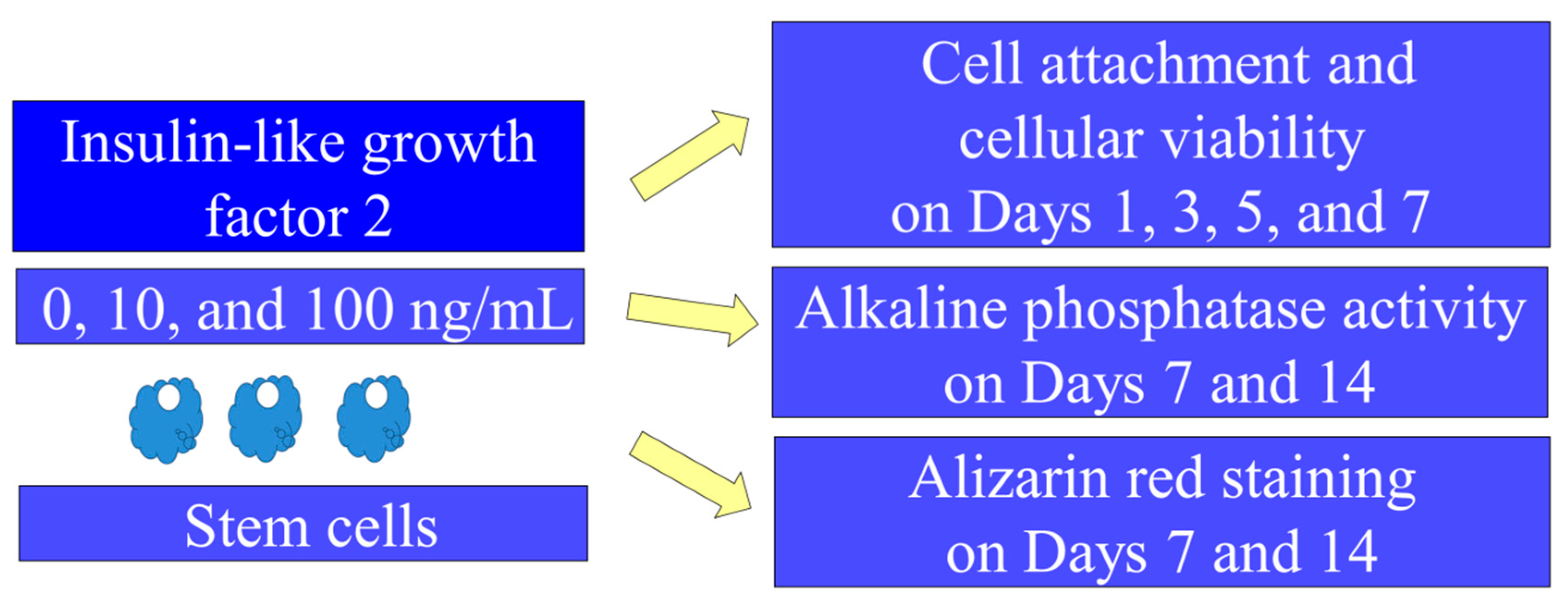
2. Materials and Methods

2.1. Culturing of Bone Marrow Mesenchymal Stem Cells on Bone

The approval of the present study was given by the Institutional Review Board of Seoul St Mary’s Hospital (KC20SISI0333). All of the experimental schemes used were carried out based on the relevant guidelines. Human bone marrow-derived mesenchymal stem cells (BMSCs; Catholic MASTER cells) were acquired from the Catholic Institute of Cell Therapy (CIC, Seoul, Korea) [14,15]. CIC verified that all of the samples showed >90% positive CD 73 and CD 90 expression.
Figure 1 indicates an overview of the study design. The stem cells were loaded onto the bone graft material (Geistlich Bio-Oss®, Geistlich Pharma AG, Wolhusen, Switzerland) and then incubated in the media. The media consist of α-minimum essential medium (MEM; Gibco; Thermo Fisher Scientific, Inc., Waltham, MA, USA), fetal bovine serum (15%; Gibco), and penicillin-streptomycin (100 U/mL and 100 µg/mL; Sigma–Aldrich; Merck KGaA, St. Louis, MO, USA). Stem cell-loaded graft was cultured in the presence of the IGF-2 at final concentrations of 0, 10, and 100 ng/mL [16].

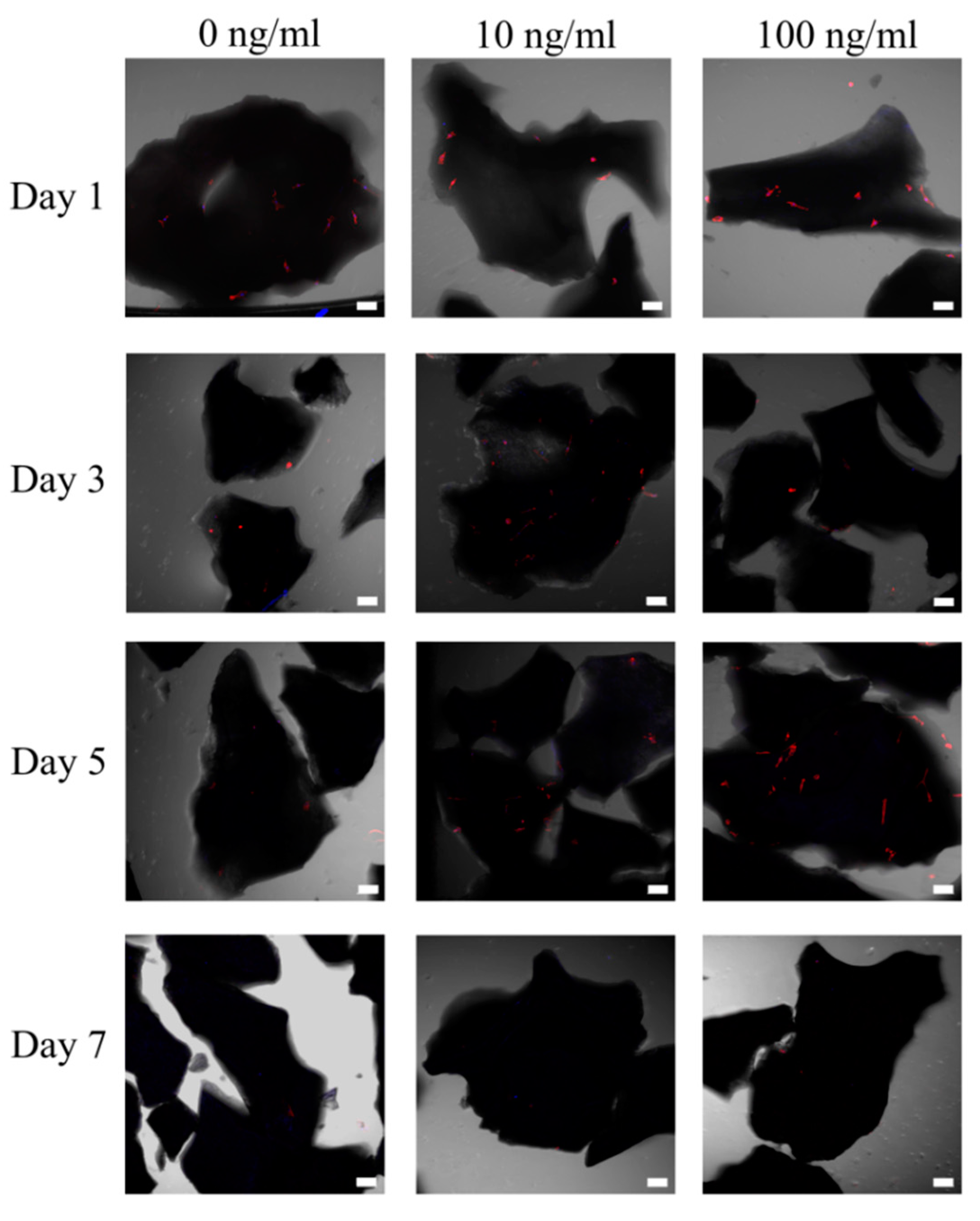
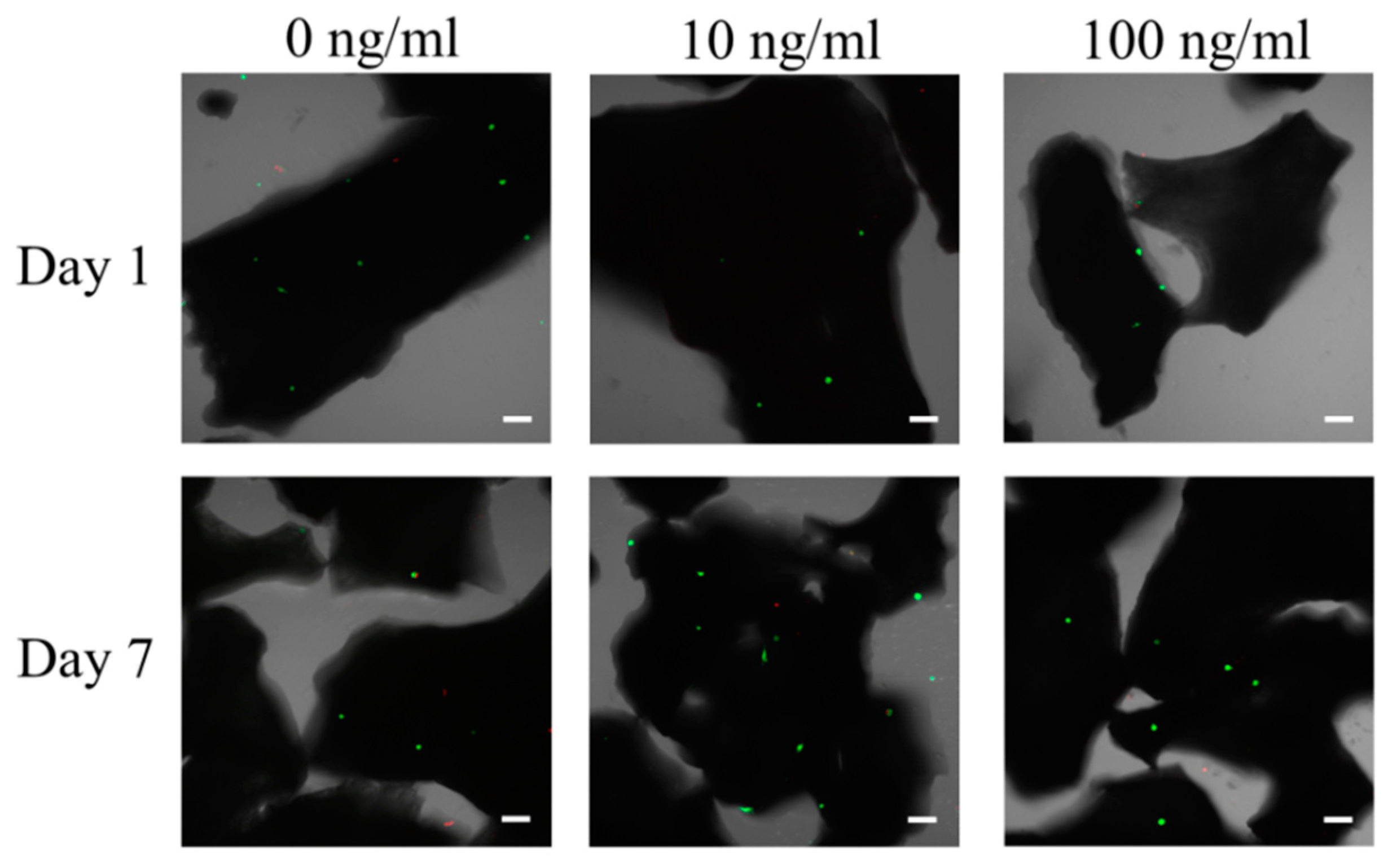
2.2. Morphologic Evaluation of Stem Cells

The morphological characteristics of the stem cells on the bone graft material was observed on Days 1, 3, 5, and 7. The culture media was then removed and the cells on the graft material were gently washed with phosphate-buffered saline. After fixation of the stem cells with 4% paraformaldehyde (Biosesang, Seongnam-si, Korea) at ambient temperature for 20 min., the cells on the bone graft material were rinsed three times with Dulbecco’s phosphate-buffered saline solution. The cells were then stained using a 1× Phalloidin conjugate working solution for 60 min. at room temperature; a 1× Phalloidin conjugate working solution can be made from 1 µL 1000× Phalloidin conjugate stock solution (ab235138, Abcam) and 1 mL of 1% BSA in phosphate-buffered saline. Small drop volumes (approximately 25 µL) of mounting medium containing nuclear stain (Vectashield, VECTOR laboratories, Burlingame, CA, USA) were applied to the cells and the stem loaded bone grafts were observed using a microscope.

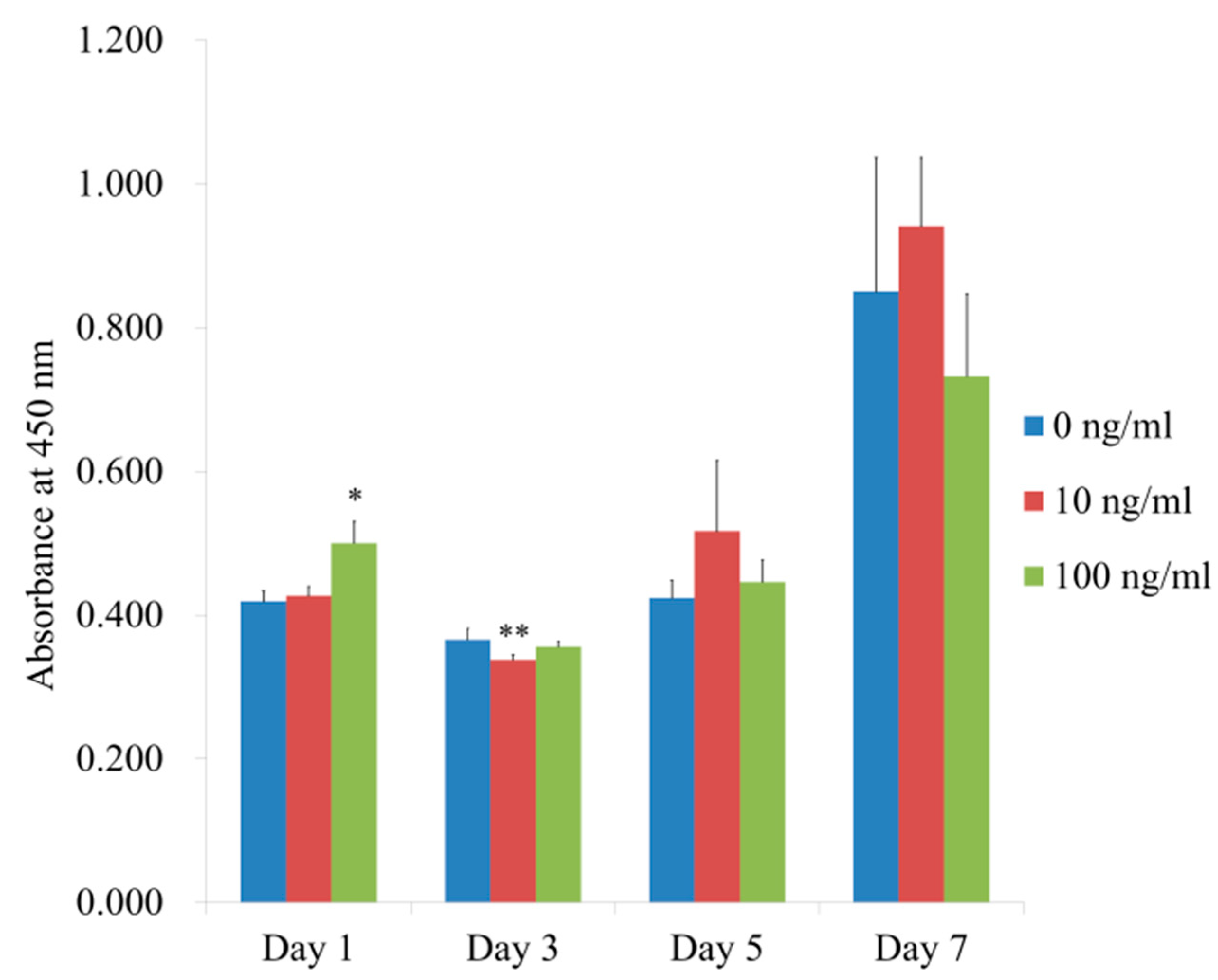
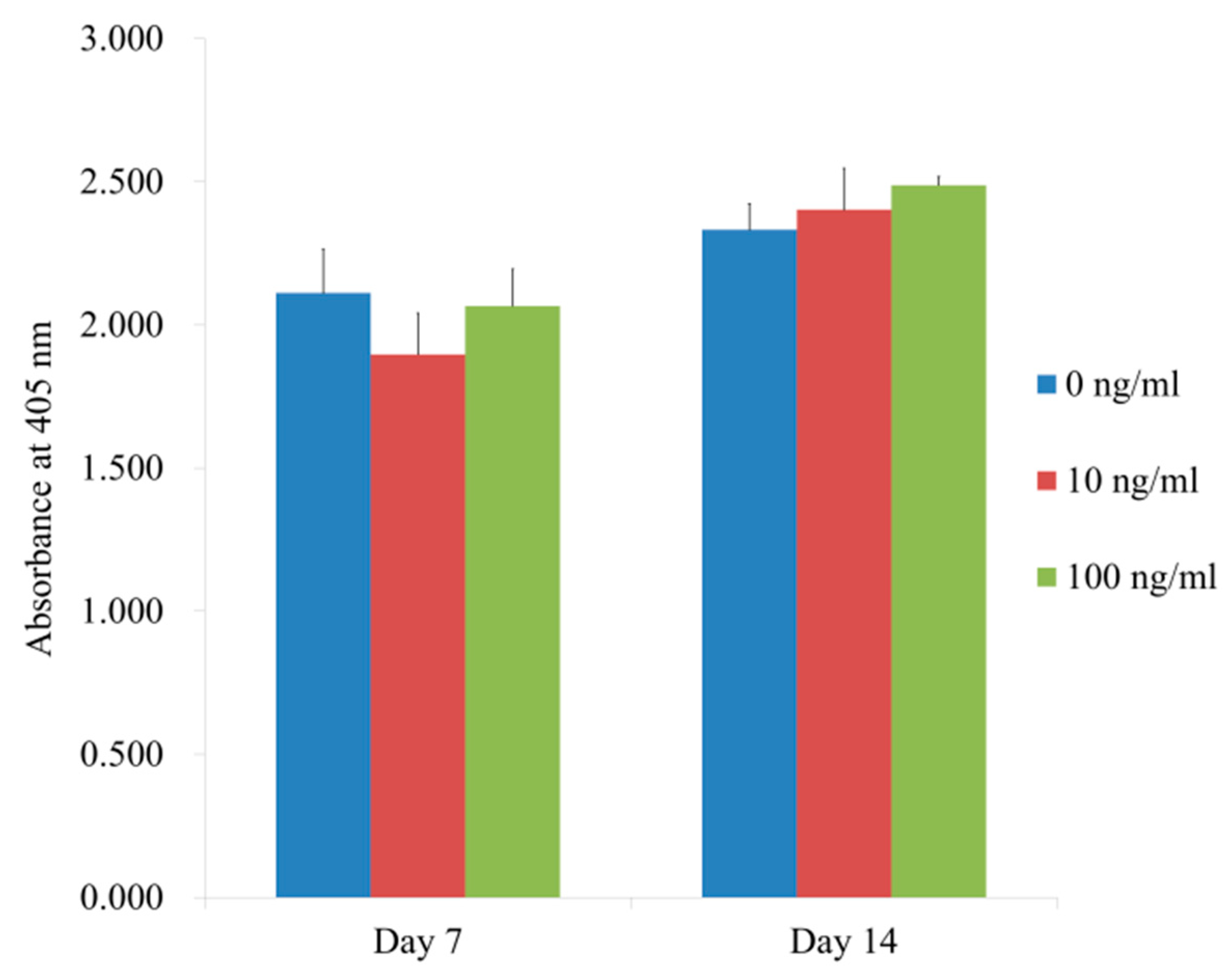
2.3. Determination of Cellular Viability

We used the commercially available two-color assay based on plasma membrane integrity and esterase activity (Live/Dead Kit assay, Molecular Probes, Eugene, OR, USA) for qualitative analysis of the stem cells that were cultured on the bone graft material on Days 1 and 7. The media was removed and then suspended in 1 mL of Dulbecco’s phosphate-buffered saline working solution (containing 2 μL of 2 mM ethidium homodimer-1 and 0.5 μL of 4 mM calcein acetoxy methyl ester) for 1 h at room temperature. We also performed a quantitative cellular viability test using an assay kit based on the water-soluble tetrazolium salt (Cell Counting Kit-8, Dojindo, Tokyo, Japan) on Days 1, 3, 5, and 7.

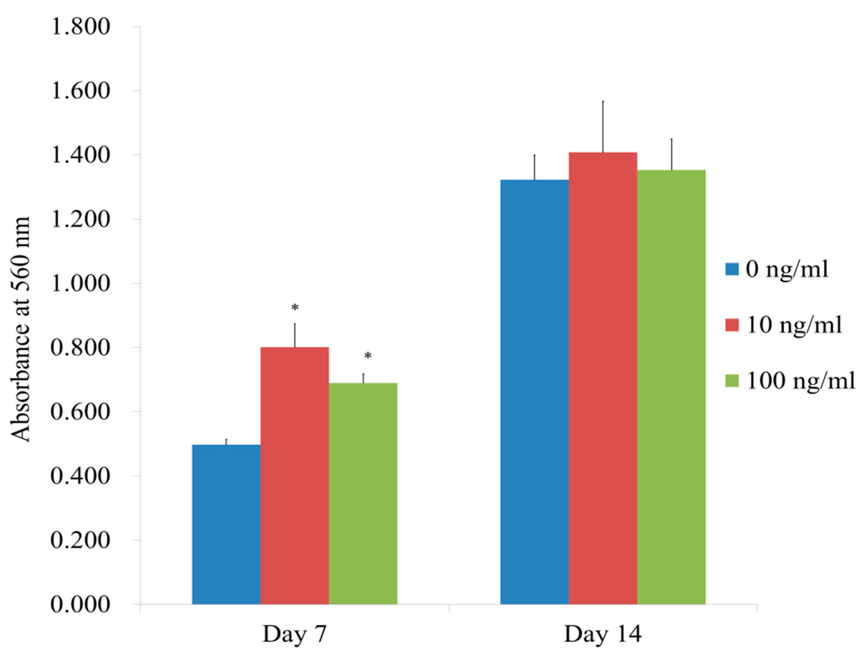
2.4. Level of Alkaline Phosphatase Activity and Calcium Deposition

The level of alkaline phosphatase activity was analyzed in order to evaluate the differentiation to osteogenic lineage on Days 7 and 14 (K412-500, BioVision, Inc., Milpitas, CA, USA). The cells on the bone mineral particle were re-suspended with an assay buffer, then decomposed with triton X-100, and then centrifuged to eliminate insoluble material. We incubated the mixture for 60 min at 4 °C after mixing the supernatant with the substrate. Subsequently, we measured the spectrophotometric absorbance at 405 nm.
We used an anthraquinone dye assay for calcium deposit evaluation on Days 7 and 14 to assess the osteogenic differentiation. After washing and fixation procedures, we stained the stem cells on the bone graft with 2% Alizarin-Red Solution (ScienCell Research Laboratories, Inc., Carlsbad, CA, USA) for 20 min. at room temperature. Ten percent cetylpyridinium chloride (Sigma-Aldrich Co.) solution was applied for 15 min at ambient temperature to solubilized the bound dye. We measured the spectrophotometric absorbance at 560 nm to perform the quantification of the bound dyes.

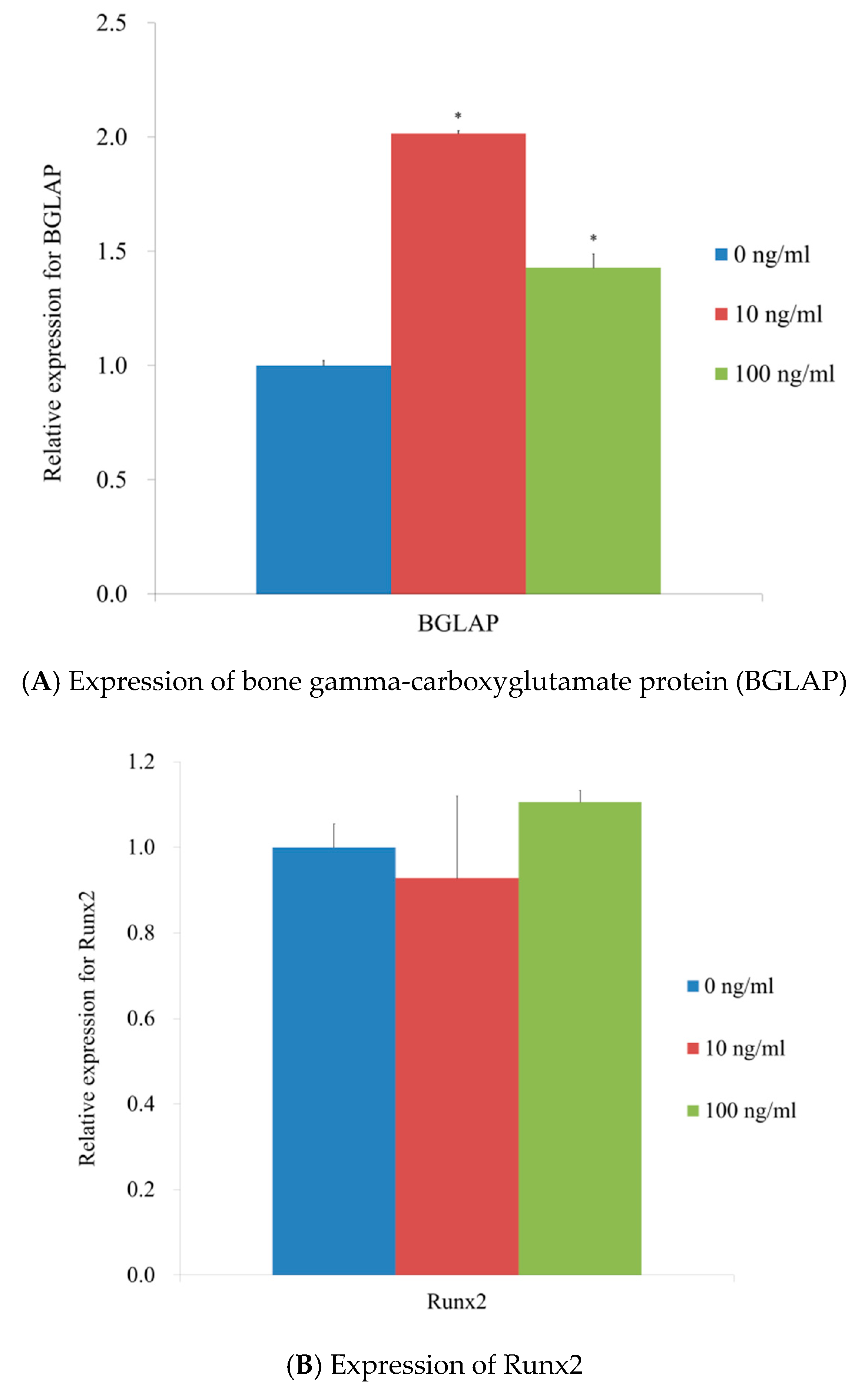
2.5. mRNA Quantification by Real-Time Polymerase Chain Reaction

We harvested the cells on Day 7 and isolated total RNA using purification kit (Thermo Fisher Scientific, Inc.), following the manufacturer’s instructions [17]. RNA quality was assessed with a bioanalyzer (Agilent 2100, Agilent Technologies) using the kit (RNA 6000 Nano Chip; Agilent Technologies, Amstelveen, The Netherlands), and the quantity was performed using the spectrophotometer (ND-2000, Thermo Fisher Scientific, Inc.) with a ratio of absorbance at 260 nm and 280 nm spectrophotometrically. We used RNA as a template for reverse transcription while using the reverse transcriptase (SuperScript II; Invitrogen, Carlsbad, CA, USA).
We detected mRNA expression by real-time polymerase chain reaction (PCR). We used GenBank to designe the sense and antisense primers. The primer sequences were as follows: bone gamma-carboxyglutamate protein (BGLAP) (forward: 5′-AATCCGGACTGTGACGAGTT-3′, reverse: 5′-CAGCAGAGCGACACCCTAGA-3′), Runx2 (forward: 5′-CAGTTCCCAAGCATTTCATCC-3′, reverse: 5′- AGGTGGCTGGATAGTGCATT-3′), and β-actin (forward: 5′-AATGCTTCTAGGCGGACTATGA-3′, reverse: 5′-TTTCTGCGCAAGTTAGGTTTT-3′). Normalization was performed by applying β-actin as a housekeeping gene. Real-time PCR was performed using SYBR Green PCR Kit (Applied Biosystems, Waltham, MA, USA) on the PCR System (StepOnePlusTM; Applied Biosystems), according to the manufacturer’s instructions.

2.6. Statistical Analysis

All of the statistical analyses were performed using the software (SPSS 12 for Windows, SPSS Inc., Chicago, IL, USA) via one-way analysis of variance followed by Tukey’s post hoc test after conducting a test of normality and the equality of variances of the samples. Three experimental repeats were evaluated for the analysis. All of the results values were presented as mean ± standard deviation with the level of significance at 0.05.

3. Results

3.1. Morphologic Evaluation and Cellular Viability of Stem Cells

The stem cells were well-attached with fibroblast morphology at Day 1 (Figure 2). Adding IGF-2 did not produce significant changes. The morphology of the cells with longer incubation showed a similar shape of fibroblast (Figure 2).
We noticed that most of the stem cells produced a high intensity of green fluorescence, meaning these were live cells (Figure 3). Low or minimal red fluorescence was noted. We did not see any noticeable changes with the longer incubation time on Day 7.
Figure 4 shows the absorbance values at 450 nm on Days 1, 3, 5, and 7. The results for cellular viability assay values for the IGF-2 groups at 0, 10, and 100 ng/mL on Day 1 were 0.419 ± 0.015, 0.427 ± 0.013, and 0.500 ± 0.030, respectively (p < 0.05).

3.2. Level of Osteogenic Differentiation and Mineralization

Figure 5 shows the results of the alkaline phosphatase activity assays. The absorbance values on Day 7 were 2.112 ± 0.152, 1.897 ± 0.144, and 2.067 ± 0.128, for IGF-2 at 0, 10, and 100 ng/mL, respectively (p > 0.05). The values at 405 nm on Day 14 were 2.332 ± 0.091, 2.403 ± 0.145, and 2.488 ± 0.030, for IGF-2 at 0, 10, and 100 ng/mL, respectively (p > 0.05).
Figure 6 shows the results of the mineralization assay at Days 7 and 14. The absorbance values at 560 nm on Day 7 for IGF-2 at 0, 10, and 100 ng/mL were 0.497 ± 0.017, 0.801 ± 0.073, and 0.690 ± 0.028, respectively (p < 0.05). The quantification results showed significantly higher values for IGF-2 groups at 10 ng/mL and 100 ng/mL concentration when compared with the control (p < 0.05). The absorbance values at 560 nm on Day 14 for IGF-2 at 0, 10, and 100 ng/mL were 1.322 ± 0.077, 1.408 ± 0.159, and 1.353 ± 0.097, respectively (p > 0.05).

3.3. Total RNA Extraction and Quantification by Real-Time Polymerase Chain Reaction

Quantitative real-time PCR revealed that the mRNA levels of BGLAP were 1.000 ± 0.022, 2.016 ± 0.011, and 1.429 ± 0.059 for IGF-2 groups at 0, 10, and 100 ng/mL, respectively. The results showed that the application of IGF-2 groups of 10 and 100 ng/mL produced a significant increase of BGLAP (Figure 7A).
Quantitative real-time PCR revealed that mRNA levels of Runx2 were 1.000 ± 0.054, 0.928 ± 0.192, and 1.106 ± 0.027 for IGF-2 groups at 0, 10, and 100 ng/mL, respectively. The results showed that the application of IGF-2 did not produce a significant change in Runx2 expressions (Figure 7B).

4. Discussion

In this report, we evaluated the effects of IGF-2 on stem cells that were cultured on deproteinized bovine bone at the concentration of 10 and 100 ng/mL. Applying IGF-2 increased alkaline phosphatase activity and mineralization of the bone marrow derived stem cells by enhancing the expression of BGLAP.
IGFs are protein hormones that share structural similarity to insulin, having affinity and specificity to IGF receptors and IGF-binding proteins [18]. IGF-1 is reported to be one of the most abundant growth factors in the bone matrix and it is involved in the maintenance of bone mass in adulthood [19]. In a previous report, IGF-1 promoted the osteogenic differentiation of stem cells and it was suggested that IGF-1 treated stem cells may serve as a candidate for bone regeneration [19]. Alternately, IGF-2 is reported to be involved in the development and function of various organs, including liver and brain [20,21]. IGF-2 is reported to be involved in reversing or preventing the aging-related memory loss in animal model [20]. IGF-2 also acted as an important mitogen regarding liver repopulation in animal model [21]. IGF-2 was reported to be at higher level in the osteoblast-conditioned medium when compared with the control medium conditioned with the stem cells [22]. The expression of IGF-2 in developing osteophyte was strongly associated with a degree of osteogenesis, with the highest expression in active osteoblasts [23]. IGF-2 was shown to produce the increase in the total protein synthesis and collagenous protein synthesis in osteoblast like cells isolated from human bone [24]. Moreover, IGF-2 is reported to be an essential component of stem cell niche and to be involved in the sproliferation and differentiation of stem cells [16]. In this study, the use of IGF-2 on stem cell loaded graft produced higher cellular viability and higher mineralization.
The various concentrations of IGF-2 were tested to evaluate/suggest the optimal concentration [16,25]. IGF-2 was used at different concentrations and concentrations ranging from 50 to 200 ng/mL promoted the proliferation of stem cells [16]. The application of IGF-2 at 200 ng/mL concentration enhanced the cellular proliferation of human marrow stromal cell line, which was shown by increased [3H]thymidine incorporation and increase in the cell number [25]. In this study, the significant increase of osteogenesis and mRNA expression was seen both at 10 and 100 ng/mL, with the highest at 10 ng/mL concentration. It was shown that IGF-2 had no effects on cell proliferation in the presence of 10% fetal bovine serum, but it promoted the cellular proliferation in the presence 1% fetal bovine serum [16]. It was shown that IGF-2 promoted the adipogenic differentiation of stem cells at 100 ng/mL concentration [26]. Likewise, the net effects or optimal dosage may differ between the studies due to culturing conditions, type, and passages of cells [27]. Moreover, IGF-2 may have additional functions. It was shown that IGF-2 promoted the adipogenic differentiation of stem cells at 100 ng/mL concentration [26]. IGFs have been shown to be involved in the chondrogenesis and the addition of IGF-2 increased the induction of chondrogenic differentiation while using the three-dimensional culture system [28,29].
The expressions of various genes were tested to evaluate the osteogenic differentiation including Runx 2 and BGLAP [17,30]. Runx2 and BGLAP are widely used biomarkers for osteogenic differentiation [30]. BGLAP was reported to play an important role in the modulation of osteogenic differentiation of mesenchymal stem cells and the maturation of mineral species [31]. In a previous report, attempts were made to achieve the increase in osteogenic potential of stem cells by applying hypoxic conditions [32]. Hypoxia condition upregulation of the various genes, including BGLAP and SPP1, but the condition did not affect Runx2 expression [32]. In this report, IGF-2 seems to enhance the expression of BGLAP without influencing the expression of Runx2.

5. Conclusions

Collectively, these results showed that the application of IGF-2 on stem cell loaded bone graft increased cellular viability, Alizarin red staining, and BGLAP expression of stem cells. This report suggests that the combined approach of stem cells and IGF-2 with scaffold may have synergistic effects on osteogenesis.

Author Contributions

Conceptualization, H.L., S.K.M., Y.-H.P. and J.-B.P.; methodology, H.L., S.K.M., Y.-H.P. and J.-B.P.; formal analysis, H.L., S.K.M., Y.-H.P. and J.-B.P.; writing—original draft preparation, H.L., S.K.M., Y.-H.P. and J.-B.P.; and writing—review and editing, H.L., S.K.M., Y.-H.P. and J.-B.P. All authors have read and agreed to the published version of the manuscript.

Funding

This study was funded by the National Research Foundation of Korea (NRF) grant funded by the Korea government (MSIT) (No. 2020R1A2C4001624). This research was also funded by Research Fund of Seoul St. Mary’s Hospital, The Catholic University of Korea.

Acknowledgments

The Catholic MASTER Cells were derived from human bone marrow donated by healthy donors after informed consent and the Catholic MASTER Cells were supplied by CIC.

Conflicts of Interest

The authors report no conflicts of interest.

References

  1. Park, J.B. Clinical and histomorphometric evaluation of staged approach using bone augmentation and autogenous masticatory mucosal graft with endosseous implant placement: A case report. J. Oral Implantol. 2008, 34, 334–338. [Google Scholar] [CrossRef]
  2. Park, J.B. Implant installation with simultaneous ridge augmentation. Report of three cases. J. Oral Implantol. 2011, 37, 595–603. [Google Scholar] [CrossRef] [PubMed]
  3. Ge, Y.; Feng, H.; Wang, L. Application of a novel resorbable membrane in the treatment of calvarial defects in rats. J. Biomater. Sci. Polym. Ed. 2011, 22, 2417–2429. [Google Scholar] [CrossRef] [PubMed] [Green Version]
  4. de Fernandez Grado, G.; Keller, L.; Idoux-Gillet, Y.; Wagner, Q.; Musset, A.M.; Benkirane-Jessel, N.; Bornert, F.; Offner, D. Bone substitutes: A review of their characteristics, clinical use, and perspectives for large bone defects management. J. Tissue Eng. 2018, 9. [Google Scholar] [CrossRef] [PubMed] [Green Version]
  5. Park, J.B. Healing of extraction socket grafted with deproteinized bovine bone and acellular dermal matrix: Histomorphometric evaluation. Implant. Dent. 2010, 19, 307–313. [Google Scholar] [CrossRef]
  6. Kang, S.H.; Park, J.B.; Kim, I.; Lee, W.; Kim, H. Assessment of stem cell viability in the initial healing period in rabbits with a cranial bone defect according to the type and form of scaffold. J. Periodontal Implant. Sci. 2019, 49, 258–267. [Google Scholar] [CrossRef]
  7. Maglione, M.; Salvador, E.; Ruaro, M.E.; Melato, M.; Tromba, G.; Angerame, D.; Bevilacqua, L. Bone regeneration with adipose derived stem cells in a rabbit model. J. Biomed. Res. 2018, 33, 38–45. [Google Scholar] [CrossRef]
  8. Park, J.B. Use of cell-based approaches in maxillary sinus augmentation procedures. J. Craniofac. Surg. 2010, 21, 557–560. [Google Scholar] [CrossRef]
  9. Park, K.H.; Kim, H.; Moon, S.; Na, K. Bone morphogenic protein-2 (BMP-2) loaded nanoparticles mixed with human mesenchymal stem cell in fibrin hydrogel for bone tissue engineering. J. Biosci. Bioeng. 2009, 108, 530–537. [Google Scholar] [CrossRef]
  10. Park, J.B.; Kim, K.Y.; Lee, W.; Kim, H.; Kim, I. Combinatorial effect of stem cells derived from mandible and recombinant human bone morphogenetic protein-2. Tissue Eng. Regen. Med. 2015, 12, 343–351. [Google Scholar] [CrossRef]
  11. Kawai, M.; Rosen, C.J. The insulin-like growth factor system in bone: Basic and clinical implications. Endocrinol. Metab. Clin. N. Am. 2012, 41, 323–333. [Google Scholar] [CrossRef] [PubMed] [Green Version]
  12. Hill, P.A.; Reynolds, J.J.; Meikle, M.C. Osteoblasts mediate insulin-like growth factor-I and -II stimulation of osteoclast formation and function. Endocrinology 1995, 136, 124–131. [Google Scholar] [CrossRef]
  13. Chen, L.; Jiang, W.; Huang, J.; He, B.C.; Zuo, G.W.; Zhang, W.; Luo, Q.; Shi, Q.; Zhang, B.Q.; Wagner, E.R.; et al. Insulin-like growth factor 2 (IGF-2) potentiates BMP-9-induced osteogenic differentiation and bone formation. J. Bone Miner. Res. Off. J. Am. Soc. Bone Miner. Res. 2010, 25, 2447–2459. [Google Scholar] [CrossRef] [PubMed] [Green Version]
  14. Jeong, C.H.; Kim, S.M.; Lim, J.Y.; Ryu, C.H.; Jun, J.A.; Jeun, S.S. Mesenchymal stem cells expressing brain-derived neurotrophic factor enhance endogenous neurogenesis in an ischemic stroke model. BioMed Res. Int. 2014, 2014, 129145. [Google Scholar] [CrossRef] [Green Version]
  15. Tae, J.Y.; Ko, Y.; Park, J.B. Evaluation of fibroblast growth factor-2 on the proliferation of osteogenic potential and protein expression of stem cell spheroids composed of stem cells derived from bone marrow. Exp. Ther. Med. 2019, 18, 326–331. [Google Scholar] [CrossRef] [PubMed]
  16. Wang, C.; Li, X.; Dang, H.; Liu, P.; Zhang, B.O.; Xu, F. Insulin-like growth factor 2 regulates the proliferation and differentiation of rat adipose-derived stromal cells via IGF-1R and IR. Cytotherapy 2019, 21, 619–630. [Google Scholar] [CrossRef]
  17. Lee, H.; Lee, H.; Na, C.B.; Park, J.B. The effects of simvastatin on cellular viability, stemness and osteogenic differentiation using 3-dimensional cultures of stem cells and osteoblast-like cells. Adv. Clin. Exp. Med. Off. Organ Wroc. Med. Univ. 2019, 28, 699–706. [Google Scholar] [CrossRef]
  18. Cohen, P.; Peehl, D.M.; Lamson, G.; Rosenfeld, R.G. Insulin-like growth factors (IGFs), IGF receptors, and IGF-binding proteins in primary cultures of prostate epithelial cells. J. Clin. Endocrinol. Metab. 1991, 73, 401–407. [Google Scholar] [CrossRef]
  19. Wang, S.; Mu, J.; Fan, Z.; Yu, Y.; Yan, M.; Lei, G.; Tang, C.; Wang, Z.; Zheng, Y.; Yu, J.; et al. Insulin-like growth factor 1 can promote the osteogenic differentiation and osteogenesis of stem cells from apical papilla. Stem Cell Res. 2012, 8, 346–356. [Google Scholar] [CrossRef] [Green Version]
  20. Steinmetz, A.B.; Johnson, S.A.; Iannitelli, D.E.; Pollonini, G.; Alberini, C.M. Insulin-like growth factor 2 rescues aging-related memory loss in rats. Neurobiol. Aging 2016, 44, 9–21. [Google Scholar] [CrossRef] [Green Version]
  21. Wang, M.J.; Chen, F.; Liu, Q.G.; Liu, C.C.; Yao, H.; Yu, B.; Zhang, H.B.; Yan, H.X.; Ye, Y.; Chen, T.; et al. Insulin-like growth factor 2 is a key mitogen driving liver repopulation in mice. Cell Death Dis. 2018, 9, 26. [Google Scholar] [CrossRef] [PubMed] [Green Version]
  22. Abu-Khader, A.; Law, K.W.; Jahan, S.; Manesia, J.K.; Pasha, R.; Hovey, O.; Pineault, N. Paracrine Factors Released by Osteoblasts Provide Strong Platelet Engraftment Properties. Stem Cells 2019, 37, 345–356. [Google Scholar] [CrossRef] [PubMed]
  23. Middleton, J.; Arnott, N.; Walsh, S.; Beresford, J. Osteoblasts and osteoclasts in adult human osteophyte tissue express the mRNAs for insulin-like growth factors I and II and the type 1 IGF receptor. Bone 1995, 16, 287–293. [Google Scholar] [CrossRef]
  24. Strong, D.D.; Beachler, A.L.; Wergedal, J.E.; Linkhart, T.A. Insulinlike growth factor II and transforming growth factor beta regulate collagen expression in human osteoblastlike cells in vitro. J. Bone Miner. Res. Off. J. Am. Soc. Bone Miner. Res. 1991, 6, 15–23. [Google Scholar] [CrossRef]
  25. Thomas, T.; Gori, F.; Spelsberg, T.C.; Khosla, S.; Riggs, B.L.; Conover, C.A. Response of bipotential human marrow stromal cells to insulin-like growth factors: Effect on binding protein production, proliferation, and commitment to osteoblasts and adipocytes. Endocrinology 1999, 140, 5036–5044. [Google Scholar] [CrossRef]
  26. Zhang, K.; Wang, F.; Huang, J.; Lou, Y.; Xie, J.; Li, H.; Cao, D.; Huang, X. Insulin-like growth factor 2 promotes the adipogenesis of hemangioma-derived stem cells. Exp. Ther. Med. 2019, 17, 1663–1669. [Google Scholar] [CrossRef] [Green Version]
  27. Yu, N.H.; Chun, S.Y.; Ha, Y.S.; Kim, H.T.; Kim, D.H.; Kim, J.; Chung, J.W.; Lee, J.N.; Song, P.H.; Yoo, E.S.; et al. Optimal Stem Cell Transporting Conditions to Maintain Cell Viability and Characteristics. Tissue Eng. Regen. Med. 2018, 15, 639–647. [Google Scholar] [CrossRef]
  28. Longobardi, L.; O’Rear, L.; Aakula, S.; Johnstone, B.; Shimer, K.; Chytil, A.; Horton, W.A.; Moses, H.L.; Spagnoli, A. Effect of IGF-I in the chondrogenesis of bone marrow mesenchymal stem cells in the presence or absence of TGF-beta signaling. J. Bone Miner. Res. Off. J. Am. Soc. Bone Miner. Res. 2006, 21, 626–636. [Google Scholar] [CrossRef]
  29. Youssef, A.; Aboalola, D.; Han, V.K. The Roles of Insulin-Like Growth Factors in Mesenchymal Stem Cell Niche. Stem Cells Int. 2017, 2017, 9453108. [Google Scholar] [CrossRef]
  30. Lv, H.; Yang, H.; Wang, Y. Effects of miR-103 by negatively regulating SATB2 on proliferation and osteogenic differentiation of human bone marrow mesenchymal stem cells. PLoS ONE 2020, 15, e0232695. [Google Scholar] [CrossRef]
  31. Tsao, Y.T.; Huang, Y.J.; Wu, H.H.; Liu, Y.A.; Liu, Y.S.; Lee, O.K. Osteocalcin Mediates Biomineralization during Osteogenic Maturation in Human Mesenchymal Stromal Cells. Int. J. Mol. Sci. 2017, 18, 159. [Google Scholar] [CrossRef] [PubMed]
  32. Fernandez-Moure, J.S.; Corradetti, B.; Chan, P.; Van Eps, J.L.; Janecek, T.; Rameshwar, P.; Weiner, B.K.; Tasciotti, E. Enhanced osteogenic potential of mesenchymal stem cells from cortical bone: A comparative analysis. Stem Cell Res. Ther. 2015, 6, 203. [Google Scholar] [CrossRef] [PubMed] [Green Version]
Figure 1. Overview of study design.
Applsci 10 05471 g001
Figure 2. Morphologic characteristics of stem cells loaded on the bone mineral particle on Day 1, 3, 5, and 7. Scale bar indicates 100 μm (original magnification: ×50).
Applsci 10 05471 g002
Figure 3. Live, dead, and merged images of stem cells on Days 1 and 7 Scale bar indicates 100 μm (original magnification: ×50).
Applsci 10 05471 g003
Figure 4. Cellular viability using Cell Counting Kit-8 on Days 1, 3, 5, and 7. * Significant differences were seen when compared to the control at Day 1 (p < 0.05). ** Significant differences were identified when the value was compared to the control at Day 3 (p < 0.05).
Applsci 10 05471 g004
Figure 5. Alkaline phosphatase activity on Days 7 and 14.
Applsci 10 05471 g005
Figure 6. Results of Alizarin Red S staining on Days 7 and 14. * Significant differences were seen when compared to the control at Day 7 (p < 0.05).
Applsci 10 05471 g006
Figure 7. Quantification by real-time polymerase chain reaction. * Statistically significant increase was seen when compared with 0 ng/mL at Day 7 (p < 0.05).
Applsci 10 05471 g007

