Communication

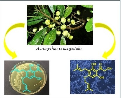
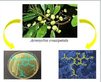
Potent Antibacterial Prenylated Acetophenones from the Australian Endemic Plant Acronychia crassipetala

1
GeneCology Research Centre, School of Science and Engineering, University of the Sunshine Coast, Maroochydore DC, Queensland 4558, Australia
2
School of Health and Sports Sciences, University of the Sunshine Coast, Maroochydore DC, Queensland 4558, Australia
3
QIMR Berghofer Medical Research Institute, Locked Bag 2000, PO Royal Brisbane Hospital, Herston, Queensland 4029, Australia
4
QBiotics Limited, PO Box 1, Yungaburra, Queensland 4884, Australia
*
Author to whom correspondence should be addressed.
Antibiotics 2020, 9(8), 487; https://doi.org/10.3390/antibiotics9080487
Submission received: 28 June 2020 / Revised: 30 July 2020 / Accepted: 4 August 2020 / Published: 6 August 2020

Abstract

:
Acronychia crassipetala is an endemic plant species in Australia. Its phytochemistry and therapeutic properties are underexplored. The hexane extract of the fruit A. crassipetala T. G. Hartley was found to inhibit the growth of the Gram-positive bacteria Staphylococcus aureus. Following bio-activity guided fractionation, two prenylated acetophenones, crassipetalonol A (1) and crassipetalone A (2), were isolated. Their structures were determined mainly by NMR and MS spectroscopic analyses. This is the first record of the isolation and structural characterisation of secondary metabolites from the species A. crassipetala. Their antibacterial and cytotoxic assessments indicated that the known compound (2) had more potent antibacterial activity than the antibiotic chloramphenicol, while the new compound (1) showed moderate cytotoxicity.

Graphical Abstract

1. Introduction

As a result of geographic isolation and a vast array of geographical and environmental habitats, Australia is one of the most megadiverse countries in the world with 84% of terrestrial plants being classified as endemic species [1]. Analysis of Australian flora from Australian tropical habitats showed these regions are particularly rich in plant diversity and species endemism [2]. Despite accounting for only 0.3% of the Australian continent, Queensland’s tropical rainforests are internationally recognised as one of the global biodiversity hotspots [3]. This unique ecological resource is a distinct and relatively untapped source of novel and new natural products with therapeutic potential [4]. For example, the Queensland tropical native species Duboisia myoporoides R. Brown is a source of tropane alkaloids including scopolamine, which is an important precursor for the synthesis of scopolamine butylbromide, an anticholinergic and antispasmodic drug with the brand name Buscopan [5]. Another example is a novel indolizidine alkaloid, grandisine A, from the Queensland rainforest tree Elaeocarpus grandis F. Muell, which exhibited binding affinity for the human δ-opioid receptor and has been considered as a potential lead for developing an analgesic agent [6]. More recently, an epoxy-tigliane diterpene, tigilanol tiglate (formerly EBC46), from the Queensland tropical endemic species Fontainea picrosperma C. T. White, has been discovered and shown to have significant anticancer activity [7]. Tigilanol tiglate has been approved by the European Medicines Authority [8] as a novel canine therapy for mast cell tumours [9] and is currently in clinical trials to assess its potential as a human anticancer therapeutic [10].
With the success of discovering and developing the novel anticancer natural compound tigilanol tiglate from QBiotics’s plant extract library (EcoLogicTM), a new drug discovery campaign has been launched to discover potent antibacterial natural products [11]. A hexane extract from the fruit of Acronychia crassipetala T. G. Hartley was shown to be active against the Gram-positive (G+ve) bacteria Staphylococcus aureus.
The genus Acronychia belonging to the family Rutaceae comprises over 40 species, which have a broad distribution from India, China, Malesia, New Caledonia to Australia [12]. Some Acronychia spp. have been used in folk medicines of indigenous Asian and Australian populations for the treatments of diarrhoea, asthma, ulcers, rheumatism, fever and parasitic infections [13,14,15]. About 60 compounds including acetophenones, flavonoids, quinoline and acridone alkaloids have been identified from approximately half of the Acronychia spp. plants [12,16]. Several compounds have been found with potential biological activities such as acronine, an anticancer agent from A. baueri [17]; acrophyllodine, an antiarrhythmic agent from A. haplophylla [18]; and acrovestone, an antityrosinase compound from A. pedunculata [19]. The species A. crassipetala is one of the 19 Acronychia spp. recognised in Australia [20]. This shrub is endemic to the wet tropical rainforests of northeastern Queensland and found exclusively at altitudes between 400 and 1250 m [20]. So far, chemical investigation of A. crassipetala has been limited to the study of essential oil extracted from A. crassipetala leaves [20].
This research reports the isolation, structural elucidation, antibacterial and cytotoxic properties of the two prenylated acetophenones from the fruit of A. crassipetala, crassipetalonol A (1) and crassipetalone A (2), of which crassipetalonol A (1) was found as a new compound (Figure 1).

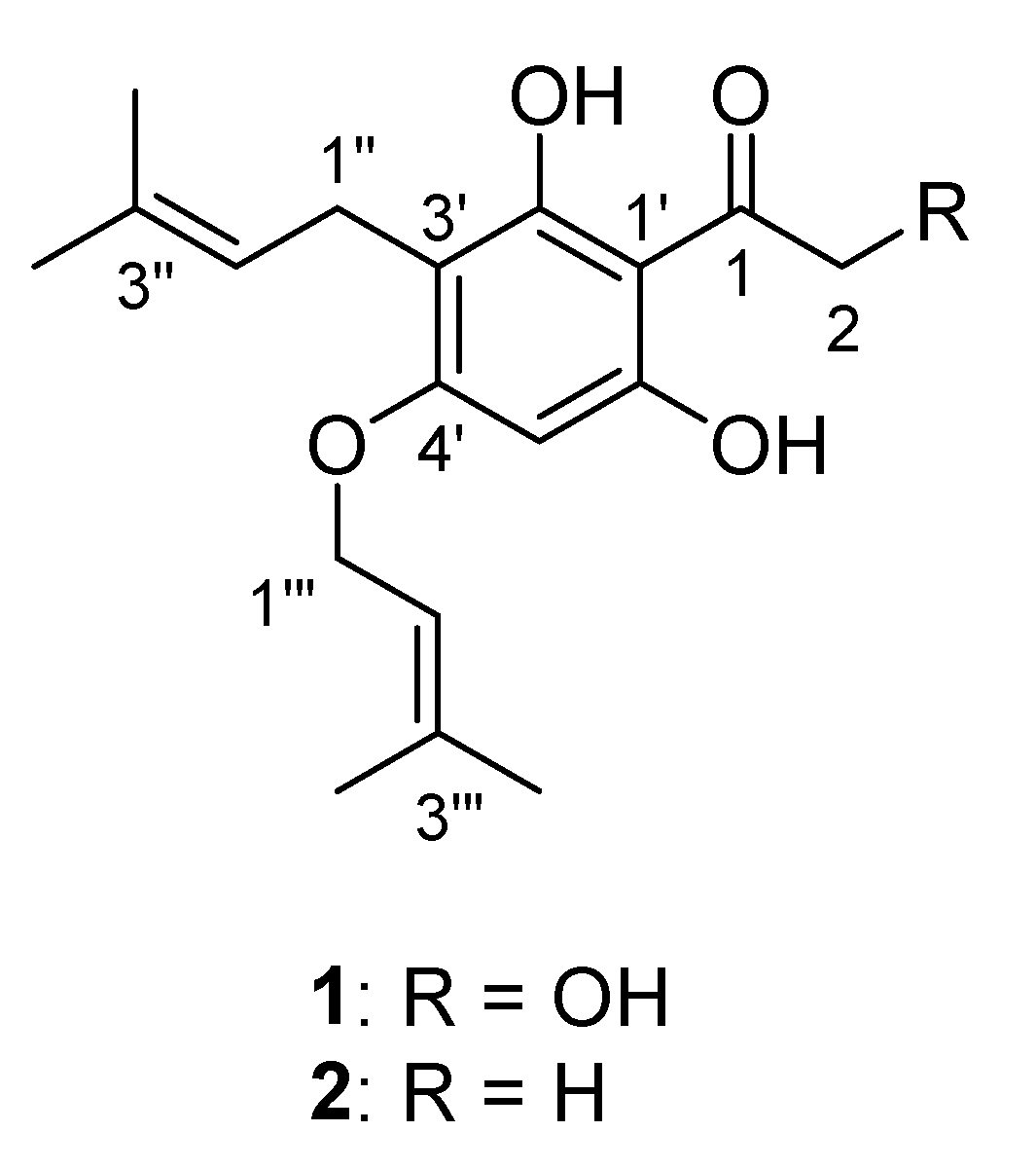
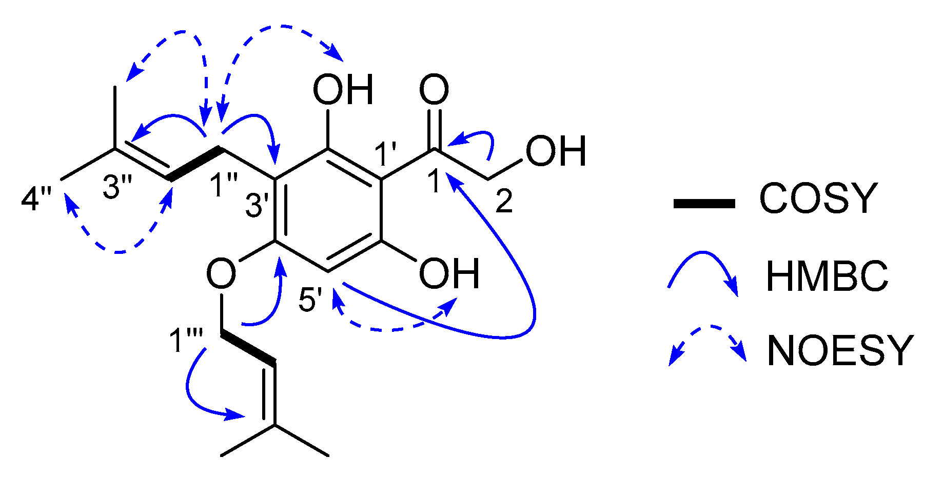
2. Results and Discussion

Compound 1 was isolated as a yellowish amorphous powder and had a molecular ion peak at (+) m/z 321.1694 in HR-ESI-MS corresponding to the molecular formula C18H24O5 with seven degrees of unsaturation. The 1H-NMR spectrum of compound 1 displayed eight singlets (δH 13.45, 10.97, 6.06, 4.63, 1.75, 1.70, 1.67 and 1.59), two doublets (δH 4.52 and 3.11), and two triplets (δH 5.40 and 5.07) (Table 1). The 13C and edited HSQC experiments confirmed 1 had 18 carbons, including 1 ketone carbonyl (δC 204.4), 3 oxygenated quaternary carbons (δC 162.5, 161.5 and 160.7), 4 olefinic quaternary carbons (δC 137.8, 129.9, 107.0 and 102.4), 3 olefinic tertiary carbons (δC 122.9, 119.2 and 91.3), 2 oxygenated methylenes (δC 68.2 and 64.6), 1 methylene (δC 20.9) and 4 methyl groups (δC 25.5, 25.4, 18.0 and 17.5) (Table 1). A spin system from the methylene at δH 3.11 (2H, d, J = 7.2 Hz, H-1″) to the olefinic proton at δH 5.07 (1H, t, J = 7.2 Hz, H-2″) showed long range HMBC correlations from H-1″ to C-3″ (δC 129.9), and H-2″ to C-4″ (δC 25.4) and C-5″ (δC 17.5), resulting in the assignment of an iso-prenyl unit. A relative orientation of the two methyl groups was defined from NOESY correlations of H-1″/H-5″ and H-2″/H-4″. A similar iso-prenyl unit was assigned for the second spin system consisting of the oxygenated methylene at δH 4.52 (2H, d, J = 6.4 Hz, H-1‴) and the olefinic proton at δH 5.40 (1H, t, J = 6.4 Hz, H-2‴). The remaining six olefinic carbons (δC 162.5, 161.5, 160.7, 107.0, 102.4 and 91.3) and four degrees of unsaturation together with HMBC correlations from H-5′ (δH 6.06) to C-1′ (δC 102.4) and C-3′ (δC 107.0) supported the establishment of a penta-substituted benzene ring system. HMBC correlations of H-1″/C-3′ and H-1‴/C-4′ enabled the first and second iso-prenyl units to connect to the benzene ring at C-3′ and C-4′, respectively. A hydroxymethyl ketone group was deduced and connected to C-1′ due to a cross-peak correlation from a hydroxymethyl H-2 to C-1 (δC 204.4) and a four-bond correlation from H-5′ to C-1. HMBC correlations of 2′-OH/C-1′, 2′-OH/C-3′, 6′-OH/C-1′ and 6′-OH/C-5′, and NOESY correlations of 2′-OH/H-1″ and 6′-OH/H-5′ confirmed the positions of the two hydroxy groups (δH 13.45 and 10.97) at C-2′ and C-6′, respectively. Therefore, compound 1 was elucidated as 1-(2′,6′-dihydroxy-3′-(3″-methylbut-2″-en-1″-yl)-4′-((3‴-methylbut-2 -en-1‴-yl)oxy)phenyl)-2-hydroxyethan-1-one with a trivial name crassipetalonol A (Figure 2).
Compound 2 was assigned as 1-(2′,6′-dihydroxy-3′-(3″-methylbut-2″-en-1″-yl)-4′-((3‴-methylbut-2‴-en-1‴-yl)oxy)phenyl)ethan-1-one (trivial name, crassipetalone A) by spectroscopic data comparisons with appropriate literature values [21]. This compound was previously identified from the Euodia lunu-ankenda T. G. Hartley root bark [22] and the Urtica dioica L. nettle leaf [23]. Crassipetalone A was reported to have a fungicidal activity against Cladosporium cladosporioides [22].
The two isolated acetophenones were tested for their antibacterial activity towards several ESCAPE pathogens (Table 2 and Table S1, Supplementary materials). While crassipetalonol A (1) was found to have low or no activity towards the pathogens at the tested concentration of 156 µM, crassipetalone A (2) potently inhibited the G+ve bacteria S. aureus and Entercoccus faecium with minimum inhibitory concentration (MIC)75 values of 2.6–20.6 µM. Importantly, compound 2 displayed 2–4 fold more inhibition against S. aureus compared to the antibiotic chloramphenicol. Replacing the acetyl in 2 by the hydroxymethyl ketone in 1 reduced potency against the S. aureus strains 30-fold. Although activity against fungi and G+ve bacteria has previously been reported for acetophenone and its derivatives [24,25,26], the presence of the phenolic hydroxy groups with acidity resulted in increased biological activity by uncoupling oxidative phosphorylation [27]. Moreover, the hydrophilic/lipophilic balance of the molecule was found to play an important role in the penetration of the antibacterial agent through a bacterial cell surface [24]. A certain degree of lipophilicity produced by the iso-prenyl and other substituents in the acetophenone molecule enhanced the antimicrobial activity [26,27]. The higher lipophilicity of compound 2 compared to 1 was predicted by their octanol–water partition coefficient (ClogP) values (4.63 of 2 versus 3.77 of 1) [28]. Therefore, compound 2 could penetrate more easily through the cell wall and exert its bactericidal activity. This study also revealed that the isolated acetophenones selectively inhibited the growth of the tested G+ve bacteria rather than the Gram-negative (G-ve) ones (Table S1, Supplementary materials). These results were in accordance with previous reports of the antibacterial activity of related prenylated acetophenones [26,29,30]. The selective activity of 2 might be related to cell wall disruption or to another specific target present only in G+ve bacteria.
Cytotoxicity of compounds 1 and 2 was evaluated using a panel of five human cell lines including immortalised keratinocyte cells (HaCaT), adult dermal fibroblast cells (HDF), neonatal foreskin fibroblast cells (NFF), immortalised embryonic kidney cells (HEK293) and hepatoma cells (HepG2) (Table 3). The data suggested that crassipetalonol A (1) was 1–5 fold less cytotoxic than crassipetalone A (2). However, comparing the antibacterial and cytotoxic activities suggested that compound 2 had more potential as an antibiotic than compound 1. This difference further supported that the acetyl group contributes significantly to the antibacterial property of the Acronychia-type acetophenone skeleton. Although the selectivity indices between human cancer cells and bacterial cells of compound 2 ranged from 1 to 5, which is relatively low, its potent inhibition against the growth of S. aureus compared to other prenylated acetophenones reported previously [26,29,30,31] warrants further investigation, including in vivo trials to confirm the value of this compound. In addition, compound 2 could be modified using medicinal chemistry approaches with an aim to further improve the activity/toxicity window.

3. Materials and Methods

3.1. General Experimental Procedures

IR spectra were obtained on a PerkinElmer Spectrum 400 FT-IR spectrometer (Waltham, MA, USA). NMR spectra were acquired on a Bruker Ascend 400 spectrometer (Billerica, MA, USA) equipped with a 5 mm room temperature probe operating at 400 MHz for 1H and 100 MHz for 13C. 1H and 13C spectra were referenced to the residual deuterated solvent peaks of DMSO-d6 at δH 2.50 and δC 39.5 ppm. HR-ESI-MS data were acquired on a Sciex X500R Q-TOF mass spectrometer (Framingham, MA, USA). HPLC purifications were performed on a preparative Agilent 1200 system equipped with a diode array detector and processed by ChemStation software (C.01.07). All solvents used for extraction and chromatography were HPLC grade and the H2O used was Mili-Q water.

3.2. Plant Material

Fruits of Acronychia crassipetala T. G. Hartley (Rutaceae) were sampled from four mature trees growing in lower montane tropical rainforest at Upper Barron, Queensland, Australia, and combined into a single collection for subsequent analysis. Voucher specimens were also collected from each individual tree and held in the QBiotics Limited herbarium (specimen numbers YA1028a to d).

3.3. Extraction and Isolation

Fresh Acronychia crassipetala fruits (220 g) were ground and sequentially extracted with n-hexane (300 × 2 mL), dichloromethane (DCM) (300 × 2 mL), methanol (MeOH) (300 × 2 mL) and water (H2O) (300 × 2 mL). The solvents were then evaporated to yield three extracts (hexane, DCM and MeOH). To 10 mg of each extract, 1 mL of DMSO was added to prepare a stock concentration of 10 mg/mL for MIC and MBC assays. The hexane extract showed antibacterial activity in MIC and MBC assays. The hexane extract (320 mg) was loaded onto a C18 Kinetex HPLC column (5 µm, 250 × 21.2 mm) and eluted by a linear gradient at a flow rate of 10 mL/min from 35% MeOH/65% H2O to 50% MeOH/50% H2O for 5 min; 50% MeOH/50% H2O to 100% MeOH over 40 min and isocratic with 100% MeOH for 15 min; 8 fractions (7.5 min each) were collected. Fraction 6 displayed the most potent antibacterial activity with MIC75 of 12.5 µg/mL and was therefore selected for further purification. Fraction 6 was fractionated on the same Kinetex HPLC column (5 µm, 250 × 21.2 mm) at a flow rate of 10 mL/min using an isocratic program with 35% MeOH (0.1% formic acid (FA))/65% H2O (0.1% FA) for 10 min, a linear gradient from 35% MeOH (0.1% FA)/65% H2O (0.1% FA) to 100% MeOH (0.1% FA) over 35 min, and isocratic with 100% MeOH (0.1% FA) for 15 min to yield compounds 1 (32 mg, tR = 34.0 min) and 2 (58 mg, tR = 36.0 min).
Crassipetalonol A (1): yellowish amorphous powder; UV (MeOH) λmax (log ε) 290 (4.26) and 240 (3.83); IR νmax 3214, 2916, 1633, 1595, 1427, 1249 and 1088 cm−1; 1H and 13C-NMR data, Table 1 and Supplementary material Figures S1–S6; (+) HR-ESI-MS m/z 321.1694 [M + H]+ (calcd for C18H25O5+, 321.1697, Δ−0.9 ppm), Supplementary material Figure S13.
Crassipetalone A (2): yellowish amorphous powder; UV (MeOH) λmax (log ε) 289 (4.41) and 240 (4.06); IR νmax 3141, 2929, 1638, 1589, 1436, 1293 and 1087 cm−1; 1H and 13C-NMR data, Supplementary material Figures S7–S12; (+) HR-ESI-MS m/z 305.1745 [M + H]+ (calcd for C18H25O4+, 305.1747, Δ−0.7 ppm), Supplementary material Figure S14.

3.4. Antibacterial Assays

3.4.1. ESKAPE Pathogens

The bacterial strains used in this study were Enterococcus faecium ATCC 35667, E. faecium C15, Staphylococcus aureus ATCC 25923, S. aureus ATCC 29247, Klebsiella pneumoniae ATCC 13883, K. pneumoniae ATCC 12657, Acinetobacter baumannii ATCC 19606, A. baumannii ATCC 17978, Pseudomonas aeruginosa ATCC 10145, P. aeruginosa ATCC 49189, Enterobacter aerogenes ATCC 13048 and E. cloacae ATCC 13047; collectively, commonly referred to as the ESCAPE pathogens.

3.4.2. Minimum Inhibitory Concentration (MIC) and Minimum Bactericidal Concentrations (MBC)

MICs and MBCs of plant extracts and fractions were evaluated using standard methodologies carried out in microdilution formats [32,33]. Bacterial isolates were grown aerobically in Mueller Hinton broth (Oxoid) overnight at 37 °C without shaking. After 16 h, the bacteria were centrifuged, the supernatant removed, and the pellet washed in 5 mL of phosphate buffered saline (PBS). The bacteria were then resuspended in Mueller Hinton broth to a concentration of approximately 1 × 10⁶ CFU/mL. One hundred microliters of serially diluted plant extract or fractions (max concentration 500 µg/mL) were added to the wells of a 96-well plate (Nunc™ MicroWell™, Sigma Aldrich, Sydney, New South Wales, Australia) containing 100 µL of bacterial suspension. Negative controls (containing 1% DMSO only) were also loaded. In addition, serially diluted plant extracts/fractions (no inoculum) were set up to determine background. The plates were then incubated for 24 h at 37 °C. Following incubation, relative bacterial growth in experimental wells compared to control wells was determined by the optical density of the solution at a wavelength of 600 nm measured by a PerkinElmer EnSpire Multimode plate reader (Waltham, MA, USA). We used MIC75 (75% inhibition), in conjunction with analysis of dose dependency as the major criteria for identifying fractions/extracts of interest. All extracts were tested in triplicate. For MBC assays, extract/bacterial combinations that displayed significant MIC75 activity were also plated onto growth media to assess viability. Chloramphenicol was used as a positive control for E. faecium 35667, E. faecium C15, S. aureus 25923, S. aureus 29247, K. pneumoniae 13883, K. pneumoniae 12657, A. baumannii 19606, A. baumannii 17978, E. aerogenes 13048 and E. cloacae 13047. Kanamycin was used as a positive control for P. aeruginosa 10145 and P. aeruginosa 49189. Each compound was assayed in triplicate with at least three biological replicates.

3.5. Cytotoxic Assays

3.5.1. Cell Culture and Reagents

HaCaT (immortalised human keratinocytes), neonatal foreskin fibroblasts (NFF) and HEK293 (immortalised human embryonic kidney cells) were cultured in RPMI media supplemented with 10% foetal calf serum (FCS). HepG2 (human hepatocellular carcinoma) were cultured in DMEM media supplemented with 10% FCS. Adult human dermal fibroblasts (HDF—ThermoFisher Scientific, Waltham, MA, USA) were cultured in Medium 106 (ThermoFisher Scientific, Waltham, MA, USA) supplemented with low serum growth supplement (LSGS) and gentamycin. Cells were maintained in a humidified incubator at 37 °C with 5% CO2 and passaged using trypsin/versene. All cell lines were confirmed mycoplasma negative prior to use, using MycoAlert (Promega, Madison, WI, USA). HDF and NFF were used between p3 and p10 for all assays.

3.5.2. Cell Growth/Survival Assays

All cells were seeded into clear 96-well plates (Corning #3595, Sigma Aldrich, Sydney, New South Wales, Australia) in 100 μL of media at the following cell concentrations: HaCaT and HEK293, 1000 cells per well; NFF and HDF, 2000 cells per well; and HepG2, 3000 cells per well. After 24 h, the media was removed and 90 μL of fresh media was inserted into each well. Compounds were prepared via serial dilution in the media (to 10× final assay concentration). Vehicle (DMSO)-only controls were also prepared. An amount of 10 μL of compound/vehicle dilutions were subsequently added to cells in duplicate and the resultant plate/s were incubated in a humidified incubator at 37 °C and 5% CO2 for either 4 (HaCaT), 5 (HEK293) or 7 (HDF, NFF and HepG2) days. Cell growth/viability was measured in each well using a CellTiter 96® AQueous One Solution Cell Proliferation Assay kit according to the manufacturer’s instructions (Promega). Absorbance values were recorded at 490 nm with a H4 Hybrid Synergy plate reader (Biotek). A media-only control was also compiled for background subtraction. Modified absorbance values from compound treated wells were normalised to vehicle-treated samples and the %growth/survival in each sample was determined. The %growth/survival was plotted against Log10[Compound] μM to generate absolute IC50 curves for each compound using PRISM 6.0. Doxorubicin (Sigma Aldrich, Sydney, New South Wales, Australia) was used as a positive control. Each compound was tested in duplicate with at least three biological replicates.

4. Conclusions

In conclusion, two prenylated acetophenones, crassipetalonol A (1) and crassipetalone A (2), were successfully isolated from the fruit of the Australian endemic plant A. crassipetala. The assessments of their biological activities indicated that the new acetophenone (1) showed relatively high levels of cytotoxicity, while the known compound (2) exhibited relatively high levels of antibacterial activity. With these findings, this study broadens our understanding of the secondary metabolites of the underexplored species A. crassipetala and the therapeutic potential of prenylated acetophenones.

Supplementary Materials

The NMR and HRMS spectra, and the inhibitory results against the Gram-negative bacteria of crassipetalonol A (1) and crassipetalone A (2) are available online at https://www.mdpi.com/2079-6382/9/8/487/s1. Figure S1: 1H Spectrum of 1 in DMSO-d6; Figure S2: 13C Spectrum of 1 in DMSO-d6; Figure S3: HSQC Spectrum of 1 in DMSO-d6; Figure S4: COSY Spectrum of 1 in DMSO-d6; Figure S5: HMBC Spectrum of 1 in DMSO-d6; Figure S6: NOESY Spectrum of 1 in DMSO-d6; Figure S7: 1H Spectrum of 2 in DMSO-d6; Figure S8: 13C Spectrum of 2 in DMSO-d6; Figure S9: HSQC Spectrum of 2 in DMSO-d6; Figure S10: COSY Spectrum of 2 in DMSO-d6; Figure S11: HMBC Spectrum of 2 in DMSO-d6; Figure S12: NOESY Spectrum of 2 in DMSO-d6; Figure S13: HRMS spectrum of 1; Figure S14: HRMS spectrum of 2; Table S1: Antibacterial activity towards Gram-negative bacteria of 12.

Author Contributions

T.D.T. performed extraction, compound isolation, structure elucidation, interpreted the results and outlined the manuscript; M.A.O. conducted the antibacterial screen and interpreted the results; J.K.C. conducted the cytotoxic screen and interpreted the results; P.W.R. collected and identified the plant sample; S.M.O., P.W.R., D.J.M. and P.G.P. conceived and designed the research. All authors have read and agreed to the published version of the manuscript.

Funding

S.M.O. was supported by EcoBiotics Ltd. and a grant from the Australian Science and Industry Endowment Fund (SIEF). T.D.T. was supported by the STEM+ Business Fellowship from SIEF.

Acknowledgments

We thank EcoBiotics Ltd., the Science and Industry Endowment Fund (SIEF) and the University of the Sunshine Coast for financial support. We thank Garry Sankowsky for providing a photo of the Acronychia crassipetala fruits to create a graphical abstract. T.D.T. acknowledges the STEM+ Business Fellowship from SIEF. We acknowledge the Australian Research Council for support towards NMR and uHPLC/QTOF-MS equipment (ARC LE140100119 and ARC LE170100192).

Conflicts of Interest

T.D.T., M.A.O. and D.J.M. have no conflict of interest to declare. EcoBiotics Ltd. partly funded this research. T.D.T. and J.K.C. are recipients of distinct fellowships co-sponsored by EcoBiotics Ltd. S.M.O. is a director and shareholder of QBiotics Group. Ltd., which is the parent company of EcoBiotics Ltd. J.K.C. is a shareholder of QBiotics Group Ltd. P.G.P. is employed by QBiotics group. P.W.R. is a director and shareholder of EcoBiotics Ltd. and QBiotics group.

References

  1. Locher, C.; Semple, S.J.; Simpson, B.S. Traditional Australian Aboriginal medicinal plants: An untapped resource for novel therapeutic compounds? Future Med. Chem. 2013, 5, 733–736. [Google Scholar] [CrossRef] [PubMed]
  2. Crisp, M.D.; Laffan, S.; Linder, H.P.; Monro, A. Endemism in the Australian flora. J. Biogeogr. 2001, 28, 183–198. [Google Scholar] [CrossRef]
  3. Williams, K.J.; Ford, A.; Rosauer, D.F.; De Silva, N.; Mittermeier, R.; Bruce, C.; Larsen, F.W.; Margules, C. Forests of East Australia: The 35th biodiversity hotspot. In Biodiversity Hotspots: Distribution and Protection of Conservation Priority Areas; Zachos, F.E., Habel, J.C., Eds.; Springer: Berlin/Heidelberg, Germany, 2011; pp. 295–310. [Google Scholar] [CrossRef]
  4. Simpson, B.S.; Bulone, V.; Semple, S.J.; Booker, G.W.; McKinnon, R.A.; Weinstein, P. Arid awakening: New opportunities for Australian plant natural product research. Rangel. J. 2016, 38, 467–478. [Google Scholar] [CrossRef]
  5. Lassak, E.V.; McCarthy, T. Australian Medicinal Plants; New Holland: Sydney, Australia, 2011. [Google Scholar]
  6. Carroll, A.R.; Arumugan, G.; Quinn, R.J.; Redburn, J.; Guymer, G.; Grimshaw, P. Grandisine A and B, novel indolizidine alkaloids with human δ-opioid receptor binding affinity from the leaves of the Australian rainforest tree Elaeocarpus grandis. J. Org. Chem. 2005, 70, 1889–1892. [Google Scholar] [CrossRef] [PubMed]
  7. Boyle, G.M.; D’Souza, M.M.; Pierce, C.J.; Adams, R.A.; Cantor, A.S.; Johns, J.P.; Maslovskaya, L.; Gordon, V.A.; Reddell, P.W.; Parsons, P.G. Intra-lesional injection of the novel PKC activator EBC-46 rapidly ablates tumors in mouse models. PLoS ONE 2014, 9, e108887. [Google Scholar] [CrossRef]
  8. Stelfonta. Available online: https://www.ema.europa.eu/en/medicines/veterinary/EPAR/stelfonta (accessed on 29 April 2020).
  9. De Ridder, T.R.; Campbell, J.E.; Burke-Schwarz, C.; Clegg, D.; Elliot, E.L.; Geller, S.; Kozak, W.; Pittenger, S.T.; Pruitt, J.B.; Riehl, J.; et al. Randomized controlled clinical study evaluating the efficacy and safety of intratumoral treatment of canine mast cell tumors with tigilanol tiglate (EBC-46). J. Vet. Intern. Med. 2020, 1–15. [Google Scholar] [CrossRef]
  10. Panizza, B.J.; de Souza, P.; Cooper, A.; Roohullah, A.; Karapetis, C.S.; Lickliter, J.D. Phase I dose-escalation study to determine the safety, tolerability, preliminary efficacy and pharmacokinetics of an intratumoral injection of tigilanol tiglate (EBC-46). EBioMedicine 2019, 50, 433–441. [Google Scholar] [CrossRef] [Green Version]
  11. Tran, T.D.; Olsson, M.A.; Choudhury, M.A.; McMillan, D.J.; Cullen, J.K.; Parsons, P.G.; Bernhardt, P.V.; Reddell, P.W.; Ogbourne, S.M. Antibacterial 5α-spirostane saponins from the fruit of Cordyline manners-suttoniae. J. Nat. Prod. 2019, 82, 2809–2817. [Google Scholar] [CrossRef]
  12. Epifano, F.; Fiorito, S.; Genovese, S. Phytochemistry and pharmacognosy of the genus Acronychia. Phytochemistry 2013, 95, 12–18. [Google Scholar] [CrossRef]
  13. Hnawia, E.; Hassani, L.; Deharo, E.; Maurel, S.; Waikedre, J.; Cabalion, P.; Bourdy, G.; Valentin, A.; Jullian, V.; Fogliani, B. Antiplasmodial activity of New Caledonia and Vanuatu traditional medicines. Pharm. Biol. 2011, 49, 369–376. [Google Scholar] [CrossRef]
  14. Wu, T.-S.; Wang, M.-L.; Jong, T.-T.; McPhail, A.T.; McPhail, D.R.; Lee, K.-H. X-Ray crystal structure of acrovestone, a cytotoxic principle from Acronychia pedunculata. J. Nat. Prod. 1989, 52, 1284–1289. [Google Scholar] [CrossRef] [PubMed]
  15. Lesueur, D.; De Rocca Serra, D.; Bighelli, A.; Minh Hoi, T.; Huy Thai, T.; Casanova, J. Composition and antimicrobial activity of the essential oil of Acronychia pedunculata (L.) Miq. from Vietnam. Nat. Prod. Res. 2008, 22, 393–398. [Google Scholar] [CrossRef]
  16. Dictionary of Natural Products 28.2 Online, Taylor & Francis Group: 2020. Available online: http://dnp.chemnetbase.com/ (accessed on 15 April 2020).
  17. Li, X.-J.; Zhang, H.-Y. Western-medicine-validated anti-tumor agents and traditional Chinese medicine. Trends Mol. Med. 2008, 1, 1–2. [Google Scholar] [CrossRef] [PubMed]
  18. Chang, G.J.; Wu, M.H.; Chen, W.P.; Kuo, S.C.; Su, M.J. Electrophysiological characteristics of antiarrhythmic potential of acrophyllidine, a furoquinoline alkaloid isolated from Acronychia halophylla. Drug Dev. Res. 2000, 50, 170–185. [Google Scholar] [CrossRef]
  19. Su, C.-R.; Kuo, P.-C.; Wang, M.-L.; Liou, M.-J.; Damu, A.G.; Wu, T.-S. Acetophenone derivatives from Acronychia pedunculata. J. Nat. Prod. 2003, 66, 990–993. [Google Scholar] [CrossRef]
  20. Brophy, J.J.; Goldsack, R.J.; Forster, P.I. Leaf essential oils of the Australian species of Acronychia (Rutaceae). J. Essent. Oil Res. 2004, 16, 597–607. [Google Scholar] [CrossRef]
  21. Tsukayama, M.; Kikuchi, M.; Kawamura, Y. Regioselective synthesis of prenylphenols. Syntheses of naturally occurring 4′-Alkenyloxy-2′, 6′-dihydroxy-3′-(3-methyl-2-butenyl) acetophenones. Chem. Lett. 1994, 23, 1203–1206. [Google Scholar] [CrossRef]
  22. Kumar, V.; Karunaratne, V.; Sanath, M.; Meegalle, K.; MacLeod, J.K. Two fungicidal phenylethanones from Euodia lunu-ankenda root bark. Phytochemistry 1990, 29, 243–245. [Google Scholar] [CrossRef]
  23. Alberte, R.S.; Roschek, W.P.; Li, D. Anti-Inflammatory and Anti-Allergy Extracts from Nettle. U.S. Patent Application 12/502,543, 14 January 2010. Available online: https://patents.google.com/patent/US20100009927A1/en (accessed on 29 April 2020).
  24. Sivakumar, P.M.; Sheshayan, G.; Doble, M. Experimental and QSAR of acetophenones as antibacterial agents. Chem. Biol. Drug Des. 2008, 72, 303–313. [Google Scholar] [CrossRef]
  25. Gul, H.I.; Denizci, A.A.; Erciyas, E. Antimicrobial evaluation of some Mannich bases of acetophenones and representative quaternary derivatives. Arzneimittelforschung 2002, 52, 773–777. [Google Scholar]
  26. Tomás-Barberán, F.; Iniesta-Sanmartín, E.; Tomás-Lorente, F.; Rumbero, A. Antimicrobial phenolic compounds from three Spanish Helichrysum species. Phytochemistry 1990, 29, 1093–1095. [Google Scholar] [CrossRef]
  27. Laks, P.E.; Pruner, M.S. Flavonoid biocides: Structure/activity relations of flavonoid phytoalexin analogues. Phytochemistry 1989, 28, 87–91. [Google Scholar] [CrossRef]
  28. Instant JChem; Instant JChem 18.8.0; ChemAxon: Budapest, Hungary, 2018.
  29. Mathekga, A.D.M.; Meyer, J.J.M.; Horn, M.M.; Drewes, S.E. An acylated phloroglucinol with antimicrobial properties from Helichrysum caespititium. Phytochemistry 2000, 53, 93–96. [Google Scholar] [CrossRef]
  30. Santander, J.; Otto, C.; Lowry, D.; Cuellar, M.; Mellado, M.; Salas, C.; Rothhammer, F.; Echiburu-Chau, C. Specific gram-positive antibacterial activity of 4-hydroxy-3-(3-methyl-2-butenyl) Acetophenone Isolated from Senecio graveolens. Br. Microbiol. Res. J. 2015, 5, 94–106. [Google Scholar] [CrossRef]
  31. Socolsky, C.; Arena, M.E.; Asakawa, Y.; Bardón, A. Antibacterial prenylated acylphloroglucinols from the fern Elaphoglossum yungense. J. Nat. Prod. 2010, 73, 1751–1755. [Google Scholar] [CrossRef] [PubMed]
  32. CLSI. Methods for Dilution Antimicrobial Susceptibility Tests for Bacteria that Grow Aerobically, Approved Standard, 10th ed.; Clinical and Laboratory Standards Institute: Wayne, PA, USA, 2015. [Google Scholar]
  33. Hettiarachchi, D.S.; Locher, C.; Longmore, R.B. Antibacterial compounds from the root of the indigenous Australian medicinal plant Carissa lanceolata R.Br. Nat. Prod. Res. 2011, 25, 1388–1395. [Google Scholar] [CrossRef]
Figure 1. Structures of bioactive compounds isolated from the fruit of A. crassipetala.
Antibiotics 09 00487 g001
Figure 2. Key COSY, HMBC and NOESY correlations of crassipetalonol A (1).
Antibiotics 09 00487 g002
Table 1. NMR Spectroscopic Data (1H 400 MHz, 13C 100 MHz) in DMSO-d6 for 1.
PositionδCmult.δH (J in Hz)NOESYHMBC
1204.4C
268.2CH24.63, s 1
1′102.4C
2′161.5C
3′107.0C
4′162.5C
5′91.3CH26.06, s6′-OH, 1‴1′, 3′, 4′, 6′, 1 b
6′160.7C
1″20.9CH23.11, d (J = 7.2)2′-OH, 5″2′, 3′, 4′, 2″, 3″
2″122.9CH5.07, t (J = 7.2)4″1″, 4″, 5″
3″129.9C
4″25.4CH31.59, s2″2″, 3″, 5″
5″17.5CH31.67, s1″2″, 3″, 4″
1‴64.6CH24.52, d (J = 6.4)5′, 4‴4′, 2‴, 3‴
2‴119.2CH5.40, t (J = 6.4)5‴4‴, 5‴
3‴137.8C
4‴18.0CH31.70, s1‴2‴, 3‴, 5‴
5‴25.5CH31.75, s2‴2‴, 3‴, 4‴
2-OH a
2’-OH 13.45, s1″1′, 2′, 3′
6’-OH 10.97, s5′1′, 5′, 6′
a Not observed. b Weak signal.
Table 2. Antibacterial activity towards G+ve bacteria crassipetalonol A (1) and crassipetalone A (2).
CompoundMIC75 (µM) cMBC (µM) d
S. aureus 29247S. aureus 25923E. faecium 35667E. faecium c15S. aureus 29247S. aureus 25923E. faecium 35667E. faecium c15
1a78.1aaaaaa
25.12.620.620.620.620.620.6a
Chloramphenicol9.79.79.79.7bbbb
a Not active at the maximum tested concentration of 50 µg/mL (approximately 160 µM). b Not active at the maximum tested concentration of 100 µg/mL (approximately 310 µM). c Minimum inhibitory concentration required to inhibit the growth of 75% of bacteria. d Minimum bactericidal concentration.
Table 3. Cytotoxic evaluation for 12.
CompoundIC50 (µM) a
HaCaTHDFNFFHEK293HepG2
115.816.729.113.421.3
28.56.413.38.69.7
Doxorubicin0.0100.0600.3600.0060.430
a Half the maximal inhibitory concentration.

