Article

Description of Komagataeibacter melaceti sp. nov. and Komagataeibacter melomenusus sp. nov. Isolated from Apple Cider Vinegar

1
Department of Biology, Faculty of Natural Sciences and Mathematics, University of Maribor, SI-2000 Maribor, Slovenia
2
BCCM/LMG Bacteria Collection, Laboratory of Microbiology, Ghent University, Faculty of Sciences, B-9000 Ghent, Belgium
3
Animal Science Department, Biotechnical Faculty, University of Ljubljana, SI-1230 Domžale, Slovenia
4
Faculty of Chemistry and Chemical Engineering, University of Maribor, SI-2000 Maribor, Slovenia
*
Author to whom correspondence should be addressed.
Microorganisms 2020, 8(8), 1178; https://doi.org/10.3390/microorganisms8081178
Submission received: 30 June 2020 / Revised: 28 July 2020 / Accepted: 30 July 2020 / Published: 3 August 2020
(This article belongs to the Special Issue Microbial Isolation and Characterization)

Abstract

:
Two novel strains AV382 and AV436 were isolated from a submerged industrial bioreactor for production of apple cider vinegar in Kopivnik (Slovenia). Both strains showed very high (≥98.2%) 16S rRNA gene sequence similarities with Komagataeibacter species, but lower 16S–23S rRNA gene internal transcribed spacer (ITS). The highest similarity of the 16S–23S rRNA gene ITS of AV382 was to Komagataeibacter kakiaceti LMG 26206T (91.6%), of AV436 to Komagataeibacter xylinus LMG 1515T (93.9%). The analysis of genome sequences confirmed that AV382 is the most closely related to K. kakiaceti (ANIb 88.2%) and AV436 to K. xylinus (ANIb 91.6%). Genome to genome distance calculations exhibit for both strains ≤47.3% similarity to all type strains of the genus Komagataeibacter. The strain AV382 can be differentiated from its closest relatives K. kakiaceti and Komagataeibacter saccharivorans by its ability to form 2-keto and 5-keto-D-gluconic acids from glucose, incapability to grow in the presence of 30% glucose, formation of C19:0 cyclo ω8c fatty acid and tolerance of up to 5% acetic acid in the presence of ethanol. The strain AV436 can be differentiated from its closest relatives K. xylinus, Komagataeibacter sucrofermentans, and Komagataeibacter nataicola by its ability to form 5-keto-D-gluconic acid, growth on 1-propanol, efficient synthesis of cellulose, and tolerance to up to 5% acetic acid in the presence ethanol. The major fatty acid of both strains is C18:1 ω7c. Based on a combination of phenotypic, chemotaxonomic and phylogenetic features, the strains AV382T and AV436T represent novel species of the genus Komagataeibacter, for which the names Komagataeibacter melaceti sp. nov. and Komagataeibacter melomenusus are proposed, respectively. The type strain of Komagataeibacter melaceti is AV382T (= ZIM B1054T = LMG 31303T = CCM 8958T) and of Komagataeibacter melomenusus AV436T (= ZIM B1056T = LMG 31304T = CCM 8959T).

1. Introduction

The genus Komagataeibacter taxonomically belongs to the family Acetobacteraceae in the class α-Proteobacteria. Additionally, it represents one of the 19 genera of a group which is classified as acetic acid bacteria [1,2]. The genus was described in 2013 by redefinition of the genus Gluconacetobacter which resulted in reclassification of the species from the Gluconacetobacter xylinus clade to a new genus Komagataeibacter [3,4]. The main phenotypic features which differentiate the genus Komagataeibacter from Gluconacetobacter are absence of flagellation, absence of water-soluble brown pigment production when grown on glucose-yeast-calcium-carbonate medium, and absence of 2,5-diketo-D-glucose production from glucose [3,5]. As accessed on 28 May 2020 from the website of List of Prokaryotic names with Standing in Nomenclature (LPSN) [6], the genus Komagataeibacter contains 17 species which were isolated from various sugar and alcohol containing substrates, such as vinegars, fruits, kombucha, coconut milk, nata de coco, and beet juice [7,8,9,10,11,12,13]. The genus Komagataeibacter gained immense interest among researchers due to the massive and efficient production of nanocellulose by members of the species Komagataeibacter xylinus, Komagataeibacter medellinensis, Komagataeibacter hansenii, Komagataeibacter nataicola, Komagataeibacter oboediens, Komagataeibacter rhaeticus, Komagataeibacter saccharivorans and Komagataeibacter pomaceti [14]. The nanocellulose can be in situ or ex situ modified to different biomedical products [15].
The phylogeny of acetic acid bacteria has been well-established on a comparison of 16S rRNA gene sequences, but especially for the genus Komagataeibacter other taxonomic approaches have also been used, such as multilocus sequence analysis of housekeeping genes [16], (GTG)5-PCR [17] and MALDI-TOF MS [18]. Additionally, sequencing of 16S-23S rRNA gene internal transcribed spacer was established as a reliable approach for species identification of acetic acid bacteria [19]. This intergenic region contains two highly conserved tRNA but also segments that remained evolutionary enough conserved to identify species [20]. It has been successfully used for tentative identification of some novel species of acetic acid bacteria [12,13].
In 2017, an industrial apple cider vinegar producing submerged bioreactor was sampled for acetic acid bacteria from different production batches aiming to establish a large pool of diverse strains for various biotechnological applications. Before starting screening of strains for specific applications, all strains were tentatively taxonomically described by phenotypic and molecular methods. In this way, strains AV382 and AV436, isolated from different batches, have been identified as potentially novel species. Both strains have been, according to their phenotypic characteristics and taxonomic gene markers, affiliated with the genus Komagataeibacter, but differed in their 16S-23S rRNA gene ITS regions significantly from each other and from the type strains of this genus. Therefore, we proposed that both strains represent novel Komagataeibacter species. Further genome sequence analysis and a detailed phenotypic analysis were performed to determine the taxonomic position of both strains.

2. Materials and Methods

2.1. Isolation and Cultivation of Strains

During industrial apple cider vinegar production in Kopivnik, a settlement in the north-eastern part of Slovenia, the vinegar probes were systematically sampled from submerged bioprocesses onto RAE medium [21] containing 1% of ethanol and 1% of acetic acid. The sampling was going on from March to May 2017. The cultures were incubated at 30 °C for 3 days at high humidity. The colonies were streaked several times to obtain pure cultures. Morphologically different colonies were preserved in medium with 20% glycerol at −80 °C for further analysis.

2.2. 16. S-23S rRNA Gene ITS and 16S rRNA Gene Sequencing

With the aim to tentatively identify the preserved strains, the isolates were revitalized from −80 °C on RAE medium with 1% of ethanol and 1% of acetic acid. A single colony from each culture was inoculated onto the same medium and grown for three days to obtain enough biomass for DNA isolation. After biomass harvesting, the DNA was isolated by the GeneJET Genomic DNA Purification Kit (Thermo Scientific, Waltham, MA, USA). The 16S–23S rRNA gene ITS regions were amplified by the primers SpaFw (5′-TGCGG(T/C)TGGATCACCTCCT-3′) and SpaRev (5′-GTGCC(A/T)AGGCATCCACCG- 3′), and the 16S rRNA gene sequence by the primers 27f (5′-AGAGTTTGATCMTGGCTCAG-3′) and rH1542 (5′-AAGGAGGTGATCCAGCCGCA-3′). The PCR-products were purified with the GenJet PCR Purification Kit (Thermo Scientific, Waltham, Massachusetts, USA) and sequenced by the Sanger method at Microsynth (Vienna, Austria).

2.3. Genome Sequencing

Genomic DNA was extracted with the NucleoSpin Tissue kit (Macherey-Nagel, Düren, Germany) using the manufacturer’s protocol. Whole genome sequencing analysis was performed at the Medical Faculty of the University of Maribor using the Illumina MiSeq Platform. At the beginning, the paired-end libraries (2 × 250 bp) were prepared with the Nextera XT DNA Library Preparation Kit (Illumina, San Diego, CA, USA) following the manufacturer’s protocol. The sequencing was performed using MiSeq Reagent Kit v. 3 (600 cycles) (Illumina, San Diego, California, USA). The raw sequencing reads were quality checked and trimmed with Trimmomatic [22] following the genome assembly of the raw reads with Velvet [23].

2.4. Phenotypic Analysis

Phenotypic characteristics of the novel strains AV382 and AV436 were compared to those of type strains of closely related species of the Komagataeibacter genus. The strains were routinely precultured on RAE medium supplemented with 1% ethanol and 1% acetic acid, with the exception of K. xylinus LMG 1515T which was grown on GY agar medium (5% glucose, 0.5% yeast extract and 1.5% agar). The plates were incubated aerobically at 30 °C for 3 days. The type strain of Gluconacetobacter entanii could not be included in the analysis since it is not available from any publicly available culture collection. All tests were performed as described previously by Škraban et al. [13] with the exception of gluconic acids identification for which a modified method of Gosselé et al. [24] was used. Briefly, the strains were grown on GY agar for 5 days, then a colony was precultured into liquid medium with 2% glucose and 2% Na-gluconate and grown with shaking (180 rpm) at 30 °C for 11 days. For separation of keto-gluconic acids, silica gel 60 thin layer chromatographic plates (Merck Millipore, Burlington, Massachusetts, USA) and a mobile phase composed of ethyl acetate, acetic acid, methanol and water in ratio 6:1.5:1.5:1 were used. After separation, the keto-acids were visualized with 2% suspension of diphenylamine.

2.5. Fatty Acid Analysis

The whole cell fatty acids of strains AV382 and AV436 were determined from cultures grown on two different media: GY agar medium and RAE medium supplemented with 1% ethanol and 1% of acetic acid. The strains were grown for 48 h at 30 °C under aerobic conditions. Inoculation and harvesting of the cells, extraction and analysis of the fatty acid methyl esters were performed according to the recommendations of the commercial identification system MIDI (Sherlock Microbial Identification System, Inc., Newark, DE, USA). The fatty acid methyl esters were separated by gas chromatography and identified using the aerobe database RTSBA6 (Sherlock v. 6.1)

2.6. Antimicrobial Resistance

For testing antimicrobial resistance of strains AV382 and AV436, a disc diffusion method was applied following a modified Kirby-Bauer method [25]. Briefly, the strains were grown on RAE medium with 1% ethanol and 1% acetic at 30 °C and high humidity for 3 days. Then the biomass was harvested, suspended in 0.9% NaCl and adjusted to a turbidity of McFarland standard 0.5 [26]. A swab was dipped into the inoculum tube and streaked over the surface of a RAE plate containing 1% ethanol and 1% acetic acid to enable confluent bacterial growth. Commercial discs impregnated with ampicillin (10 µg), chloramphenicol (30 µg), gentamicin (10 µg) and gentamicin (30 µg) were placed on the surface of agar. The plate was incubated for 2 days at 30 °C and high humidity before reading the inhibition zones.

2.7. Bioinformatics

Komagataeibacter phylogeny was constructed using concatenated core genes, 16S-23S rRNA gene ITS regions and 16S rRNA gene sequences.
The procedure started with local annotation of genome sequences by Prokka [27], followed by alignment at the minimum BlastP [28] identity of 90% by Roary [29]. From the concatenated core genes the phylogeny was inferred with the maximum likelihood algorithm included in PhyML [30] using the GTR nucleotide substitution model and 1000 bootstrap replicates.
When using 16S rRNA gene sequences and 16S-23S rRNA gene ITS, the homologous sequences of Komagataeibacter type strains and strains AV386 and AV436 were aligned by ClustalX [31]. The aligned sequences were used for phylogenetic tree construction by MEGA X [32]. All positions containing gaps were eliminated. Maximum-likelihood phylogenies were constructed based on the Tamura–Nei model [33] and 1000 bootstrap replicates.
Genes and operons important for the Komagataeibacter ecology, such as alcohol and aldehyde dehydrogenase genes (adhA, aldh), acetic acid resistance conferring genes (aarC, azr1, aatA) and extracellular polysaccharide synthesis operons (bcs, ace, rfb), were searched for by using BlastP [28]. The queries were protein products of these genes which were already identified in other Komagateibacter species spanning the diversity in this genus [13]. The results were checked manually for high similarity, coverage, operon structure as well as protein family membership included in Pfam [34] and CAZy [35].
Average nucleotide identity (ANI) values were calculated by the programme JSpecies [36]. For each sequenced genome, contigs longer than 500 bp were extracted and used for ANI calculation.
Genome distances between strains AV382, AV436 and type strains of species of Komagataeibacter were calculated by the Genome-to-Genome Distance Calculator 2.1. The method reliably mimics conventional DNA-DNA hybridization [37].

3. Results and Discussion

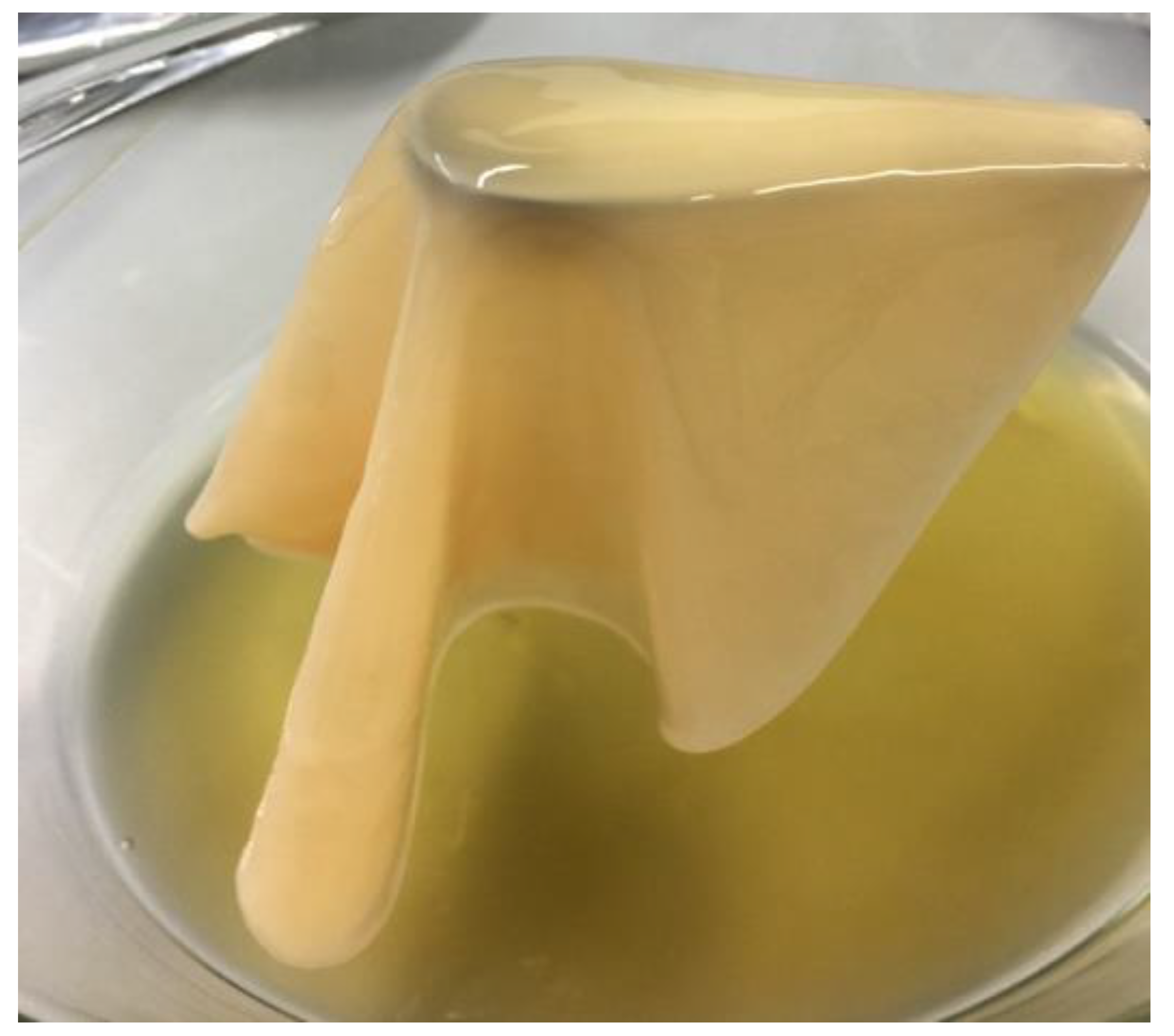
3.1. Isolation and Basic Characterization of Strains AV382 and AV436

Since vinegar is the most well-known product of acetic acid bacteria, it is not surprising that out of 17 type species of the genus Komagataeibacter, seven species, namely Komagataeibacter europaeus [38], K. hansenii [39], Komagataeibacter kakiaceti [40], Komagataeibacter maltaceti [12], Komagataeibacter medellinensis [8], Komagataeibacter oboediens [41] and K. pomaceti [13] have been isolated from vinegars. The novel strains AV382 and AV436 were also isolated from vinegar on RAE medium supplemented with 1% of acetic acid and 1% of ethanol after sampling the same bioreactor, but different batches for apple cider vinegar production. During apple cider vinegar production apple cider prepared from different varieties of apples is added to the bioreactor as a substrate. The apples are carriers of different acetic acid bacterial strains, which proceed to the vinegar producing bioreactors after resisting stress conditions during ethanol fermentation. This may be a reason of isolating the strains AV382 and AV436 from different batches but the same vinegar producing bioreactor. Although isolation and cultivation of acetic acid bacteria from industrial bioreactors for vinegar production have very low efficiency [20,21,38,42], both strains grew very well on RAE medium, but also on media without acetic acid and ethanol, such as GY and mannitol agar. The nutrient agar medium without any sugar supplement enabled only very poor and slow growth of both strains, which finally resulted in growth arrest. Both strains oxidize ethanol to acid and further on to CO2 and H2O on Carr and Frateur agar media. This is one of the typical characteristics of the genus Komagataeibacter [43]. Moreover, both strains exhibit high acetic acid tolerance by persisting up to 5% of acetic acid in liquid RAE medium with 1% and 3% of ethanol, which is comparable to the acidity during apple cider vinegar production. Both strains preserved the acetic acid tolerance after storage at −80 °C in the presence of 20% glycerol for at least one and half year.
The classical microbiological analysis showed that strains AV382 and AV436 are Gram-staining negative, catalase positive, oxidase negative and strictly aerobic bacteria. On RAE medium with 1% of acetic acid and 1% of ethanol both strains are forming round shaped colonies of irregular size between 0.9–3.0 mm in diameter after three days of cultivation. Microscopical examination showed small rods for both strains (Supplementary Figure S1). Cells of AV436 typically form short chains of 3–10 cells. The formation of chains was not observed for strain AV382.

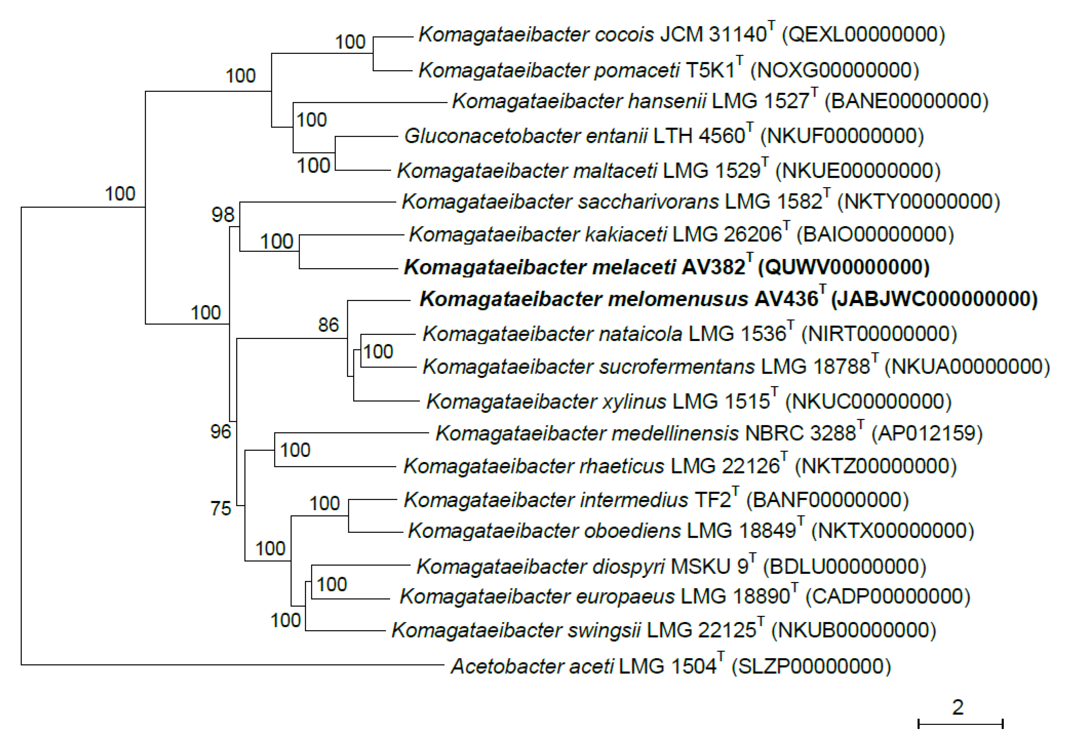
3.2. Phylogenetic Affiliation of Strains AV382 and AV436

The 16S rRNA gene sequencing confirmed the affiliation of AV382 and AV436 to the genus Komagataeibacter (Supplementary Figure S2). Besides, the nucleotide identity of both strains showed very high 16S rRNA gene sequence similarity to the closest species, representing just a few nucleotide differences. The 16S rRNA gene sequence of strain AV436 showed the highest similariry (99.4–99.8%) to the sequences of species Komagataeibacter intermedius, Komagataeibacter oboediens, Komagataeibacter swingsii, K. nataicola, Komagataeibacter sucrofermentans, Komagataeibacter europaeus, Komagataeibacter diospyri and K. xylinus and the sequence of strain AV382 the highest similarity (99.3–99.5%) to species K. swingsii, K. europaeus, K. sucrofermentans, K. nataicola, Komagataeibacter kakiaceti, K. saccharivorans, K. oboediens and K. intermedius. Since this high sequence identity represents only 2–9 nucleotide differences, we proceeded with the analysis of 16S–23S rRNA gene ITS regions which revealed more nucleotide differences to type strains of species of the genus Komagataeibacter. The closest relatives based on comparison of 16S–23S rRNA gene ITS was Komagataeibacter kakiaceti (91.6%) for strain AV382, and species K. xylinus (93.9%), K. sucrofermentans (92.7%) and K. nataicola (94.6%) for strain AV436. The phylogenetic relation among all type strains of the species of Komagataeibacter and strains AV382 and AV436, based on 16S–23S rRNA gene ITS is shown in Figure 1.
The genome sequencing of strains AV382 and AV436 enabled us to perform further phylogenetic analysis. We constructed a phylogenetic tree based on core genes (Figure 2) which confirmed the classification of strain AV382 close to K. kakiaceti and strain AV436 close to K. xylinus, K. sucrofermentans and K. nataicola. Moreover, the ANI values calculated from the comparison of the entire genome sequences of both strains to the type strains species of the Komagataeibacter genus (Supplementary Table S1) revealed that strains AV382 and AV436 represent novel species of this genus since the highest ANI value (91.6%) was below the 94% cut-off value for species delineation [44]. The novel species status of both strains AV382 and AV436 was confirmed by in-silico DNA-DNA hybridization calculations (Supplementary Table S2) since both strains showed ≤47.3% identity to type strains of species of the Komagataeibacter genus which is below the accepted value of 70% for species delineation [45].

3.3. Phenotypic Characterization of Strains AV382 and AV436

The fatty acid profile of both strains, AV382 and AV436 was analyzed from cells cultured on RAE and GY medium (Table 1). In both cases the major fatty acid was cis-vaccenic acid (C18:1 ω7c) with 34.4–36.9 mol% for strain AV382 and 31.4–43.4 mol% for strain AV436. This fatty acid was reported as the predominant fatty acid for all acetic acid bacteria, with the exception of the recently described species Komagataeibacter cocois JCM 31140T [9]. However, the fatty acid analysis of this type strain cultured on GY and RAE medium could not confirm the published results (Table 1). Interestingly, in the strain AV382, a second predominant fatty acid was a 19-carbon cyclopropane analogue of cis-vaccenic acid, i.e., C19:0 cyclo ω8c. This fatty acid represents 13 mol% of the total fatty acids when the strain was grown on RAE medium and 16.7 mol% when grown on GY medium. Cyclopropane fatty acids as the major fatty acids are not typical for the genus Komagataeibacter and also not for other genera of acetic acid bacteria [46], with the exception of the genera Swaminathania [47], Saccharibacter [48] and Bombella [49]. The presence of cyclopropane fatty acid in bacteria favours tolerance to various stressors, such as osmotic and low pH [50]. The fatty acid C19:0 cyclo ω8c is synthesized from monounsaturated fatty acids; for example, from cis-vaccenic acid [51]. The high amount of this cyclopropane fatty acid in the novel strain AV382 can be used as a differentiation marker from other species of the genus Komagataeibacter.
Both strains AV382 and AV436 showed resistance against chloramphenicol (30 µg), the strain AV436 additionally to gentamicin (10 µg). However, both strains showed inhibition zones around ampicillin (10 µg) and gentamicin (30 µg). Using the Comprehensive Antibiotic Resistance Database [52] for analysis of possible antibiotic resistance molecular determinants in genomes of both strains, chloramphenicol acetyltransferase homologue were identified: RFD20163 for strain AV382 and WP_172158671 for strain AV436. These enzymes are structurally modifying the antibiotic and thus inhibiting chloramphenicol catalytic activity [53], which may explain resistance against chloramphenicol of both strains. No homologue to proteins coding for aminoglycoside acetyltransferases which are responsible for aminoglycoside antibiotic inactivation, such as gentamicin [54], could be identified in both genomes. However, in both genomes some putative efflux resistance-modulation-cell (RND) permease transporters, DHA2 family efflux major facilitator superfamily (MFS) permease transporters and two multidrug efflux small multidrug resistance (SMR) transporters have been identified (Supplementary Table S3). These proteins may be involved in transport of antibiotics out of the cell [55]. The presence of antibiotic resistance determinants in food grade bacteria, such as acetic acid bacteria, is not problematic by itself, but when this genetic information is transferred to human pathogenic bacteria, it may become of immense concern. The probability of this scenario is presently increasing with a trend of preparing and consuming nonfiltered biomass containing apple cider vinegar.
Further phenotypic analysis showed that both strains produce 2-keto-D-gluconic and 5-keto-D-gluconic acids from glucose which differentiates strain AV382 from its closest relative K. kakiaceti and strain AV436 from one of the three most closest relatives K. sucrofermentans (Table 2). None of the strains produced 2,5-diketo-D-gluconic acid. Both strains formed 2-keto-D-gluconic acid and 5-keto-D-gluconic acid during cultivation in glucose rich liquid medium. In contrast to strain AV382, 2-keto-D-gluconic acid could not be detected any more in the growth medium of strain AV436 at our second sampling point of cultivation, suggesting that the acid was transported back into the cytoplasm and used as an energy source. This kind of metabolic action was observed in some other acetic acid bacteria [56]. None of the strains AV382 and AV436 grew in the presence of 30% of glucose. This characteristic obviously differentiates strain AV382 from both closest species K. kakiaceti and K. saccharivorans (Table 2). Strain AV436 can well use D-ribose, sorbitol, D-mannitol and glycerol as carbon source and also 1-propanol, albeit with weak growth. In contrast, K. nataicola cannot grow on 1-propanol. Strain AV382 can grow on Hoyer-Frateur medium with ethanol as carbon source, but both of its closest relatives K. kakiaceti and K. saccharivorans cannot. In contrast to AV382 and K. xylinus, strain AV436 cannot grow on Hoyer-Frateur medium with ethanol as carbon source. Strain AV382 can also grow on Asai medium with ethanol, but its closest relative K. kakiaceti cannot. The pellet of strain AV436 did not dissolve after cooking in 5M NaOH, which shows that the strain is a cellulose producer. Cellulose production was not observed for the strain AV382. The tolerance to 5% acetic acid differentiates strain AV382 from K. kakiaceti and K. saccharivorans which tolerate 4% of acetic acid. The 5% acetic acid tolerance of AV436 differentiates it from K. xylinus and K. nataicola, which tolerate 4% of acetic acid in the presence of 1% ethanol and 3% of acetic acid in the presence of 3% ethanol.

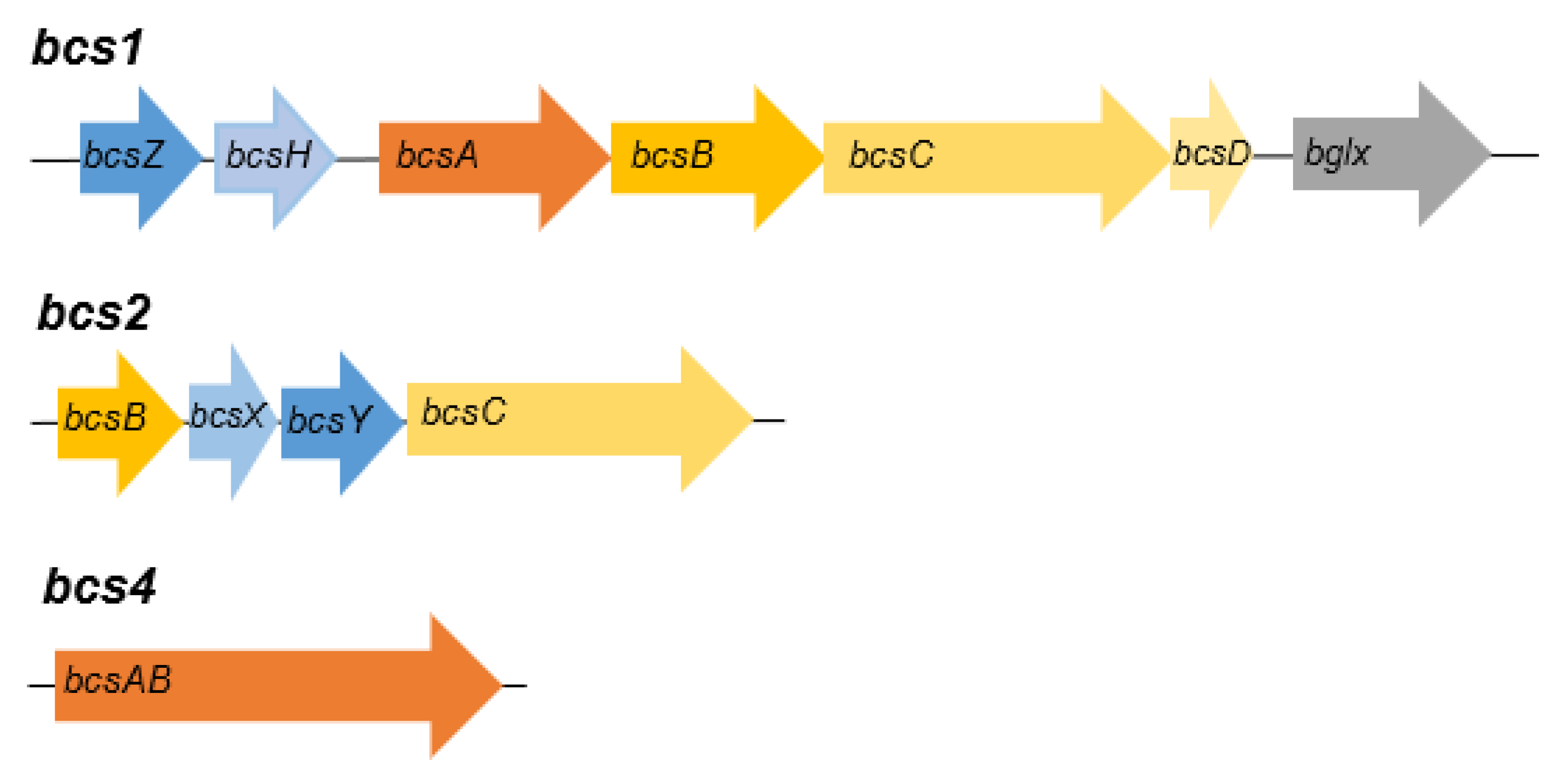
3.4. Genome analysis of strains AV382 and AV436

Acetic acid bacteria in vinegar are under high acid stress, and thus they evolved tools for coping with acid. Among them is a transporter pumping acetic acid out of the cell, extracellular polysaccharide formation to block entrance of acid in the cytoplasm and major alcohol and aldehyde dehydrogenases embedded into the cytoplasmic membrane to oxidize ethanol to acetic acid and parallelly producing a big energy pool [57]. Using the available information on molecular determinants relevant to these characteristics [13], we searched the genome sequences of strains AV382 and AV436 for homologues.
The genome sequence of AV436 is coding for homologues to proteins encoded by cellulose operon bcs1 [58], bcs2 [59], bcs4 [60], but not to bcs3 [61]. Genes of operon bcs1 (Figure 3) are coding for putative endo-1,4-beta-glucanase (NPC65873), cellulose-complementing protein A (NPC65872), cellulose synthase catalytic subunit A (NPC65871), cellulose synthase subunit B (NPC65870), cellulose synthase subunit C (NPC65869), cellulose synthase subunit D (NPC65868) and β-glucosidase (NPC65867). Except for NPC65872, all other ORFs of putative AV436 bcs1 are of similar size and almost identical to proteins encoded by bcs1 of Komagataeibacter xylinus E25 [58]. The cellulose-complementing protein A acts on expression levels of BcsB and BcsC, interacts with BcsD and probably assists in arrangement of glucan chains into crystalline ribbons [62]. Since the NPC65872 and its homologue AHI24409 in K. xylinus E25 share only 36.7% identity, this may affect the quantity and the quality of cellulose production in each species. The AV436 bcs2 operon (Figure 3) is encoding putative cellulose synthase subunit C (NPC65045), hydrolase (NPC65044) and acyltransferase (NPC65046). Besides the two operons, a catalytic subunit cellulose synthase (NPC67849) encompassing operon bcs4 (Figure 3) has been identified at another chromosomal location of AV436. All these ORFs support our phenotypic observation on strong cellulose production by strain AV436. The strain produces cellulose layer of approximate size 25 cm × 15 cm × 0.3 cm on 500 mL RAE medium in 5 days (Figure 4). In the genome sequence of AV382, homologues to some of the proteins encoded by operon bcs1 and bcs2 were identified but again no homologues to proteins of operon bcs3. Genes coding for acetan, the second most well-known polysaccharide exported by some acetic acid bacteria [63,64], were then searched in AV382 and AV436. Homologues to some of the proteins encoded by acetan operon have been identified in strain AV382 (glycosyltransferase: WP_11670167, flipase: WP_116701683), but not in strain AV436. Both strains also possess homologues to proteins encoded by rfbABCD operon, which is involved in dTDP-rhamnose synthesis pathway and most probably forms capsular polysaccharides attached to the cell [65,66]. In strain AV436 the putative rfbA coding for glucose-1-phosphate thymidylyltransferase (WP_172156247) and rfbB coding for dTDP-glucose 4,6-dehydratase (WP_172156248) are part of an operon, the rfbC coding for dTDP-4-dehydrorhamnose 3,5-epimerase (WP_172156300) and rfbD (WP_172156302) coding for dTDP-4-dehydrorhamnose reductase are part of another operon. In strain AV382 the genes coding for the putative proteins RfbA (WP_116702174) and RfbB (WP_116702134) are not part of a transcriptional unit. In contrast to rfbA and rfbB, the genes coding for putative RfbC (WP_116702180) and RfbD (WP_116702181) are part of an operon.
AV382 and AV436 both possess AarC, RFD20455 and NPC66889, respectively, which is a homologue to succinyl-coenzyme A (CoA): acetate CoA transferase. This enzyme replaces succinyl-CoA synthetase and converts succinyl-CoA and acetate to acetyl-CoA and succinate, and thus enables an efficient assimilation of acetic acid [67]. In strain AV436, one MFS transporter (WP_172156362) shows some similarities to plasma membrane protein of Saccharomyces cerevisiae (Azr1), which is contributing cell adaptation to weak acids [68]. Further studies should reveal its potential involvement in acetic acid tolerance in Komagataeibacter melomenusus.
Of three putative types of a catalytic subunit of PQQ-dependent alcohol dehydrogenase identified in some Komagataeibacter species [13], only homologues to AdhA-1 have been identified in AV382 (RFD19113) and AV436 (NPC67328). In both strains, the homologues to AldH, a catalytic subunit of aldehyde dehydrogenase, have also been identified: WP_116702452 for AV382 and WP_172154413 for AV436.
In acetic acid bacteria, 2-ketogluconate and 5-ketogluconate are mainly produced from D-gluconate with membrane bound FAD-dependent gluconate dehydrogenase (FAD-GADH) and PQQ-dependent glycerol dehydrogenase (PQQ-GLDH), respectively [69]. Some strains have also PQQ-dependent gluconate dehydrogenase (PQQ-GADH) which converts D-gluconate to 2,5-diketogluconate [70]. In genomes of both strains, AV382 and AV436 the putative large and small subunits of FAD-GADH (AV382: WP_116702246, WP_116702271; AV436: WP_172157429, WP_172157431) and PQQ-GLDH (AV382: WP_116701675, WP_116701676; AV436: WP_172158295, WP_172158288) but not PQQ-GADH were identified which support the phenotypic data described above.

4. Conclusions

In this study we described two novel species from the group of acetic acid bacteria. Their main features are summarized below. The study was focused on taxonomic characterization of both novel strains, but it also confirms that the microbiota of apple cider vinegar is heterogenous as described in previous studies using different molecular approaches [71,72,73,74]. Additionally, the genetic potential of both strains confirms that the acetic acid bacteria are biotechnologically important microorganisms, which will remain in focus of further scientific studies.

4.1. Description of Komagataeibacter melaceti sp. nov.

Komagataeibacter melaceti (mel.a.ce’ti. G. n. melon, apple; L. n. acetum, vinegar, N.L. n. melaceti, of apple vinegar from where the type strain was isolated).
Cells are Gram-staining negative, rod shaped, 1.9–5.0 µm long, 1.1–1.5 µm wide, occurring singly. Colonies are cream to light brown, circular, smooth, forming convex circled shapes with diameters of about 1.0–3.0 mm on RAE medium supplemented with 1% ethanol and 1% acetic acid after 3 days of incubation at high humidity. Cells are obligately aerobic, catalase positive and oxidase negative. The optimum growth temperature is 28–30 °C. Ethanol is oxidized to acetic acid and further on to CO2 and H2O. Both 2- and 5-keto-D-gluconic acids are formed but not 2,5-diketo-D-gluconic acid from D-glucose. Bacterial cellulose is not produced from D- glucose. Growth is observed in the presence of 1–5% (v/v) acetic acid and 1% or 3% ethanol (v/v), but acetic acid is not required for growth. Growth is observed on glucose-yeast agar and mannitol agar. Growth is observed on D-ribose, D-glucose, D-mannitol, D-sorbitol, glycerol and 1-propanol as sole carbon source. The strain can utilize ammoniacal nitrogen in Hoyer-Frateur medium with D-glucose, D-mannitol and ethanol, and in Asai medium with D-glucose and D-mannitol, but not with ethanol. The major cellular fatty acid is C18:1 ω7c and a second predominant fatty acid is C19:0 cyclo ω8c.
The type strain, AV382T (= ZIM B1054T = LMG 31303T = CCM 8958T), was isolated from apple cider vinegar in Kopivnik, Slovenia.
The GenBank/ENA/DDBJ accession number for the 16S rRNA gene sequence, 16S–23S rRNA gene ITS sequence and complete genome sequence of the type strain are MT422125, MT423516 and QUWV00000000, respectively.

4.2. Description of Komagataeibacter melomenusus sp. nov.

Komagataeibacter melomenusus (mel.o.me.nu’sus. G. n. melon, apple; Gr. v. meno to reside, stay, live [masc. pres. part. menusus]; N.L. masc. adj. melomenusus from apple [cider] as the niche of these bacteria).
Cells are Gram-staining negative, rod shaped, 1.5–2.8 µm long, 0.8–1.0 µm wide, occurring in chains of 3–10 cells. Colonies are cream to light brown, circular, smooth, forming convex circle shapes with diameter of about 0.9–3.0 mm on RAE medium supplemented with 1% ethanol and 1% acetic acid after 3 days of incubation at high humidity. Cells are obligately aerobic, catalase positive and oxidase negative. The optimum growth temperature is 28–30 °C. Ethanol is oxidized to acetic acid and further on to CO2 and H2O. Both 2- and 5-keto-D-gluconic acids are formed but not 2,5-diketo-D-gluconic acid from D-glucose. Bacterial cellulose is produced from D- glucose. Growth is observed in the presence of 1–5% (v/v) acetic acid and 1% or 3% ethanol (v/v), but acetic acid is not required for growth. Growth is observed on glucose-yeast agar and mannitol agar. Growth is observed on D-ribose, D-glucose, D-mannitol, D-sorbitol and glycerol as sole carbon source. Growth on 1-propanol is weak. The strain can utilize ammoniacal nitrogen in Hoyer-Frateur medium with D-glucose and D-mannitol, but not with ethanol, in Asai medium with D-glucose and D-mannitol, but not with ethanol. The major cellular fatty acid is C18:1 ω7c.
The type strain, AV436T (= ZIM B1056T = LMG 31304T = CCM 8959T), was isolated from apple cider vinegar in Kopivnik, Slovenia.
The GenBank/ENA/DDBJ accession number for the 16S rRNA gene sequence, 16S–23S rRNA gene ITS sequence and complete genome sequence of the type strain are MT422127, MT423518 and JABJWC000000000, respectively.

Supplementary Materials

The following are available online at https://www.mdpi.com/2076-2607/8/8/1178/s1, Figure S1: Microscopic images of bacterial cells after Gram-staining at 1000-fold magnification. A: Komagataeibacter melaceti AV382T, B: Komagataeibacter melomenusus AV436T, Figure S2: Phylogenetic reconstruction based on 16S rRNA gene sequences of the type strains of the species of Komagataeibacter genus. The tree was constructed using the maximum-likelihood method. Bootstrap values are indicated at branching (1000 replicates). Acetobacter aceti is included as outgroup. GenBank accession numbers are given in brackets and the scale bar represents the number of substitutions per nucleotide position, Table S1: ANIb (A) and ANIm (B) values for strains AV382 and AV436 in comparison to the type strains of species of the Komagataeibacter genus, Table S2: Digital DNA-DNA hybridization values of strains AV382 and AV436 in comparison to type strains of species of genus Komagataeibacter, Table S3: List of putative efflux protein subunits in genomes of strains AV382 and AV436.

Author Contributions

L.M. performed the isolation and phenotypic characterization of strains AV382 and AV436. L.M., T.A., I.C., P.V. and J.T. carried out the phylogenetic and genomic analyses. J.T. designed and managed the experiments. All authors contributed to the preparation of the manuscript. All authors have read and agreed to the published version of the manuscript.

Funding

This research was funded by the Slovenian Research Agency through program P2-0006. Financial support from the Ministry of Education, Science and Sport and the European Social Fund under the scheme Practical Knowledge through Creative Pathways is gratefully acknowledged. The BCCM/LMG Bacteria Collection is supported by the Federal Public Planning Service - Science Policy, Belgium.

Acknowledgments

We thank Lijana Fanedl from the Biotechnical Faculty University of Ljubljana for help with the fatty acid analysis. Lora Klanfar, Kim Fijok and Jure Škraban from the Faculty of Natural Sciences and Mathematics University of Maribor are acknowledged for technical assistance with antibiotic resistance analysis, for photography of cellulose product and the initial involvement in isolation of strains AV382 and AV436, respectively.

Conflicts of Interest

The authors declare no conflict of interest.

References

  1. Trček, J.; Barja, F. Updates on quick identification of acetic acid bacteria with a focus on the 16S-23S rRNA gene internal transcribed spacer and the analysis of cell proteins by MALDI-TOF mass spectrometry. Int. J. Food Microbiol. 2015, 196, 137–144. [Google Scholar] [CrossRef]
  2. Cleenwerck, I.; De Vos, P. Polyphasic taxonomy of acetic acid bacteria: An overview of the currently applied methodology. Int. J. Food Microbiol. 2008, 125, 2–14. [Google Scholar] [CrossRef] [PubMed]
  3. Yamada, Y.; Yukphan, P.; Vu, H.T.L.; Muramatsu, Y.; Ochaikul, D.; Tanasupawat, S.; Nakagawa, Y. Description of Komagataeibacter gen. nov., with proposals of new combinations (Acetobacteraceae). J. Gen. Appl. Microbiol. 2012, 58, 397–404. [Google Scholar] [CrossRef] [PubMed] [Green Version]
  4. Euzéby, J. List of new names and new combinations previously effectively, but not validly, published. Int. J. Syst. Evol. Microbiol. 2013, 63, 1–5. [Google Scholar] [CrossRef]
  5. Yamada, Y.; Yukphan, P.; Vu, H.T.L.; Muramatsu, Y.; Ochaikul, D.; Nakagawa, Y. Subdivision of the genus Gluconacetobacter Yamada, Hoshino and Ishikawa 1998: The proposal of Komagatabacter gen. nov., for strains accommodated to the Gluconacetobacter xylinus group in the α-Proteobacteria. Ann. Microbiol. 2012, 62, 849–859. [Google Scholar] [CrossRef]
  6. Parte, A.C. LPSN—List of prokaryotic names with standing in nomenclature (Bacterio.net), 20 years on. Int. J. Syst. Evol. Microbiol. 2018, 69, 1825–1829. [Google Scholar] [CrossRef] [PubMed]
  7. Boesch, C.; Trček, J.; Sievers, M.; Teuber, M. Acetobacter intermedius, sp. nov. Syst. Appl. Microbiol. 1998, 21, 220–229. [Google Scholar] [CrossRef]
  8. Castro, C.; Cleenwerck, I.; Trček, J.; Zuluaga, R.; de Vos, P.; Caro, G.; Aguirre, R.; Putaux, J.-L.; Gañán, P. Gluconacetobacter medellinensis sp. nov., cellulose- and non-cellulose-producing acetic acid bacteria isolated from vinegar. Int. J. Syst. Evol. Microbiol. 2013, 63, 1119–1125. [Google Scholar] [CrossRef] [Green Version]
  9. Liu, L.X.; Liu, S.X.; Wang, Y.M.; Bi, J.C.; Chen, H.M.; Deng, J.; Zhang, C.; Hu, Q.S.; Li, C.F. Komagataeibacter cocois sp. nov., a novel cellulose-producing strain isolated from coconut milk. Int. J. Syst. Evol. Microbiol. 2018, 68, 3125–3131. [Google Scholar] [CrossRef]
  10. Naloka, K.; Yukphan, P.; Matsutani, M.; Matsushita, K.; Theeragool, G. Komagataeibacter diospyri sp. nov., a novel species of thermotolerant bacterial nanocellulose-producing bacterium. Int. J. Syst. Evol. Microbiol. 2020, 70, 251–258. [Google Scholar] [CrossRef]
  11. Lisdiyanti, P.; Navarro, R.R.; Uchimura, T.; Komagata, K. Reclassification of Gluconacetobacter hansenii strains and proposals of Gluconacetobacter saccharivorans sp. nov. and Gluconacetobacter nataicola sp. nov. Int. J. Syst. Evol. Microbiol. 2006, 56, 2101–2111. [Google Scholar] [CrossRef] [PubMed] [Green Version]
  12. Slapšak, N.; Cleenwerck, I.; De Vos, P.; Trček, J. Gluconacetobacter maltaceti sp. nov., a novel vinegar producing acetic acid bacterium. Syst. Appl. Microbiol. 2013, 36, 17–21. [Google Scholar] [CrossRef] [PubMed]
  13. Škraban, J.; Cleenwerck, I.; Vandamme, P.; Fanedl, L.; Trček, J. Genome sequences and description of novel exopolysaccharides producing species Komagataeibacter pomaceti sp. nov. and reclassification of Komagataeibacter kombuchae (Dutta and Gachhui 2007) Yamada et al., 2013 as a later heterotypic synonym of Komagataeibacter hansenii (Gosselé et al. 1983) Yamada et al., 2013. Syst. Appl. Microbiol. 2018, 41, 581–592. [Google Scholar] [CrossRef] [PubMed]
  14. Gorgieva, S.; Trček, J. Bacterial cellulose: Production, modification and perspectives in biomedical applications. Nanomaterials 2019, 9, 1352. [Google Scholar] [CrossRef] [PubMed] [Green Version]
  15. Gorgieva, S. Bacterial cellulose as a versatile platform for research and development of biomedical materials. Processes 2020, 8, 624. [Google Scholar] [CrossRef]
  16. Cleenwerck, I.; De Vos, P.; De Vuyst, L. Phylogeny and differentiation of species of the genus Gluconacetobacter and related taxa based on multilocus sequence analyses of housekeeping genes and reclassification of Acetobacter xylinus subsp. sucrofermentans as Gluconacetobacter sucrofermentans (Toyosaki et al. 1996) sp. nov., comb. nov. Int. J. Syst. Evol. Microbiol. 2010, 60, 2277–2283. [Google Scholar] [CrossRef] [Green Version]
  17. De Vuyst, L.; Camu, N.; De Winter, T.; Vandemeulebroecke, K.; Van de Perre, V.; Vancanneyt, M.; De Vos, P.; Cleenwerck, I. Validation of the (GTG)5-rep-PCR fingerprinting technique for rapid classification and identification of acetic acid bacteria, with a focus on isolates from Ghanaian fermented cocoa beans. Int. J. Food Microbiol. 2008, 125, 79–90. [Google Scholar] [CrossRef]
  18. Andrés-Barrao, C.; Benagli, C.; Chappuis, M.; Ortega Pérez, R.; Tonolla, M.; Barja, F. Rapid identification of acetic acid bacteria using MALDI-TOF mass spectrometry fingerprinting. Syst. Appl. Microbiol. 2013, 36, 75–81. [Google Scholar] [CrossRef]
  19. Trček, J.; Teuber, M. Genetic and restriction analysis of the 16S-23S rDNA internal transcribed spacer regions of the acetic acid bacteria. FEMS Microbiol. Lett. 2002, 208, 69–75. [Google Scholar] [CrossRef]
  20. Trcek, J. Quick identification of acetic acid bacteria based on nucleotide sequences of the 16S-23S rDNA internal transcribed spacer region and of the PQQ-dependent alcohol dehydrogenase gene. Syst. Appl. Microbiol. 2005, 28, 735–745. [Google Scholar] [CrossRef]
  21. Sokollek, S.J.; Hammes, W.P. Description of a starter culture preparation for vinegar fermentation. Syst. Appl. Microbiol. 1997, 20, 481–491. [Google Scholar] [CrossRef]
  22. Bolger, A.M.; Lohse, M.; Usadel, B. Trimmomatic: A flexible trimmer for Illumina sequence data. Bioinformatics 2014, 30, 2114–2120. [Google Scholar] [CrossRef] [PubMed] [Green Version]
  23. Zerbino, D.R.; Birney, E. Velvet: Algorithms for de novo short read assembly using de Bruijn graphs. Genome Res. 2008, 18, 821–829. [Google Scholar] [CrossRef] [PubMed] [Green Version]
  24. Gosselé, F.; Swings, J.; De Ley, J. A rapid, simple and simultaneous detection of 2-keto-, 5-keto-and 2,5-diketogluconic acids by thin-layer chromatography in culture media of acetic acid bacteria. Zentralblatt für Bakteriol. I. Abt. Orig. C Allg. angew. und ökologische Mikrobiol. 1980, 1, 178–181. [Google Scholar] [CrossRef]
  25. Hudzicki, J. Kirby-Bauer Disk Diffusion Susceptibility Test Protocol Author Information; American Society for Microbiology: Washington, DC, USA, 2009; pp. 1–13. [Google Scholar]
  26. European Society of Clinical Microbiology and Infectious Diseases. Antimicrobial Susceptibility Testing EUCAST Disk Diffusion Method, Version 8.0, January 2020; European Society of Clinical Microbiology and Infectious Diseases: Basel, Switzerland, 2020; pp. 1–21. [Google Scholar]
  27. Seemann, T. Prokka: Rapid prokaryotic genome annotation. Bioinformatics 2014, 30, 2068–2069. [Google Scholar] [CrossRef]
  28. Camacho, C.; Coulouris, G.; Avagyan, V.; Ma, N.; Papadopoulos, J.; Bealer, K.; Madden, T.L. BLAST+: Architecture and applications. BMC Bioinform. 2009, 10, 421. [Google Scholar] [CrossRef] [Green Version]
  29. Page, A.J.; Cummins, C.A.; Hunt, M.; Wong, V.K.; Reuter, S.; Holden, M.T.G.; Fookes, M.; Falush, D.; Keane, J.A.; Parkhill, J. Roary: Rapid large-scale prokaryote pan genome analysis. Bioinformatics 2015, 31, 3691–3693. [Google Scholar] [CrossRef]
  30. Guindon, S.; Dufayard, J.F.; Lefort, V.; Anisimova, M.; Hordijk, W.; Gascuel, O. New algorithms and methods to estimate maximum-likelihood phylogenies: Assessing the performance of PhyML 3.0. Syst. Biol. 2010, 59, 307–321. [Google Scholar] [CrossRef] [Green Version]
  31. Larkin, M.A.; Blackshields, G.; Brown, N.P.; Chenna, R.; McGettigan, P.A.; McWilliam, H.; Valentin, F.; Wallace, I.M.; Wilm, A.; Lopez, R.; et al. ClustalW2 and ClustalX version 2.0. Bioinfomatics 2007, 23, 2947–2948. [Google Scholar] [CrossRef] [Green Version]
  32. Kumar, S.; Stecher, G.; Li, M.; Knyaz, C.; Tamura, K. MEGA X: Molecular evolutionary genetics analysis across computing platforms. Mol. Biol. Evol. 2018, 35, 1547–1549. [Google Scholar] [CrossRef]
  33. Tamura, K.; Nei, M. Estimation of the number of nucleotide substitutions in the control region of mitochondrial DNA in humans and chimpanzees. Mol. Biol. Evol. 1993, 10, 512–526. [Google Scholar] [CrossRef] [PubMed]
  34. El-Gebali, S.; Mistry, J.; Bateman, A.; Eddy, S.R.; Luciani, A.; Potter, S.C.; Qureshi, M.; Richardson, L.J.; Salazar, G.A.; Smart, A.; et al. The Pfam protein families database in 2019. Nucleic Acids Res. 2019, 47, D427–D432. [Google Scholar] [CrossRef] [PubMed]
  35. Lombard, V.; Golaconda Ramulu, H.; Drula, E.; Coutinho, P.M.; Henrissat, B. The carbohydrate-active enzymes database (CAZy) in 2013. Nucleic Acids Res. 2014, 42, D490–D495. [Google Scholar] [CrossRef] [PubMed] [Green Version]
  36. Arahal, D.R. Whole-genome analyses: Average nucleotide identity. In Methods in Microbiology: New Approaches to Prokaryotic Systematics; Goodfellow, M., Sutcliffe, I.C., Chun, J., Eds.; Elsevier: London, UK, 2014; Volume 41, pp. 103–122. [Google Scholar]
  37. Meier-Kolthoff, J.P.; Auch, A.F.; Klenk, H.P.; Göker, M. Genome sequence-based species delimitation with confidence intervals and improved distance functions. BMC Bioinform. 2013, 14, 60. [Google Scholar] [CrossRef] [Green Version]
  38. Sievers, M.; Sellmer, S.; Teuber, M. Acetobacter europaeus sp. nov., a main component of industrial vinegar fermenters in central Europe. Syst. Appl. Microbiol. 1992, 15, 386–392. [Google Scholar] [CrossRef]
  39. Gosselé, F.; Swings, J.; Kersters, K.; Pauwels, P.; De Ley, J. Numerical analysis of phenotypic features and protein gel electrophoregrams of a wide variety of Acetobacter strains. Proposal for the improvement of the taxonomy of the genus Acetobacter Beijerinck 1898, 215. Syst. Appl. Microbiol. 1983, 4, 338–368. [Google Scholar] [CrossRef]
  40. Iino, T.; Suzuki, R.; Tanaka, N.; Kosako, Y.; Ohkuma, M.; Komagata, K.; Uchimura, T. Gluconacetobacter kakiaceti sp. nov., an acetic acid bacterium isolated from a traditional Japanese fruit vinegar. Int. J. Syst. Evol. Microbiol. 2012, 62, 1465–1469. [Google Scholar] [CrossRef]
  41. Sokollek, S.J.; Hertel, C.; Hammes, W.P. Description of Acetobacter oboediens sp. nov. and Acetobacter pomorum sp. nov., two new species isolated from industrial vinegar fermentations. Int. J. Syst. Bacteriol. 1998, 48, 935–940. [Google Scholar] [CrossRef] [Green Version]
  42. Schüller, G.; Hertel, C.; Hammes, W.P. Gluconacetobacter entanii sp. nov., isolated from submerged high-acid industrial vinegar fermentations. Int. J. Syst. Evol. Microbiol. 2000, 50, 2013–2020. [Google Scholar] [CrossRef] [Green Version]
  43. Komagata, K.; Iino, T.; Yamada, Y. The family Acetobacteraceae. In The Prokaryotes: Alphaproteobacteria and Betaproteobacteria; Rosenberg, E., De Long, E.F., Lory, S., Stackebrandt, E., Thompson, F., Eds.; Springer: Berlin/Heidelberg, Germany, 2014; pp. 3–78. [Google Scholar]
  44. Richter, M.; Rosselló-Móra, R. Shifting the genomic gold standard for the prokaryotic species definition. Proc. Natl. Acad. Sci. USA 2009, 106, 19126–19131. [Google Scholar] [CrossRef] [Green Version]
  45. Thompson, C.C.; Chimetto, L.; Edwards, R.A.; Swings, J.; Stackebrandt, E.; Thompson, F.L. Microbial genomic taxonomy. BMC Genomics 2013, 14, 913. [Google Scholar] [CrossRef] [PubMed] [Green Version]
  46. Yamada, Y.; Nunoda, M.; Ishikawa, T.; Tahara, Y. The cellular fatty acid composition in acetic acid bacteria. J. Gen. Appl. Microbiol. 1981, 27, 405–417. [Google Scholar] [CrossRef]
  47. Loganathan, P.; Nair, S. Swaminathania salitolerans gen. nov., sp. nov., a salt-tolerant, nitrogen-fixing and phosphate-solubilizing bacterium from wild rice (Porteresia coarctata Tateoka). Int. J. Syst. Evol. Microbiol. 2004, 54, 1185–1190. [Google Scholar] [CrossRef] [PubMed]
  48. Jojima, Y.; Mihara, Y.; Suzuki, S.; Yokozeki, K.; Yamanaka, S.; Fudou, R. Saccharibacter floricola gen. nov., sp. nov., a novel osmophilic acetic acid bacterium isolated from pollen. Int. J. Syst. Evol. Microbiol. 2004, 54, 2263–2267. [Google Scholar] [CrossRef] [PubMed]
  49. Li, L.; Praet, J.; Borremans, W.; Nunes, O.C.; Manaia, C.M.; Cleenwerck, I.; Meeus, I.; Smagghe, G.; De Vuyst, L.; Vandamme, P. Bombella intestini gen. nov., sp. nov., an acetic acid bacterium isolated from bumble bee crop. Int. J. Syst. Evol. Microbiol. 2015, 65, 267–273. [Google Scholar] [CrossRef] [PubMed]
  50. Li, Y.; Yan, P.; Lei, Q.; Li, B.; Sun, Y.; Li, S.; Lei, H.; Xie, N. Metabolic adaptability shifts of cell membrane fatty acids of Komagataeibacter hansenii HDM1-3 improve acid stress resistance and survival in acidic environments. J. Ind. Microbiol. Biotechnol. 2019, 46, 1491–1503. [Google Scholar] [CrossRef]
  51. Brown, J.L.; Ross, T.; McMeekin, T.A.; Nichols, P.D. Acid habituation of Escherichia coli and the potential role of cyclopropane fatty acids in low pH tolerance. Int. J. Food Microbiol. 1997, 37, 163–173. [Google Scholar] [CrossRef]
  52. Alcock, B.P.; Raphenya, A.R.; Lau, T.T.Y.; Tsang, K.K.; Bouchard, M.; Edalatmand, A.; Huynh, W.; Nguyen, A.L.V.; Cheng, A.A.; Liu, S.; et al. CARD 2020: Antibiotic resistome surveillance with the comprehensive antibiotic resistance database. Nucleic Acids Res. 2020, 48, D517–D525. [Google Scholar] [CrossRef]
  53. Schwarz, S.; Kehrenberg, C.; Doublet, B.; Cloeckaert, A. Molecular basis of bacterial resistance to chloramphenicol and florfenicol. FEMS Microbiol. Rev. 2004, 28, 519–542. [Google Scholar] [CrossRef] [Green Version]
  54. Shaw, K.J.; Rather, P.N.; Hare, R.S.; Miller, G.H. Molecular genetics of aminoglycoside resistance genes and familial relationships of the aminoglycoside-modifying enzymes. Microbiol. Rev. 1993, 57, 138–163. [Google Scholar] [CrossRef]
  55. Blanco, P.; Hernando-Amado, S.; Reales-Calderon, J.; Corona, F.; Lira, F.; Alcalde-Rico, M.; Bernardini, A.; Sanchez, M.; Martinez, J. Bacterial multidrug efflux pumps: Much more than antibiotic resistance determinants. Microorganisms 2016, 4, 14. [Google Scholar] [CrossRef] [PubMed] [Green Version]
  56. Saichana, N.; Matsushita, K.; Adachi, O.; Frébort, I.; Frebortova, J. Acetic acid bacteria: A group of bacteria with versatile biotechnological applications. Biotechnol. Adv. 2015, 33, 1260–1271. [Google Scholar] [CrossRef] [PubMed]
  57. Škraban, J.; Trček, J. Comparative genomics of Acetobacter and other acetic acid bacteria. In Acetic Acid Bacteria: Fundamentals and Food Applications; Sengun, I.Y., Ed.; CRC Press, Taylor and Francis Group: Boca Raton, FL, USA, 2017; pp. 44–70. [Google Scholar]
  58. Kubiak, K.; Kurzawa, M.; Jedrzejczak-Krzepkowska, M.; Ludwicka, K.; Krawczyk, M.; Migdalski, A.; Kacprzak, M.M.; Loska, D.; Krystynowicz, A.; Bielecki, S. Complete genome sequence of Gluconacetobacter xylinus E25 strain-Valuable and effective producer of bacterial nanocellulose. J. Biotechnol. 2014, 176, 18–19. [Google Scholar] [CrossRef] [PubMed]
  59. Zhang, H.; Xu, X.; Chen, X.; Yuan, F.; Sun, B.; Xu, Y.; Yang, J.; Sun, D. Complete genome sequence of the cellulose-producing strain Komagataeibacter nataicola RZS01. Sci. Rep. 2017, 7, 4431. [Google Scholar] [CrossRef]
  60. Liu, M.; Liu, L.; Jia, S.; Li, S.; Zou, Y.; Zhong, C. Complete genome analysis of Gluconacetobacter xylinus CGMCC 2955 for elucidating bacterial cellulose biosynthesis and metabolic regulation. Sci. Rep. 2018, 8, 1–10. [Google Scholar] [CrossRef]
  61. Pfeffer, S.; Santos, R.; Ebels, M.; Bordbar, D.; Brown, R.M. Complete genome sequence of Komagataeibacter hansenii LMG 23726T. Genome Announc. 2017, 5, e00168-17. [Google Scholar] [CrossRef] [Green Version]
  62. Römling, U.; Galperin, M.Y. Bacterial cellulose biosynthesis: Diversity of operons, subunits, products, and functions. Trends Microbiol. 2015, 23, 545–557. [Google Scholar] [CrossRef] [Green Version]
  63. Griffin, A.M.; Morris, V.J.; Gasson, M.J. Genetic analysis of the acetan biosynthetic pathway in Acetobacter xylinum: Nucleotide sequence analysis of the aceB aceC aceD and aceE genes. Mitochondrial DNA 1996, 6, 275–284. [Google Scholar] [CrossRef]
  64. Brandt, J.U.; Jakob, F.; Wefers, D.; Bunzel, M.; Vogel, R.F. Characterization of an acetan-like heteropolysaccharide produced by Kozakia baliensis NBRC 16680. Int. J. Biol. Macromol. 2018, 106, 248–257. [Google Scholar] [CrossRef]
  65. Deeraksa, A.; Moonmangmee, S.; Toyama, H.; Adachi, O.; Matsushita, K. Conversion of capsular polysaccharide, involved in pellicle formation, to extracellular polysaccharide by galE deletion in Acetobacter tropicalis. Biosci. Biotechnol. Biochem. 2006, 70, 2536–2539. [Google Scholar] [CrossRef] [Green Version]
  66. Deeraksa, A.; Moonmangmee, S.; Toyama, H.; Yamada, M.; Adachi, O.; Matsushita, K. Characterization and spontaneous mutation of a novel gene, polE, involved in pellicle formation in Acetobacter tropicalis SKU1100. Microbiology 2005, 151, 4111–4120. [Google Scholar] [CrossRef] [Green Version]
  67. Mullins, E.A.; Francois, J.A.; Kappock, T.J. A specialized citric acid cycle requiring succinyl-coenzyme A (CoA): Acetate CoA-transferase (AarC) confers acetic acid resistance on the acidophile Acetobacter aceti. J. Bacteriol. 2008, 190, 4933–4940. [Google Scholar] [CrossRef] [Green Version]
  68. Tenreiro, S.; Rosa, P.C.; Viegas, C.A.; Sá-Correia, I. Expression of the AZR1 gene (ORF YGR224w), encoding a plasma membrane transporter of the major facilitator superfamily, is required for adaptation to acetic acid and resistance to azoles in Saccharomyces cerevisiae. Yeast 2000, 6, 1469–1481. [Google Scholar] [CrossRef]
  69. Matsushita, K.; Fujii, Y.; Ano, Y.; Toyama, H.; Shinjoh, M.; Tomiyama, N.; Miyazaki, T.; Sugisawa, T.; Hoshino, T.; Adachi, O. 5-keto-D-gluconate production is catalyzed by a quinoprotein glycerol dehydrogenase, major polyol dehydrogenase, in Gluconobacter species. Appl. Environ. Microbiol. 2003, 69, 1959–1966. [Google Scholar] [CrossRef] [Green Version]
  70. Saichana, I.; Moonmangmee, D.; Adachi, O.; Matsushita, K.; Toyama, H. Screening of thermotolerant Gluconobacter strains for production of 5-keto-D-gluconic acid and disruption of flavin adenine dinucleotide-containing D-gluconate dehydrogenase. Appl. Environ. Microbiol. 2009, 75, 4240–4247. [Google Scholar] [CrossRef] [PubMed] [Green Version]
  71. Mariette, I.; Schwarz, E.; Vogel, R.F.; Hammes, W.P. Characterization by plasmid profile analysis of acetic acid bacteria from wine, spirit and cider acetators for industrial vinegar production. J. Appl. Bacteriol. 1991, 71, 134–138. [Google Scholar] [CrossRef]
  72. Sievers, M.; Andresen, A.; Teuber, M. Plasmid profiles as tools to characterize the microflora of industrial vinegar fermenters. Food Biotechnol. 1990, 4, 555. [Google Scholar]
  73. Štornik, A.; Skok, B.; Trček, J. Comparison of cultivable acetic acid bacterial microbiota in organic and conventional apple cider vinegar. Food Technol. Biotechnol. 2016, 54, 113–119. [Google Scholar] [CrossRef]
  74. Trček, J.; Mahnič, A.; Rupnik, M. Diversity of the microbiota involved in wine and organic apple cider submerged vinegar production as revealed by DHPLC analysis and next-generation sequencing. Int. J. Food Microbiol. 2016, 223, 57–62. [Google Scholar] [CrossRef]
Figure 1. Phylogenetic reconstruction based on 16S–23S rRNA gene internal transcribed spacer (ITS) sequences of the type strains of species of the Komagataeibacter genus. Strains AV382 and AV436 representing novel spcies are in bold. The tree was constructed using the maximum-likelihood method. Bootstrap values are indicated at branching (1000 replicates). GenBank accession numbers are given in brackets and the scale bar represents the number of substitutions per nucleotide position.
Microorganisms 08 01178 g001
Figure 2. Phylogenetic reconstruction based on core genes (46 at minimum BlastP identity of 90%) of the type strains of species of the Komagataeibacter genus. Strains AV382 and AV436 representing novel species are in bold. The tree was constructed using the maximum-likelihood method. Bootstrap values are indicated at branching (1000 replicates). GenBank accession numbers are given in brackets and the scale bar represents 2% estimated sequence differences.
Microorganisms 08 01178 g002
Figure 3. Structure of operons bcs1, bcs2 and bcs4 of Komagataeibacter melomenusus AV436T. The gene sizes are presented relatively to each other. Gene symbols are taken from publication of Römling and Galperin [62]. The gene symbols correspond to the following product: bcsZ, endoglucanase; bcsH, cellulose-complementing protein; bcsA, cellulose synthase catalytic subunit A; bcsB, periplasmic cellulose synthase subunit Bc; bcsC, cellulose synthase subunit C, spans periplasm and outer membrane; bcsD, periplasmic cellulose synthase subunit D; bglx, β-glucosidase; bcsX, acyltransferase; bcsY, hydrolase.
Microorganisms 08 01178 g003
Figure 4. Production of cellulose with strain AV436 on RAE medium.
Microorganisms 08 01178 g004
Table 1. Cellular fatty acid profiles of strains AV382, AV436 and Komagataeibacter cocois JCM 31140T.
Fatty AcidAV382 (mol%)AV436 (mol%)K. cocois JCM 31140T (mol%)
C14:0 2-OH5.9/6.513.2/1.79.5/5.5
C16:09.2/9.016.4/2.013.8/1.7
C16:0 2-OH7.1/7.415.2/2.68.0/6.3
C16:0 3-OH1.1/1.75.4/2.01.7/1.7
C17:08.2/6.70.8/1.0nd/1.9
C17:1 ω6c6.6/2.6nd/0.4nd/0.9
C18:1 ω7c36.9/34.431.4/43.458.9/61.9
C19:1 cyclo ω8c13.0/16.75.0/4.5nd/nd
For each fatty acid two numbers are presented: first one for biomass harvested from RAE agar with 1% acetic acid and 1% ethanol, the second one for biomass harvested from GY agar. The fatty acids with more than 3 mol% in at least one of the strains are presented. nd, not detectable.
Table 2. Differential phenotypic characteristics of strains AV382 and AV436 from their closest species.
Phenotypic Features1234567
Formation from D-Glucose
2-keto-D-Gluconic acid+++ a+ a+ b+ a- c
5-keto-D-Gluconic acid+++ a- a+ b+ a- c
Growth on carbon sources:
D-Ribose++++w+w
Sorbitol++++w+w
D-Mannitol+++++++
Glycerol++++++w
1-Propanol+w++-w+
Growth in the presence of 30% D-glucose-----++
Utilization of ammoniacal nitrogen in:
Hoyer-Frateur medium with:
Ethanol+-+----
Asai medium with:
D-Mannitol++++++-
Growth on RAE medium in the presence of 1% ethanol and acetic acid at:
4%+++++++
5%++-+--+
Growth on RAE medium in the presence of 3% ethanol and acetic acid at:
4%++-+-++
5%++-+---
Strains: 1, AV382T; 2, AV436T; 3, Komagataeibacter xylinus LMG 1515T; 4, Komagataeibacter sucrofermentans LMG 18788T; 5, Komagataeibacter nataicola LMG 1536T; 6, Komagataeibacter saccharivorans LMG 1582T; 7, Komagataeibacter kakiaceti LMG 26206T. Data marked with letters were obtained from publications: a, Castro et al. [8]; b, Slapšak et al. [12] and c, Iino et al. [40]. Abbreviations: +, positive growth; - negative growth; w, weak growth.

