Article

NDAT Targets PI3K-Mediated PD-L1 Upregulation to Reduce Proliferation in Gefitinib-Resistant Colorectal Cancer

by
Tung-Yung Huang
1,2,†,
Tung-Cheng Chang
3,4,†,
Yu-Tang Chin
5,
Yi-Shin Pan
1,2,
Wong-Jin Chang
1,2,
Feng-Cheng Liu
6,
Ema Dwi Hastuti
7,
Shih-Jiuan Chiu
7,
Shwu-Huey Wang
8,9,
Chun A. Changou
9,10,
Zi-Lin Li
1,2,
Yi-Ru Chen
1,2,
Hung-Ru Chu
1,2,
Ya-Jung Shih
1,2,
R. Holland Cheng
11,
Alexander Wu
12,*,
Hung-Yun Lin
1,13,14,15,16,17,*,
Kuan Wang
2,
Jacqueline Whang-Peng
1,13,15,
Shaker A Mousa
17 and
Paul J. Davis
17,18
add Show full author list remove Hide full author list
1
Graduate Institute of Cancer Biology and Drug Discovery, College of Medical Science and Technology, Taipei Medical University, Taipei 11031, Taiwan
2
Graduate Institute of Nanomedicine and Medical Engineering, College of Medical Engineering, Taipei Medical University, Taipei 11031, Taiwan
3
Division of Colorectal Surgery, Department of Surgery, Taipei Medical University Shuang Ho Hospital, New Taipei City 235041, Taiwan
4
Division of Colorectal Surgery, Department of Surgery, School of Medicine, College of Medicine, Taipei Medical University, Taipei 11031, Taiwan
5
School of Dentistry, Taipei Medical University, Taipei 11031, Taiwan
6
Division of Rheumatology, Immunology, and Allergy, Tri-Service General Hospital, Taipei 114, Taiwan
7
School of Pharmacy, College of Pharmacy, Taipei Medical University, Taipei 11031, Taiwan
8
Department of Biochemistry and Molecular Cell Biology, College of Medicine, Taipei Medical University, Taipei 11031, Taiwan
9
Core Facility Center, Department of Research Development, Taipei Medical University, Taipei 11031, Taiwan
10
TMU Research Center of Cancer Translational Medicine, Taipei Medical University, Taipei 11031, Taiwan
11
Department of Molecular and Cellular Biology, College of Biological Sciences, University of California, Davis, CA 95616, USA
12
The Ph.D. Program for Translational Medicine, College of Medical Science and Technology, Taipei Medical University, Taipei 110, Taiwan
13
Graduate Institute for Cancer Molecular Biology and Drug Discovery, College of Medical Science and Technology, Taipei Medical University, Taipei 11031, Taiwan
14
Integrated Laboratory, Center of Translational Medicine, Taipei Medical University, Taipei 11031, Taiwan
15
Cancer Center, Wan Fang Hospital, Taipei Medical University, Taipei 11031, Taiwan
16
Traditional Herbal Medicine Research Center of Taipei Medical University Hospital, Taipei Medical University, Taipei 11031, Taiwan
17
Pharmaceutical Research Institute, Albany College of Pharmacy and Health Sciences, Albany, NY 12208, USA
18
Department of Medicine, Albany Medical College, Albany, NY 12208, USA
*
Authors to whom correspondence should be addressed.
These authors contributed equally to this study.
Cells 2020, 9(8), 1830; https://doi.org/10.3390/cells9081830
Submission received: 29 May 2020 / Revised: 23 July 2020 / Accepted: 31 July 2020 / Published: 3 August 2020
(This article belongs to the Special Issue Role of KRAS Mutations in Colorectal Cancer)

Abstract

:
The property of drug-resistance may attenuate clinical therapy in cancer cells, such as chemoresistance to gefitinib in colon cancer cells. In previous studies, overexpression of PD-L1 causes proliferation and metastasis in cancer cells; therefore, the PD-L1 pathway allows tumor cells to exert an adaptive resistance mechanism in vivo. Nano-diamino-tetrac (NDAT) has been shown to enhance the anti-proliferative effect induced by first-line chemotherapy in various types of cancer, including colorectal cancer (CRC). In this work, we attempted to explore whether NDAT could enhance the anti-proliferative effect of gefitinib in CRC and clarified the mechanism of their interaction. The MTT assay was utilized to detect a reduction in cell proliferation in four primary culture tumor cells treated with gefitinib or NDAT. The gene expression of PD-L1 and other tumor growth-related molecules were quantified by quantitative polymerase chain reaction (qPCR). Furthermore, the identification of PI3K and PD-L1 in treated CRC cells were detected by western blotting analysis. PD-L1 presentation in HCT116 xenograft tumors was characterized by specialized immunohistochemistry (IHC) and the hematoxylin and eosin stain (H&E stain). The correlations between the change in PD-L1 expression and tumorigenic characteristics were also analyzed. (3) The PD-L1 was highly expressed in Colo_160224 rather than in the other three primary CRC cells and HCT-116 cells. Moreover, the PD-L1 expression was decreased by gefitinib (1 µM and 10 µM) in two cells (Colo_150624 and 160426), but 10 µM gefitinib stimulated PD-L1 expression in gefitinib-resistant primary CRC Colo_160224 cells. Inactivated PI3K reduced PD-L1 expression and proliferation in CRC Colo_160224 cells. Gefitinib didn’t inhibit PD-L1 expression and PI3K activation in gefitinib-resistant Colo_160224 cells. However, NDAT inhibited PI3K activation as well as PD-L1 accumulation in gefitinib-resistant Colo_160224 cells. The combined treatment of NDAT and gefitinib inhibited pPI3K and PD-L1 expression and cell proliferation. Additionally, NDAT reduced PD-L1 accumulation and tumor growth in the HCT116 (K-RAS mutant) xenograft experiment. (4) Gefitinib might suppress PD-L1 expression but did not inhibit proliferation through PI3K in gefitinib-resistant primary CRC cells. However, NDAT not only down-regulated PD-L1 expression via blocking PI3K activation but also inhibited cell proliferation in gefitinib-resistant CRCs.

1. Introduction

In recent years, colorectal cancer (CRC) has been recognized as the third most prevalent and fourth most lethal cancer worldwide [1]. The incidence of this cancer has rapidly increased in the past 10 years in Taiwan (4576 cases/106 population in 2002 vs. 9299 cases/106 population in 2012) [2]. New therapeutic approaches for metastatic colon cancer are needed. Mutations of Adenomatous Polyposis Coli (APC), K-RAS, and β-catenin genes have been proposed as early events in the tumorigenesis of CRC [3,4], but whether relationships exist among such events is unclear. The APC-mediated initiation of intestinal tumorigenesis requires normal epidermal growth factor receptor (EGFR) activity for the establishment of intestinal tumors [5]. Aberrant activation of EGFR stimulates several intracellular signaling pathways—Phosphoinositide 3-kinase (PI3K)/AKT, RAS/RAF/MEK/ERK, Src/signal transducer and activator of transcription (STAT)—Which in turn cause augmented cell proliferation and other oncogenic characteristics in cancers [6]. On the other hand, low EGFR expression in CRC cells is correlated clinically with low tumor metastasis risk and better survival [4,7].
In addition to EGFR, thyroid hormone (thyroxine, T4) has been shown to be involved in CRC progression [8,9,10,11]. Thyroid hormone can also induce expression/activation of the programmed cell death protein 1 (PD-1)/programmed death-ligand 1 (PD-L1) immune checkpoints in different types of cancers [10,12]. PD-L1 expression has a positive correlation with cancer progression and decreased patient survival [13,14,15]. PD-L1 can be induced by EGF [16] and by interferon-γ (IFN-γ) [17]. PD-1/PD-L1 is an essential regulator of the interactions between T cells and tumor cells [18,19,20] and protects tumor cells against immune system-mediated destruction (apoptosis). Mechanisms involved in thyroid hormone-induced expression of PD-L1 are not fully understood. However, the thyroid hormone enhances oxidation, a consequence of which is constitutive of PD-L1 expression. In addition, the activation of ERK1/2 is required for T4-induced PD-L1 expression [12], and the activation of the PI3K/AKT pathway regulates the expression of PD-L1 in triple-negative breast cancer cells [21].
Gefitinib is a tyrosine kinase inhibitor (TKI) anticancer agent with multiple mechanisms of action. It promotes cell cycle arrest and decreases the expression of cancer metastasis-related proteins, such as basic fibroblast growth factor (bFGF) and matrix metalloproteinases-2 (MMP-2) and MMP-9 [22]. Gefitinib has been combined with other chemotherapeutic agents to manage a variety of cancers [23,24,25,26]. On the other hand, a deaminated L-thyroxine analog, tetraiodothyroacetic acid (tetrac), and its nanoparticulate analog nano-diamino-tetrac (NDAT) have been shown to inhibit proliferation of cancer cells in vitro and in vivo [8,9,10,27,28,29]. The anticancer properties of NDAT include inhibition of cancer cell proliferation, angiogenesis, metastasis, and immune-escape mechanisms of cancer cells, and it induces apoptosis of cancer cells [27,28,30,31]. In addition, NDAT has been shown to inhibit immune-escape mechanisms of cancer cells [12]. Recently, we demonstrated that NDAT enhances gefitinib-induced anti-proliferation in K-RAS mutant CRC HCT116 cells in vitro and in murine xenografts [9]. However, the mechanisms involved in the potentiating effect of NDAT on gefitinib-induced anticancer activity in xenografts and primary CRC cell lines have not been defined.
In the current study, we investigated the mechanisms of NDAT-induced anti-proliferation in gefitinib-resistant primary CRC cell cultures and xenografts. We studied the role of activated PI3K on the PD-L1 expression and cancer growth in CRC primary cultures and K-RAS mutant HCT116 cell xenografts. The results indicated that the inactivation of PI3K by NDAT or the PI3K inhibitor was able to inhibit PD-L1 accumulation and cell proliferation. On the other hand, gefitinib didn’t inhibit PD-L1 expression in gefitinib-resistant primary CRC cells. Therefore, NDAT may be useful to compensate for the therapeutic effect in gefitinib-resistant patients.

2. Materials and Methods

2.1. Cell Line

Human colorectal cancer cell line HT-29 (ATCC® HTB-38TM) and HCT116 (ATCC® CCL-247TM) were purchased from the American Type Culture Collection (ATCC, Manassas, VA, USA) by the Bioresource Collection and Research Center (BCRC, Hsinchu, Taiwan) and maintained in RPMI-1640 (Life Technologies Corp., Carlsbad, CA, USA), supplemented with 10% FBS. The incubation conditions were 5% CO2 at 37 °C.

2.2. Tissue Specimen Source of Primary Cultures of Tumor Cells

CRC patients were admitted to the Division of Colorectal Surgery, Department of Surgery, Shuang-Ho Hospital (Taipei Medical University, Taipei, Taiwan) and were included in this study according to standardized diagnostic criteria. All patients provided informed consent to the protocol approved by the Taipei Medical University Joint Institutional Review Board (TMU-JIRB number: N201603078, duration of validity was from 30 November 2017 to 29 November 2018). Samples of resected CRCs were collected from patients. The enrolled patients received no chemotherapy or radiation therapy prior to surgery. The histopathology of each specimen was carefully evaluated.

2.3. Specimen Preparation and Tumor Cell Isolation

The isolation and culture procedures for primary cultures of human CRC cells were modified from previous studies [32,33]. Four primary human CRC cell samples (Colo_150624, Colo_150812-2, Colo_160224, and Colo_160426) were isolated and cultured in RPMI 1640 medium with 10% FBS and antibiotics (penicillin 100 IU/mL, streptomycin 100 μg/mL, amphotericin B 2.5 μg/mL) until use. Before these treatments, cells were placed in serum-free medium for 24 h starvation. The detailed information is described in the Supplementary Materials.

2.4. Cell Viability Assay

The four established primary cultures of human CRC cells (Colo_150624, Colo_150812-2, Colo_160224, and Colo_160426) (5 × 10³ cells per well) were cultured in 96-well plates, then treated with NDAT (0.01 and 0.1 μM) (NanoPharmaceuticals LLC, Rensselaer, NY, USA), gefitinib (0.1, 1, and 10 μM) (ZD1839; Selleck Chemicals, Houston, TX, USA) and combination treatment for six days. Cell proliferation was examined by the MTT assay as described previously [8,34].

2.5. Quantitative RT-PCR (qPCR)

As described previously [8,9], the total RNA was extracted by using the Illustra RNAspin Mini RNA Isolation Kit (GE Healthcare Life Sciences, Buckinghamshire, UK). One μg of DNase I-treated total RNA was reverse-transcribed into cDNA and used as the template for real-time PCR reactions and analysis. The real-time PCR reactions were performed using the QuantiNovaTM SYBR® Green PCR Kit (QIAGEN, Germantown, MD) on a CFX Connect™ Real-Time PCR Detection System (Bio-Rad Laboratories, Inc., Hercules, CA, USA). The primer sequences were listed in Table 1. The fidelity of thre qPCR reaction was determined by melt curve analysis. The relative gene expression (normalized to 18S gene) was calculated by the ΔΔCT method.

2.6. Western Blotting

To test the signaling pathways involved in the anti-proliferative effects of NDAT, gefitinib, and their combination, we applied western blot to quantify the protein expression levels in the total cell lysates of the primary cultures of human CRC cells (Colo_160224 cells); the cells were treated with NDAT, gefitinib, or in combination for 24 h. Protein samples were resolved by 10% sodium dodecyl sulfate-polyacrylamide gel. The resolved proteins were transferred to Millipore Immobilon-PSQ Transfer PVDF membranes (Millipore, Billerica, MA, USA) by the Mini Trans-Blot Cell (Bio-Rad Laboratories, Inc.). The membranes were blocked with 5% skim milk in TBST, and incubated with primary antibodies against PD-L1 and GAPDH (GeneTex International Corp., Hsinchu City, Taiwan) at 4 °C overnight. HRP-conjugated secondary antibodies and the Immobilon TM Western Chemiluminescent HRP Substrate (WBKLS0500, Millipore, Billerica, MA, USA) were used to detect the target antigen. Western blots were visualized and recorded with the BioSpectrum Imaging System (UVP, LLC, Upland, CA, USA). The densitometric analyses of western blots were analyzed with the ImageJ 1.5 software (NIH, Bethesda, MD, USA).

2.7. Confocal Microscopy

Exponentially growing primary colorectal cancer cells and HT-29 were seeded on sterilized cover glasses (Paul Marienfeld, LaudaKönigshofen, Germany). After treatment of NDAT (0.1 μM) or LY294002 (10 μM) for 24 h, cells were fixed with 4% paraformaldehyde in phosphate-buffered saline (PBS) for 30 min and then permeabilized in 0.06% Triton X-100 for 30 min. Cells were incubated with monoclonal rabbit anti-PD-L1 antibody, followed by an Alexa-647-labeled goat anti-rabbit antibody (Abcam, Cambridge, MA, USA) and mounted in EverBrite Hardset mounting medium with DAPI (Biotium, Fremont, CA). The fluorescent signals were recorded and analyzed with the TCS SP5 Confocal Spectral Microscope Imaging System (Leica Microsystems). The figures shown are representative of at least four fields for each experimental condition.

2.8. Xenografts

Forty nude mice (BALB/cAnN.Cg-Foxn1nu/CrlNarl, male) were purchased from the National Laboratory Animal Center (Taipei, Taiwan) and were housed in a reserved, pathogen-free facility, and were treated following the protocols approved by the Institutional Animal Care and Use Committee of the National Defense Medical Center, Taipei, Taiwan (IACUC-15-340). The detailed information of studies was described previously [8,9] and in the Supplementary Materials.

2.9. Immunohistochemical (IHC) Staining

Serial sections of 5 μm thickness were sliced from paraffin-embedded xenograft tissue samples for IHC staining. The IHC procedure was carried out according to the NovolinkTM max polymer detection system (RE7280-K, Leica Biosystems Newcastle Ltd). Briefly, antigen-retrieved sections were neutralized with a peroxidase block and blocked with a protein block for 5 min each. The primary antibody, rabbit anti-PD-L1 antibodies (Cell Signaling Technology, Inc., Beverly, MA, USA), 200-folds diluted in PBST containing 1% BSA was added to the sections and incubated at 4 °C overnight. After TBS washing, sections were primed with post-primary and subsequent Novolink polymer for 30 min each. The DAB substrate kit was used to visualize the PD-L1 levels (Ab64238, Abcam, Cambridge, UK). Cell nuclei were counterstained by hematoxylin. Sections were examined under the Nikon Eclipse ci optical microscope imaging system (Nikon Eclipse ci, Nikon Instruments, Tokyo, Japan). Low-power (40×) and high-power (400×) microscopic fields were randomly chosen from each slide to demonstrate the positive staining of PD-L1 with brown staining.

2.10. Statistical Analysis

The fold changes in gene expression in qPCR, in western blot protein densities, and in tumor volume were evaluated with the IBM SPSS Statistics software version 19.0 (SPSS Inc., Chicago, IL, USA). Student’s t-test was conducted, and changes were considered significant at p < 0.05 (*, #, &, $), 0.01 (**, ##, &&, $$) and 0.001 (***, ###, &&&, $$$). One-way analysis of variance (ANOVA) with the Duncan’s post-hoc test was used to analyze the difference of basal PD-L1 expression levels among HCT116 cells and the four established primary cultures of human CRC cells (Colo_150624, Colo_150812-2, Colo_160224, and Colo_160426).

3. Results

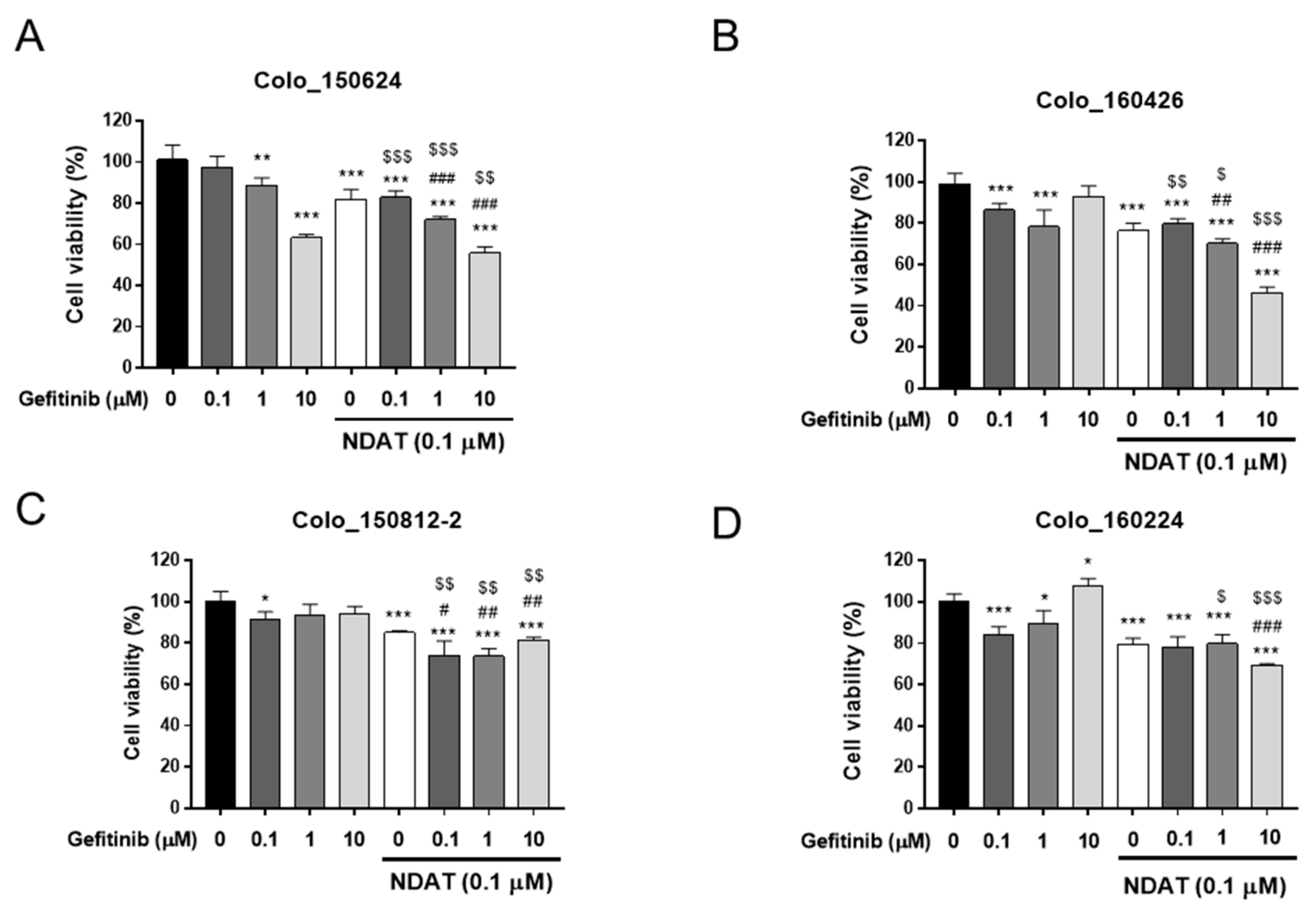
3.1. NDAT Enhances Gefitinib-Induced Anti-Proliferation in Primary Cultures of Human CRC Cells

NDAT inhibits PD-L1 expression and cell proliferation stimulated by thyroid hormone in cancer cells [12,35]. Gefitinib has been shown to suppress cell proliferation in K-RAS wild type HT-29 cells, but not in K-RAS-mutant HCT116 cells [9]. NDAT potentiates a gefitinib-induced anti-proliferative effect in both types of cancer cells [9]. We investigated the effects of NDAT and gefitinib in primary CRC cell lines. Gefitinib significantly inhibited cell proliferation in Colo_150624 (Figure 1A) and Colo_160426 cells (Figure 1B) in a concentration-dependent manner although 10 µM gefitinib did not inhibit Colo_160426 cancer growth. On the other hand, gefitinib lacked such activity in Colo_150812-2 (Figure 1C) and Colo_160224 cells (Figure 1D). In the latter, a high concentration of gefitinib (10 μM) significantly enhanced cell proliferation in Colo_160224 cells (Figure 1D), but not in Colo_160426 (Figure 1B) and Colo_150812-2 (Figure 1C) cells. A low concentration of NDAT (0.01 μM) inhibited the proliferation of these primary cultures of CRC cells but not in Colo_150812-2 (Figure S1). The treatment of NDAT (0.1 μM) significantly suppressed cell proliferation and showed a potentiated effect when combined with gefitinib in these cells (Figure 1).

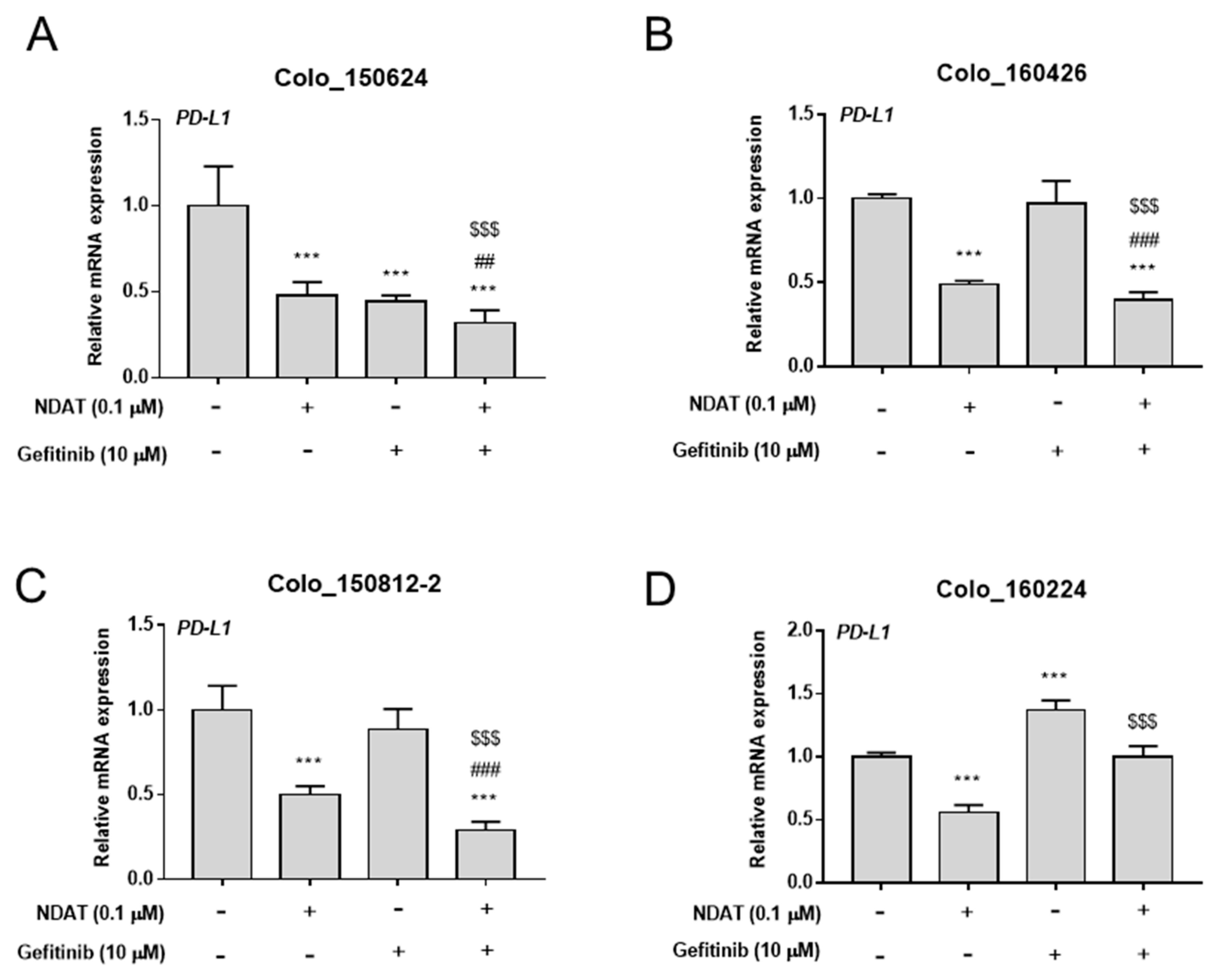
3.2. NDAT Enhances Gefitinib-Inhibited PD-L1 Expression in Primary Cultures of CRC Cells

PD-L1 expression may affect cancer cell proliferation [35,36]; therefore, we examined the possible linkage among PD-L1, cell proliferation, and gefitinib resistance. Gefitinib at a concentration of 10 μM significantly inhibited PD-L1 expression only in Colo_150624 cells (Figure 2A), whereas it significantly enhanced PD-L1 expression in Colo_160224 cells (Figure 2D). On the other hand, NDAT (0.1 μM) significantly potentiated gefitinib-repressed PD-L1 expression in primary cultures of CRC cells (Figure 2). The basal expression levels of PD-L1 mRNA in HCT116 cells and four established primary cultures of human CRC cells were evaluated. Expression of PD-L1 in gefitinib-resistant Colo_160224 cells was meaningfully higher than that in other primary CRC cancer cell lines and HCT116 cells (Figure S2). NDAT significantly repressed PD-L1 expression in these primary cultures of CRC cells at the concentration of 0.1 μM (Figure 2). Expression of PD-L1 was significantly inhibited by 1 μM gefitinib in Colo_150624 (Figure S2B), Colo_150812-2 (Figure S2D), and Colo_160224 (Figure S2E) cells.

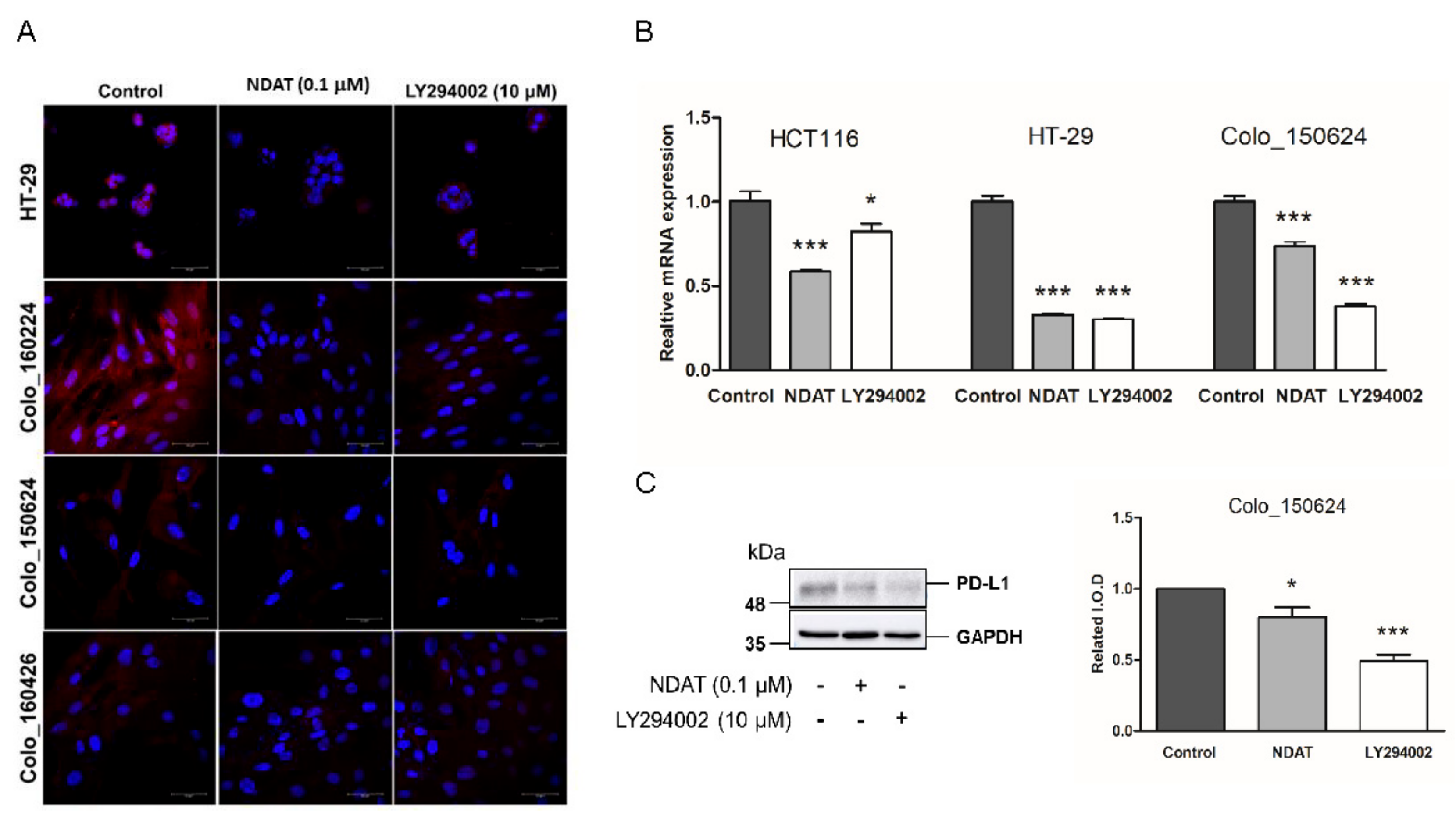
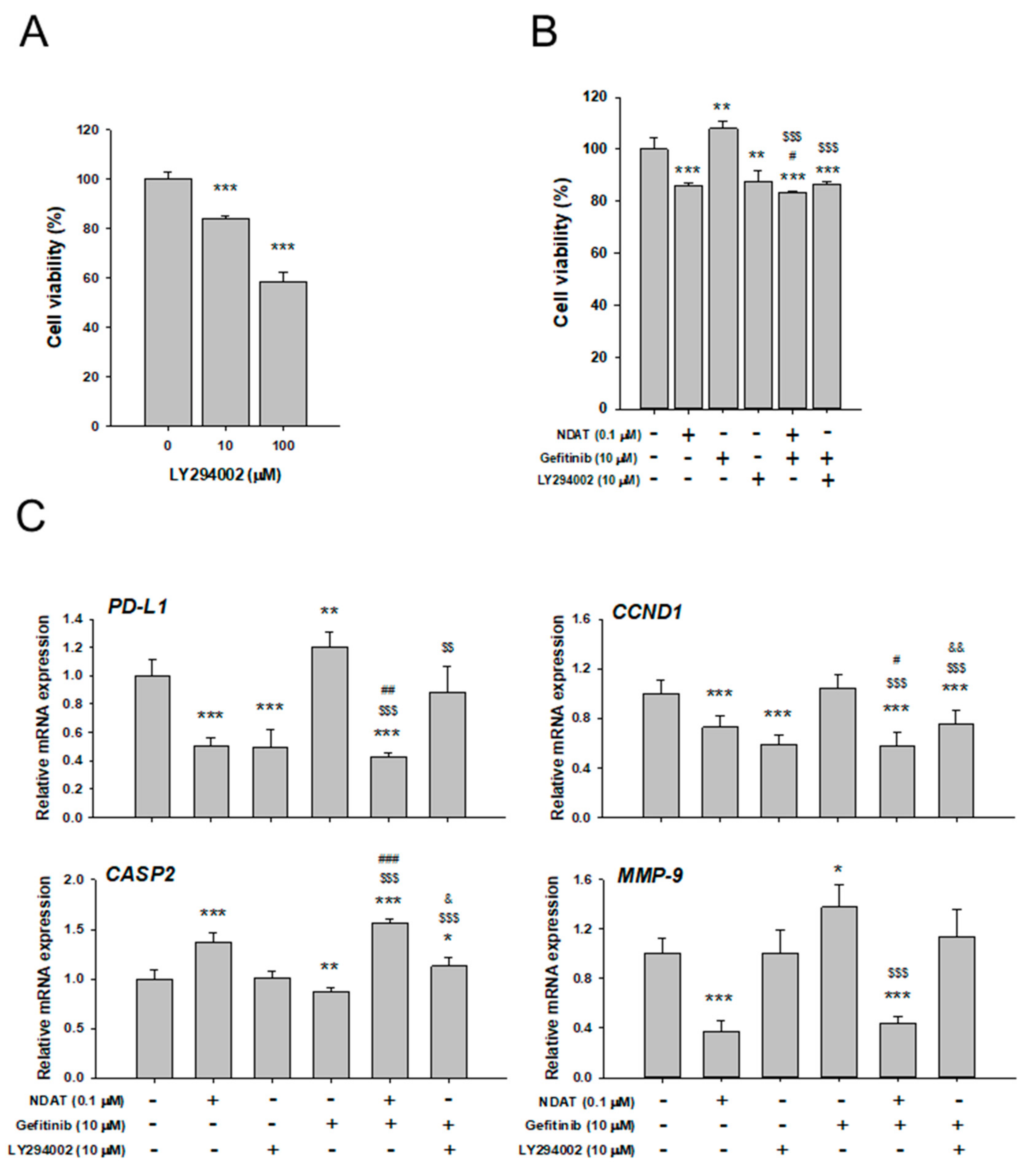
3.3. NDAT But Not Gefitinib Functions as PI3K Inhibitor to Inhibit PD-L1 Expression and Cell Proliferation in Gefitinib-Resistant Primary CRC

Constitutive activation of PI3K/Akt and RAS/ERK pathways is associated with gefitinib-resistance in certain cancers, such as non-small cell lung cancer (NSCLC) cells [37]. Confocal microscopy revealed that both NDAT and PI3K inhibitor, LY294002 inhibited the abundance of PD-L1 protein in the established HT-29 CRC cell and three established primary cultures of CRC cells (Colo_160224, Colo_150624, and Colo_160426) (Figure 3A). The gene expression and protein accumulation of PD-L1 suppressed significantly by NDAT and the PI3K inhibitor, LY294002, were demonstrated in Colo_150624 primary CRC cell culture (Figure 3B and 3C). These results suggested that blockage of PI3K activity could inhibit PD-L1 accumulation. NDAT also demonstrated its anti-PI3K activity [9]. Furthermore, the PI3K inhibitor, LY294002, induced anti-proliferation in gefitinib-resistant primary CRC cells, Colo_160224 primary CRC cell cultures (Figure 4A).
To investigate the effect of PI3K activation in the NDAT- or gefitinib-induced anti-proliferation in gefitinib-resistant primary CRC cells, Colo_160224 primary CRC cell cultures were treated with 0.1 μM NDAT, 10 µM gefitinib, 10 µM LY294002, or their combination in the medium with reagents changed daily for six days. Both NDAT and LY294002 inhibited cell proliferation significantly (Figure 4B). On the other hand, gefitinib stimulated cell proliferation significantly (Figure 4B), as observed in Figure 1. The combined NDAT and gefitinib or gefitinib in the presence of LY294002 inhibited cell growth significantly (Figure 4B).
To address the effect of combinations of agents on gefitinib-resistant CRC proliferation, we examined the effects of NDAT, LY294002, or gefitinib and their combination on the expression of PD-L1 and related genes in Colo_160224 cells (Figure 4C). After 24 h of treatment, cells were harvested, and RNA was extracted. The qPCR assay was conducted for PD-L1, CCND1 (proliferation), CASP2 (anti-proliferation), and MMP-9 (metastasis). NDAT significantly suppressed the expression of PD-L1, CCND1, and MMP-9, and enhanced that of CASP2 in Colo_160224 cells, as we have reported previously in K-RAS mutant CRC HCT116 cells [9,10,12]. LY294002 significantly reduced the expression of PD-L1 and CCND1, but not that of CASP2 and MMP-9. However, gefitinib stimulated the expression of PD-L1 and MMP-9 and suppressed CASP2 expression. The combined treatment of NDAT with gefitinib showed a significantly potentiated effect on CASP2 expression and inhibited more significantly the expression of PD-L1, CCND1, and MMP-9. The combination of LY294002 with gefitinib reduced the expression of CCND1 significantly as compared untreated control or gefitinib treatment alone, although the effect was less than LY294002 treatment alone (Figure 4C). The combined treatment of LY294002 with gefitinib inhibited PD-L1 expression significantly as compared with gefitinib treatment alone. Also, the same phenomenon appeared in the combined treatment of NDAT and gefitinib.

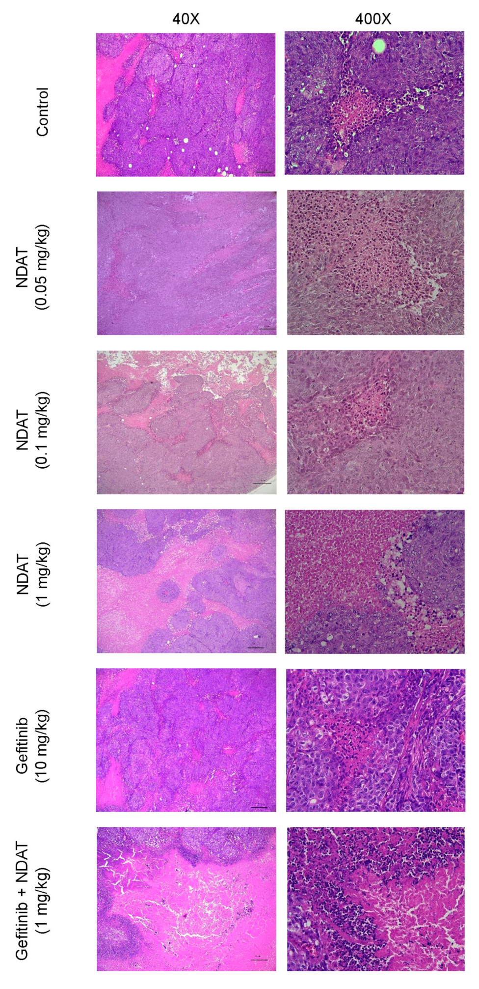
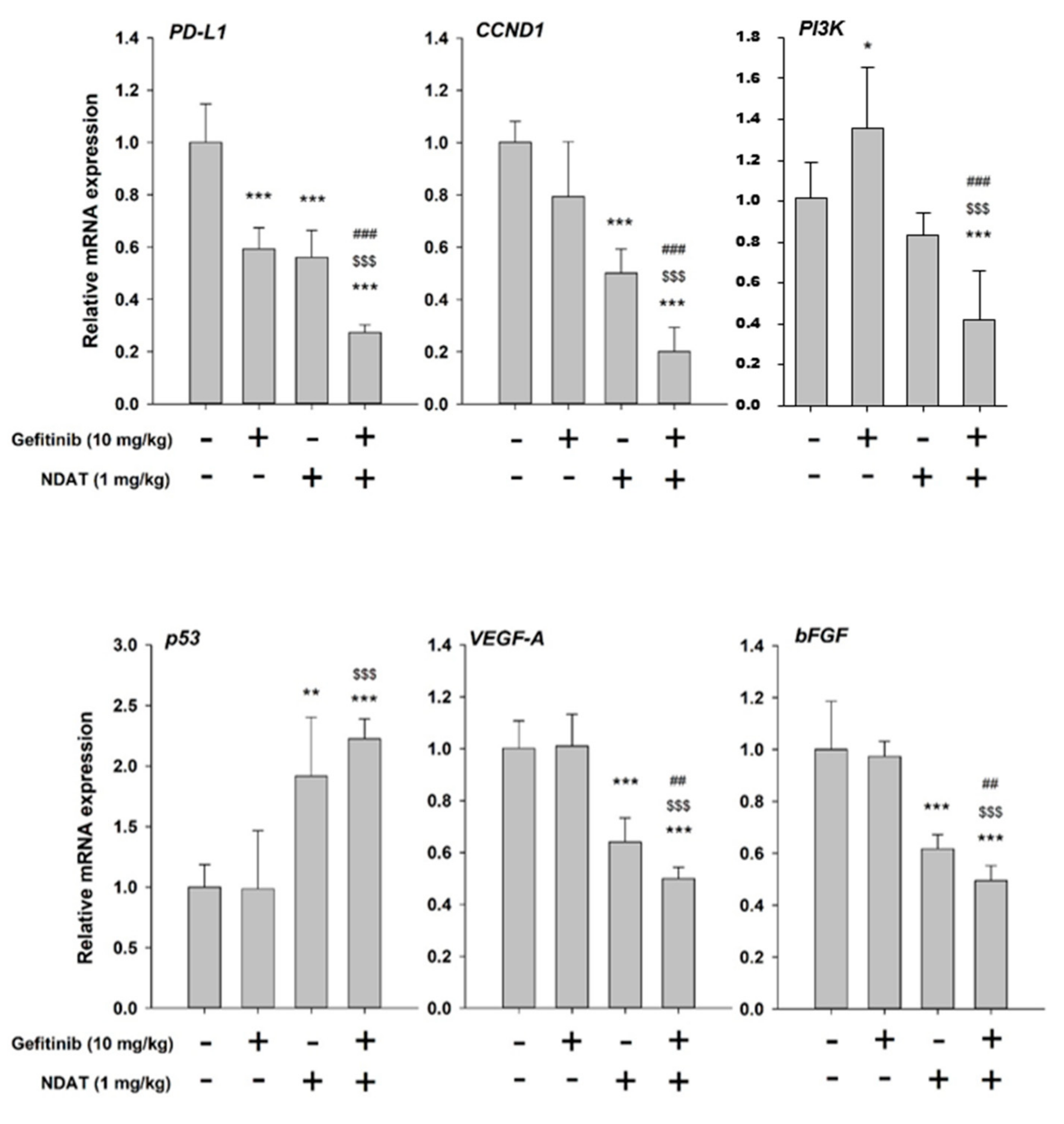
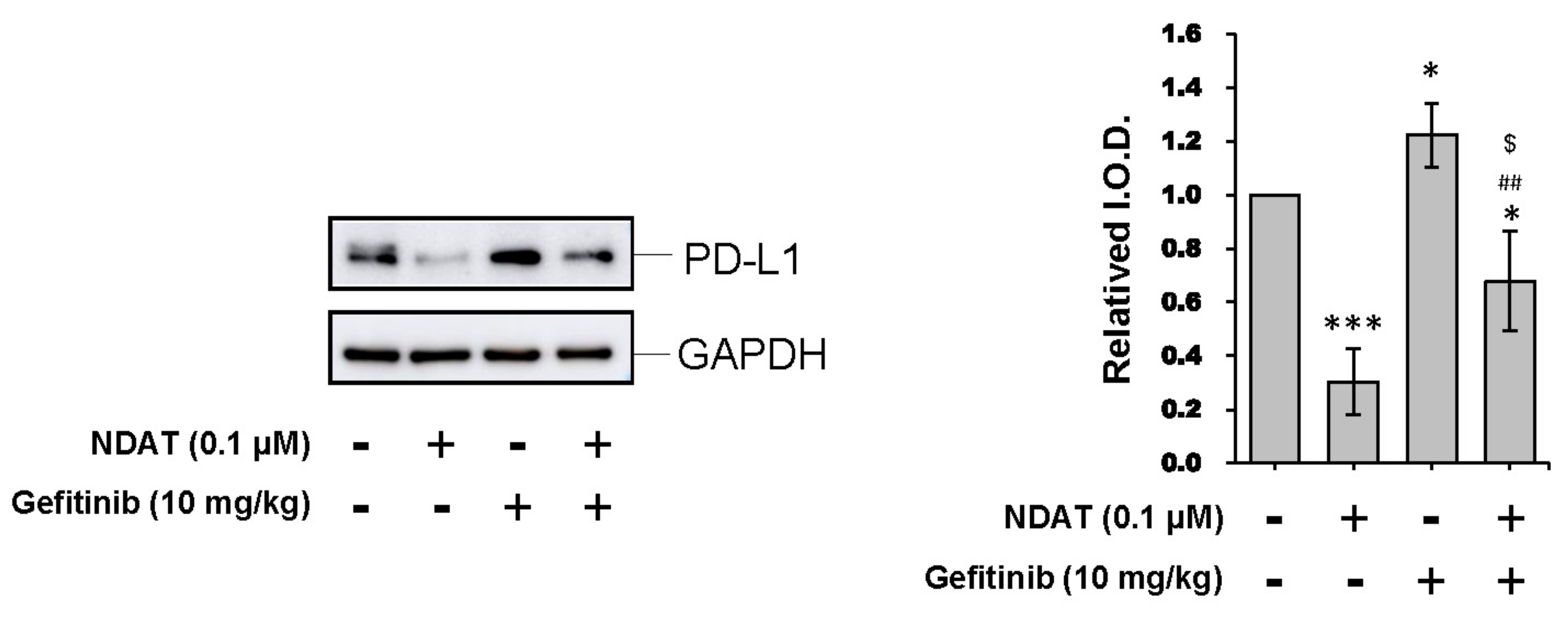
3.4. NDAT Inhibits PD-L1 Accumulation and Tumor Growth in HCT116 Cell Mouse Xenografts

In order to confirm our observation that NDAT suppressed PI3K to downregulate PD-L1 accumulation and cell proliferation in gefitinib-resistant CRC cells, xenograft of gefitinib-resistant HCT116 cells was conducted. The tumor size was assessed in BALB/C nude mice with gefitinib and NDAT treatment (Figure S3). NDAT had a more significant inhibitory effect on tumor growth than gefitinib. Then immunohistochemical staining of PD-L1 was apparently primarily on the outer cells of the tumor masses at five weeks. Expression of PD-L1 was decreased in xenografts of mice treated with gefitinib (10 mg/kg) and in the tumors of mice that received the combination of NDAT (1 mg/kg) and gefitinib (10 mg/kg) (Figure 5).
In a previous study, we demonstrated that NDAT enhanced the anti-proliferative activity of gefitinib in gefitinib-resistant CRC cells by inhibiting ST6Gal1 activity and PI3K activation. NDAT also enhanced the anticancer activity of gefitinib in HCT116 CRC cell xenografts [9]. In the current experiments, we further investigated the histological changes in the HCT116 cell-derived xenograft tissues after these treatments of NDAT, gefitinib, and their combination. The morphological changes of cancer cells with various treatments in xenografts were shown in Figure 6. Typical tumor morphology, such as a high nucleus/cytoplasm ratio and active mitosis, was observed in tissues from the control xenografts. The structure of the tumor masses was loosened, and the tumor cells were not packed together tightly after NDAT treatment. However, this was not observed in the gefitinib-treated group. The combination of NDAT (1 mg/kg) with gefitinib (10 mg/kg) showed destruction of tumor masses, disordered cell morphology, and extensive necrosis characterized by pyknosis. As a result, the non-tumor space was occupied by connective tissue. Reagent-treated groups reduced the nucleus-to-cytoplasm ratio compared to the control group. The combination of NDAT (1 mg/kg) and gefitinib (10 mg/kg) had less compact tumor mass and more connective tissue filling.
qPCR studies were carried out in tumor samples harvested from xenografts to confirm the suppressive effects of NDAT on PI3K-related signal transduction pathway in gefitinib-resistant CRC. The PI3K expression was inhibited by NDAT. Alternatively, gefitinib stimulated PI3K expression. However, the combination of NDAT and gefitinib inhibited PI3K expression significantly (Figure 7). Additionally, the expression of PD-L1, reduced by gefitinib, was further downregulated by NDAT co-administration. Also, NDAT significantly increased the expression of the pro-apoptotic gene p53 and reduced the expression of CCND1, VEGF-A, and bFGF but these were not observed in gefitinib-treated animals (Figure 7). Western blot analyses showed that NDAT but not gefitinib inhibited PD-L1 accumulation significantly in HCT116 xenograft tissues (Figure 8). In summary, although gefitinib may affect PD-L1 expression, NDAT inhibited PI3K expression and PD-L1 accumulation to inhibit cell proliferation in gefitinib-resistant CRC proliferation in vitro and in vivo.

4. Discussion

PD-1 belongs to the cluster of differentiation (CD) 28/CTL-associated antigen (CTLA)-4/inducible T cell costimulator (ICOS) family. It is categorized as an inhibitory receptor expressed by T cells, dendritic cells, natural killer cells, macrophages, and B cells [38]. PD-1-mediated T cell signaling depends upon binding of its ligands, PD-L1 (B7-H1) or PD-L2 (B7-DC) to alter cytotoxic killing, cytokine production, and T cell proliferation [39]. Expression of PD-L1 and PD-L2 are up-regulated in many human solid tumors, including CRC [14,40,41]. Blockade of PD-1 or PD-L1 activities may provide an optional management approach for PD-L1-overexpressing tumors, e.g., NSCLC with EGFR mutation, that has EGFR-TKI resistance [19]. Recently, we demonstrated that NDAT non-immunologically downregulates both gene expression and protein accumulation of basal and T4-induced PD-L1 in cancer cells, such as oral cancer cells, breast cancer cells, and CRC cells [10,12].
In this study, NDAT inhibited protein accumulation of PD-L1 in a concentration-dependent manner. In addition, gefitinib decreased the protein accumulation of PD-L1 in HCT116 cell xenografts. This finding is consistent with previous reports that gefitinib decreased PD-L1 expression in NSCLC cell lines [42,43]. The combination of NDAT and gefitinib further inhibited PD-L1 protein accumulation in xenografts. The PD-1/PD-L1 axis can modulate nature killer cells’ actions on tumor cells [44,45]. Gefitinib was reported to immuno-regulate lung cancer cells and NK cells that could enhance the interaction between them [43]. As a result, the combination of NDAT and gefitinib in our studies showed enhanced action on tumor size reduction in HCT116 cell xenografts [9].
The combination of NDAT and gefitinib also reduced viability and inhibited expression of PD-L1 in primary cultures of human CRC cells. One of the mechanisms for tumor cell-stimulated expression of PD-L1 is the activation of the EGFR, ERK1/2, PI3K-Akt, or Janus kinase 2/STAT1 signaling pathways [46]. In our studies, gefitinib did not inhibit the activation of PI3K but instead activated it in one primary culture (Colo_160224 cells). NDAT not only reduced PD-L1 expression but also inhibited PI3K activate by gefitinib (Figure S4). As a result, gefitinib (10 μM) induced the gene expression of PD-L1 in these cells (Figure 2D) and improved the viability in these cells (Figure 1D). On the other hand, NDAT not only inhibited the activation of PI3K but also decreased the expression of PI3K. Thus, PI3K appears to play an essential role in the inhibitory effect on NDAT-blocked PD-L1 expression in CRC cells.
Patients with phosphorylated EGFR-dependent cancer cell proliferation and metastasis are well-suited for therapy with EGFR TKIs [47]. However, the mutation of EGFR or downstream signaling pathways linked to EGFRs reduce the efficacy of such targeting therapy. Gefitinib is the first generation of EGFR-TKI to target EGFR-19del and EGFR-L858R mutation [19], but it has been less effective in CRC management than in other types of cancer [48,49,50]. In contrast to its efficacy in NSCLC treatment, gefitinib used in CRC patients in Phase I trials attained stable disease without an objective response in tumor size. This was the case although it may be effective in higher doses for lung cancer patients [48,51]. However, gefitinib, in combination with other EGFR-targeted agents, has been found to have antitumor effects that were more impressive than those obtained with the administration of a single EGFR inhibitors [52].
Atorvastatin has been shown to enhance its anticancer effects in combination with gefitinib. At a dose of 5 μM, atorvastatin enhanced the anticancer activity of gefitinib, apparently through concomitant inhibition of AKT and ERK signaling activities [53]. In current preclinical studies, NDAT improved the anti-proliferative effectiveness of gefitinib in primary cultures of CRC cells. The combined use of these two agents in studies of primary human CRC cells suppressed the transcription of genes regarded as biomarkers of proliferation, anti-proliferation, and angiogenesis.
In gefitinib-resistant HCT116 xenograft studies, NDAT suppressed the expression of PI3K and PD-L1 (Figure 7). On the other hand, gefitinib only suppressed the expression of PD-L1 but not PI3K (Figure 7). However, their combination further downregulated the expression of PD-L1 and PI3K (Figure 7). These observations suggested that NDAT could compensate gefitinib-regulated gene expression in gefitinib-resistant CRC cells. The accumulation of PD-L1 in xenograft studies further confirmed NDAT but not gefitinib-reduced PD-L1 accumulation (Figure 8). Although it is not clear what mechanisms are involved in gefitinib-resistance in Colo_160224 primary cell culture; however, NDAT suppressed PI3K activation and PD-L1 accumulation to inhibit cancer growth in Colo_160224 cells and K-RAS mutant HCT116 CRC cells.

5. Conclusions

NDAT was shown to enhance the anti-proliferative effects of gefitinib in four primary cultures of human CRC cells. In all four cell lines, NDAT alone suppressed PD-LI expression and proliferation. The combination treatment of NDAT and gefitinib further enhanced this effect. However, different mechanisms were involved in gefitinib- and NDAT-induced inhibition of PD-L1 accumulation. Gefitinib-inhibited PD-L1 accumulation was not inactive PI3K-dependent; however, NDAT inhibited PD-L1 accumulation via PI3K inactivation. These results affirm certain anticancer actions of NDAT and suggest that the combination of NDAT and gefitinib may be useful clinically in the setting of human gefitinib-resistant CRC.

Supplementary Materials

The following are available online at https://www.mdpi.com/2073-4409/9/8/1830/s1, Figure S1: NDAT augments gefitinib-stimulated anti-proliferation in primary cultures of human CRC cells, Figure S2: NDAT attenuate expression of PD-L1 regulated by gefitinib in human CRC primary cell cultures, Figure S3: NDAT and gefitinib inhibit tumor growth of HCT116 xenograft, Figure S4: NDAT inhibited PD-L1 accumulation in Colo_160224.

Author Contributions

Conception and design, T.-Y H., T.-C.C., Y.-T.C., K.W., H.Y. Lin and P.J. Davis.; Development of methodology, T.-C.C., Y.-T.C., T.Y. Huang, W.-J.C., S.-H.W., and Y.-S.P.; Acquisition of data: T.-Y.H., Y.-T.C., Z.-L.L., Y.-S.P., Y.-R.C., and Y.-J.S.; Analysis and interpretation of data: Y.-S.P., E.D.H., F.-C.L., and P.J. Davis.; Writing, review, and revision of the manuscript: T.-C.C., Y.-T.C., H.-R.C., H.Y. Lin, and P.J. Davis.; Technical support and advice: S.-H.W., A.W., R.H.C., C.A.C., and K.W.; Study supervision: S.-J.C., K.W., H.-Y.L., J.W.-P., S.A.M., and P.J.D. All authors have read and agreed to the published version of the manuscript.

Funding

This research was funded by a grant from Shuang-Ho Hospital Collaborating Program (T.-C.C. and A.Wu, 102TMU-SHH-05), the Ministry of Education (MOE) in Taiwan (DP2-107-20000), and by a general grant of Ministry of Science and Technology Taiwan (MOST 107-2314-B-038-017 MOST108-2314-B-038-050 to HYL and MOST108-2119-038-001 to JWP). Study is also supported partly by the National Institute of Food and Agriculture (CA-D*-MCB-7399-H) by the NIH grants (UG3TR002866, AI095382, EB021230, CA198880) to R.H.C.

Acknowledgments

Authors would like to send their appreciation to Joyce Peng for her excellent admission operation (Graduate Institute of Nanomedicine and Medical Engineering, Taipei Medical University) and a gift from Paul J. Davis (Albany Medical College, and Pharmaceutical Research Institute at Albany College of Pharmacy and Health Sciences) to Albany College of Pharmacy and Health Sciences.

Conflicts of Interest

Co-authors Dr. Shaker A Mousa and Dr. Paul J. Davis are co-inventors of NDAT and stockholders in a company (NanoPharmaceuticals LLC) that is commercializing the agent. All other co-authors declare no competing financial interests.

Abbreviations

APC, Adenomatous Polyposis Coli; bFGF, basic fibroblast growth factor; CCND1, cyclin D1; CRC, colorectal cancer; EGFR, epidermal growth factor receptor; FGF2, fibroblast growth factor 2; IFN-γ, interferon-γ; MMP-2, matrix metalloproteinases-2; matrix metalloproteinases-9; NDAT, nano-diamino-tetrac; PD-1, programmed cell death protein 1; PD-L1, programmed death-ligand 1; tetrac, tetraiodothyroxyacetic acid; TKI, tyrosine kinase inhibitor; VEGF-A, vascular endothelial growth factor A.

References

  1. Ferlay, J.; Soerjomataram, I.; Dikshit, R.; Eser, S.; Mathers, C.; Rebelo, M.; Parkin, D.M.; Forman, D.; Bray, F. Cancer incidence and mortality worldwide: Sources, methods and major patterns in GLOBOCAN 2012. Int. J. Cancer 2015, 136, E359–E386. [Google Scholar] [CrossRef] [PubMed]
  2. Chiang, C.J.; Lo, W.C.; Yang, Y.W.; You, S.L.; Chen, C.J.; Lai, M.S. Incidence and survival of adult cancer patients in Taiwan, 2002–2012. J. Formos. Med. Assoc. 2016, 115, 1076–1088. [Google Scholar] [CrossRef] [PubMed] [Green Version]
  3. Conlin, A.; Smith, G.; Carey, F.A.; Wolf, C.R.; Steele, R.J. The prognostic significance of K-ras, p53, and APC mutations in colorectal carcinoma. Gut 2005, 54, 1283–1286. [Google Scholar] [CrossRef] [PubMed] [Green Version]
  4. Li, J.; Kleeff, J.; Giese, N.; Büchler, M.W.; Korc, M.; Friess, H. Gefitinib (‘Iressa’, ZD1839), a selective epidermal growth factor receptor tyrosine kinase inhibitor, inhibits pancreatic cancer cell growth, invasion, and colony formation. Int. J. Oncol. 2004, 25, 203–210. [Google Scholar] [CrossRef] [PubMed]
  5. Spano, J.P.; Fagard, R.; Soria, J.C.; Rixe, O.; Khayat, D.; Milano, G. Epidermal growth factor receptor signaling in colorectal cancer: Preclinical data and therapeutic perspectives. Ann. Oncol. 2005, 16, 189–194. [Google Scholar] [CrossRef]
  6. Logue, J.S.; Morrison, D.K. Complexity in the signaling network: Insights from the use of targeted inhibitors in cancer therapy. Genes Dev. 2012, 26, 641–650. [Google Scholar] [CrossRef] [Green Version]
  7. Hu, T.; Li, C. Convergence between Wnt-β-catenin and EGFR signaling in cancer. Mol. Cancer 2010, 9, 236. [Google Scholar] [CrossRef] [Green Version]
  8. Nana, A.W.; Chin, Y.T.; Lin, C.Y.; Ho, Y.; Bennett, J.A.; Shih, Y.J.; Chen, Y.R.; Changou, C.A.; Pedersen, J.Z.; Incerpi, S.; et al. Tetrac downregulates β-catenin and HMGA2 to promote the effect of resveratrol in colon cancer. Endocr. Relat. Cancer 2018, 25, 279–293. [Google Scholar] [CrossRef] [Green Version]
  9. Chang, T.C.; Chin, Y.T.; Nana, A.W.; Wang, S.H.; Liao, Y.M.; Chen, Y.R.; Shih, Y.J.; Changou, C.A.; Yang, Y.S.; Wang, K.; et al. Enhancement by Nano-Diamino-Tetrac of Antiproliferative Action of Gefitinib on Colorectal Cancer Cells: Mediation by EGFR Sialylation and PI3K Activation. Horm. Cancer 2018, 9, 420–432. [Google Scholar] [CrossRef] [Green Version]
  10. Nana, A.W.; Wu, S.Y.; Yang, Y.S.; Chin, Y.T.; Cheng, T.M.; Ho, Y.; Li, W.S.; Liao, Y.M.; Chen, Y.R.; Shih, Y.J.; et al. Nano-Diamino-Tetrac (NDAT) Enhances Resveratrol-Induced Antiproliferation by Action on the RRM2 Pathway in Colorectal Cancers. Horm. Cancer 2018, 9, 349–360. [Google Scholar] [CrossRef]
  11. Lee, Y.S.; Chin, Y.T.; Shih, Y.J.; Nana, A.W.; Chen, Y.R.; Wu, H.C.; Yang, Y.S.H.; Lin, H.Y.; Davis, P.J. Thyroid Hormone Promotes β-Catenin Activation and Cell Proliferation in Colorectal Cancer. Horm. Cancer 2018, 9, 156–165. [Google Scholar] [CrossRef] [PubMed]
  12. Lin, H.Y.; Chin, Y.T.; Nana, A.W.; Shih, Y.J.; Lai, H.Y.; Tang, H.Y.; Leinung, M.; Mousa, S.A.; Davis, P.J. Actions of l-thyroxine and Nano-diamino-tetrac (Nanotetrac) on PD-L1 in cancer cells. Steroids 2016, 114, 59–67. [Google Scholar] [CrossRef] [PubMed] [Green Version]
  13. Xiang, X.; Yu, P.C.; Long, D.; Liao, X.L.; Zhang, S.; You, X.M.; Zhong, J.H.; Li, L.Q. Prognostic value of PD-L1 expression in patients with primary solid tumors. Oncotarget 2018, 9, 5058–5072. [Google Scholar] [CrossRef] [PubMed] [Green Version]
  14. Lee, K.S.; Kim, B.H.; Oh, H.K.; Kim, D.W.; Kang, S.B.; Kim, H.; Shin, E. Programmed cell death ligand-1 protein expression and CD274/PD-L1 gene amplification in colorectal cancer: Implications for prognosis. Cancer Sci. 2018, 109, 2957–2969. [Google Scholar] [CrossRef] [PubMed] [Green Version]
  15. Enkhbat, T.; Nishi, M.; Takasu, C.; Yoshikawa, K.; Jun, H.; Tokunaga, T.; Kashihara, H.; Ishikawa, D.; Shimada, M. Programmed Cell Death Ligand 1 Expression Is an Independent Prognostic Factor in Colorectal Cancer. Anticancer Res. 2018, 38, 3367–3373. [Google Scholar] [CrossRef] [PubMed]
  16. Akbay, E.A.; Koyama, S.; Carretero, J.; Altabef, A.; Tchaicha, J.H.; Christensen, C.L.; Mikse, O.R.; Cherniack, A.D.; Beauchamp, E.M.; Pugh, T.J.; et al. Activation of the PD-1 pathway contributes to immune escape in EGFR-driven lung tumors. Cancer Discov. 2013, 3, 1355–1363. [Google Scholar] [CrossRef] [PubMed] [Green Version]
  17. Abiko, K.; Matsumura, N.; Hamanishi, J.; Horikawa, N.; Murakami, R.; Yamaguchi, K.; Yoshioka, Y.; Baba, T.; Konishi, I.; Mandai, M. IFN-gamma from lymphocytes induces PD-L1 expression and promotes progression of ovarian cancer. Br. J. Cancer 2015, 112, 1501–1509. [Google Scholar] [CrossRef] [Green Version]
  18. Schmidt, L.H.; Kummel, A.; Gorlich, D.; Mohr, M.; Brockling, S.; Mikesch, J.H.; Grunewald, I.; Marra, A.; Schultheis, A.M.; Wardelmann, E.; et al. PD-1 and PD-L1 Expression in NSCLC Indicate a Favorable Prognosis in Defined Subgroups. PLoS ONE 2015, 10, e0136023. [Google Scholar] [CrossRef] [Green Version]
  19. Chen, N.; Fang, W.; Zhan, J.; Hong, S.; Tang, Y.; Kang, S.; Zhang, Y.; He, X.; Zhou, T.; Qin, T.; et al. Upregulation of PD-L1 by EGFR Activation Mediates the Immune Escape in EGFR-Driven NSCLC: Implication for Optional Immune Targeted Therapy for NSCLC Patients with EGFR Mutation. J. Thorac. Oncol. 2015, 10, 910–923. [Google Scholar] [CrossRef] [Green Version]
  20. Page, D.B.; Postow, M.A.; Callahan, M.K.; Allison, J.P.; Wolchok, J.D. Immune modulation in cancer with antibodies. Ann. Rev. Med. 2014, 65, 185–202. [Google Scholar] [CrossRef]
  21. Mittendorf, E.A.; Philips, A.V.; Meric-Bernstam, F.; Qiao, N.; Wu, Y.; Harrington, S.; Su, X.; Wang, Y.; Gonzalez-Angulo, A.M.; Akcakanat, A.; et al. PD-L1 expression in triple-negative breast cancer. Cancer Immunol. Res. 2014, 2, 361–370. [Google Scholar] [CrossRef] [PubMed] [Green Version]
  22. Song, J.; Zhu, J.; Zhao, Q.; Tian, B. Gefitinib causes growth arrest and inhibition of metastasis in human chondrosarcoma cells. J. Buon. 2015, 20, 894–901. [Google Scholar] [PubMed]
  23. Tebbutt, N.; Pedersen, M.W.; Johns, T.G. Targeting the ERBB family in cancer: Couples therapy. Nat. Rev. Cancer 2013, 13, 663–673. [Google Scholar] [CrossRef] [PubMed]
  24. Toda, D.; Ota, T.; Tsukuda, K.; Watanabe, K.; Fujiyama, T.; Murakami, M.; Naito, M.; Shimizu, N. Gefitinib decreases the synthesis of matrix metalloproteinase and the adhesion to extracellular matrix proteins of colon cancer cells. Anticancer Res. 2006, 26, 129–134. [Google Scholar] [PubMed]
  25. Koizumi, F.; Kanzawa, F.; Ueda, Y.; Koh, Y.; Tsukiyama, S.; Taguchi, F.; Tamura, T.; Saijo, N.; Nishio, K. Synergistic interaction between the EGFR tyrosine kinase inhibitor gefitinib ("Iressa") and the DNA topoisomerase I inhibitor CPT-11 (irinotecan) in human colorectal cancer cells. Int. J. Cancer 2004, 108, 464–472. [Google Scholar] [CrossRef] [PubMed]
  26. Giaccone, G.; González-Larriba, J.L.; van Oosterom, A.T.; Alfonso, R.; Smit, E.F.; Martens, M.; Peters, G.J.; van der Vijgh, W.J.; Smith, R.; Averbuch, S.; et al. Combination therapy with gefitinib, an epidermal growth factor receptor tyrosine kinase inhibitor, gemcitabine and cisplatin in patients with advanced solid tumors. Ann. Oncol. 2004, 15, 831–838. [Google Scholar] [CrossRef]
  27. Lin, H.Y.; Chin, Y.T.; Yang, Y.C.; Lai, H.Y.; Wang-Peng, J.; Liu, L.F.; Tang, H.Y.; Davis, P.J. Thyroid Hormone, Cancer, and Apoptosis. Compr. Physiol. 2016, 6, 1221–1237. [Google Scholar]
  28. Lee, Y.S.; Chin, Y.T.; Yang, Y.S.H.; Wei, P.L.; Wu, H.C.; Shih, A.; Lu, Y.T.; Pedersen, J.Z.; Incerpi, S.; Liu, L.F.; et al. The combination of tetraiodothyroacetic acid and cetuximab inhibits cell proliferation in colorectal cancers with different K-ras status. Steroids 2016, 111, 63–70. [Google Scholar] [CrossRef]
  29. Schmohl, K.A.; Müller, A.M.; Wechselberger, A.; Rühland, S.; Salb, N.; Schwenk, N.; Heuer, H.; Carlsen, J.; Göke, B.; Nelson, P.J.; et al. Thyroid hormones and tetrac: New regulators of tumour stroma formation via integrin αvβ3. Endocr. Relat. Cancer 2015, 22, 941–952. [Google Scholar] [CrossRef] [Green Version]
  30. Davis, P.J.; Glinsky, G.V.; Lin, H.Y.; Leith, J.T.; Hercbergs, A.; Tang, H.Y.; Ashur-Fabian, O.; Incerpi, S.; Mousa, S.A. Cancer Cell Gene Expression Modulated from Plasma Membrane Integrin αvβ3 by Thyroid Hormone and Nanoparticulate Tetrac. Front. Endocrinol. 2014, 5, 240. [Google Scholar]
  31. Sudha, T.; Bharali, D.J.; Yalcin, M.; Darwish, N.H.; Coskun, M.D.; Keating, K.A.; Lin, H.Y.; Davis, P.J.; Mousa, S.A. Targeted delivery of cisplatin to tumor xenografts via the nanoparticle component of nano-diamino-tetrac. Nanomedicine 2017, 12, 195–205. [Google Scholar] [CrossRef] [PubMed] [Green Version]
  32. Failli, A.; Consolini, R.; Legitimo, A.; Spisni, R.; Castagna, M.; Romanini, A.; Crimaldi, G.; Miccoli, P. The challenge of culturing human colorectal tumor cells: Establishment of a cell culture model by the comparison of different methodological approaches. Tumori 2009, 95, 343–347. [Google Scholar] [CrossRef] [PubMed]
  33. Ali, M.Y.; Anand, S.V.; Tangella, K.; Ramkumar, D.; Saif, T.A. Isolation of Primary Human Colon Tumor Cells from Surgical Tissues and Culturing Them Directly on Soft Elastic Substrates for Traction Cytometry. J. Vis. Exp. 2015, e52532. [Google Scholar] [CrossRef] [PubMed] [Green Version]
  34. Yang, S.H.; Lin, H.Y.; Chang, V.H.; Chen, C.C.; Liu, Y.R.; Wang, J.; Zhang, K.; Jiang, X.; Yen, Y. Lovastatin overcomes gefitinib resistance through TNF-α signaling in human cholangiocarcinomas with different LKB1 statuses in vitro and in vivo. Oncotarget 2015, 6, 23857–23873. [Google Scholar] [CrossRef] [PubMed]
  35. Lin, S.J.; Chin, Y.T.; Ho, Y.; Chou, S.Y.; Sh Yang, Y.C.; Nana, A.W.; Su, K.W.; Lim, Y.T.; Wang, K.; Lee, S.Y.; et al. Nano-diamino-tetrac (NDAT) inhibits PD-L1 expression which is essential for proliferation in oral cancer cells. Food Chem. Toxicol. 2018, 120, 1–11. [Google Scholar] [CrossRef] [PubMed]
  36. Clark, C.A.; Gupta, H.B.; Sareddy, G.; Pandeswara, S.; Lao, S.; Yuan, B.; Drerup, J.M.; Padron, A.; Conejo-Garcia, J.; Murthy, K.; et al. Tumor-Intrinsic PD-L1 Signals Regulate Cell Growth, Pathogenesis, and Autophagy in Ovarian Cancer and Melanoma. Cancer Res. 2016, 76, 6964–6974. [Google Scholar] [CrossRef] [Green Version]
  37. Janmaat, M.L.; Rodriguez, J.A.; Gallegos-Ruiz, M.; Kruyt, F.A.; Giaccone, G. Enhanced cytotoxicity induced by gefitinib and specific inhibitors of the Ras or phosphatidyl inositol-3 kinase pathways in non-small cell lung cancer cells. Int. J. Cancer 2006, 118, 209–214. [Google Scholar] [CrossRef]
  38. Boussiotis, V.A. Molecular and Biochemical Aspects of the PD-1 Checkpoint Pathway. New Engl. J. Med. 2016, 375, 1767–1778. [Google Scholar] [CrossRef] [Green Version]
  39. Tseng, S.Y.; Otsuji, M.; Gorski, K.; Huang, X.; Slansky, J.E.; Pai, S.I.; Shalabi, A.; Shin, T.; Pardoll, D.M.; Tsuchiya, H. B7-DC, a new dendritic cell molecule with potent costimulatory properties for T cells. J. Exp. Med. 2001, 193, 839–846. [Google Scholar] [CrossRef]
  40. Dong, H.; Strome, S.E.; Salomao, D.R.; Tamura, H.; Hirano, F.; Flies, D.B.; Roche, P.C.; Lu, J.; Zhu, G.; Tamada, K.; et al. Tumor-associated B7-H1 promotes T-cell apoptosis: A potential mechanism of immune evasion. Nat. Med. 2002, 8, 793–800. [Google Scholar] [CrossRef]
  41. Wilke, C.M.; Wei, S.; Wang, L.; Kryczek, I.; Kao, J.; Zou, W. Dual biological effects of the cytokines interleukin-10 and interferon-γ. Cancer Immunol. Immunother. 2011, 60, 1529–1541. [Google Scholar] [CrossRef] [PubMed]
  42. Zhang, N.; Zeng, Y.; Du, W.; Zhu, J.; Shen, D.; Liu, Z.; Huang, J.A. The EGFR pathway is involved in the regulation of PD-L1 expression via the IL-6/JAK/STAT3 signaling pathway in EGFR-mutated non-small cell lung cancer. Int. J. Oncol. 2016, 49, 1360–1368. [Google Scholar] [CrossRef] [PubMed] [Green Version]
  43. He, S.; Yin, T.; Li, D.; Gao, X.; Wan, Y.; Ma, X.; Ye, T.; Guo, F.; Sun, J.; Lin, Z.; et al. Enhanced interaction between natural killer cells and lung cancer cells: Involvement in gefitinib-mediated immunoregulation. J. Transl. Med. 2013, 11, 186. [Google Scholar] [CrossRef] [PubMed] [Green Version]
  44. Hsu, J.; Hodgins, J.J.; Marathe, M.; Nicolai, C.J.; Bourgeois-Daigneault, M.C.; Trevino, T.N.; Azimi, C.S.; Scheer, A.K.; Randolph, H.E.; Thompson, T.W.; et al. Contribution of NK cells to immunotherapy mediated by PD-1/PD-L1 blockade. J. Clin. Investig. 2018, 128, 4654–4668. [Google Scholar] [CrossRef] [PubMed]
  45. Benson, D.M., Jr.; Bakan, C.E.; Mishra, A.; Hofmeister, C.C.; Efebera, Y.; Becknell, B.; Baiocchi, R.A.; Zhang, J.; Yu, J.; Smith, M.K.; et al. The PD-1/PD-L1 axis modulates the natural killer cell versus multiple myeloma effect: A therapeutic target for CT-011, a novel monoclonal anti-PD-1 antibody. Blood 2010, 116, 2286–2294. [Google Scholar] [CrossRef]
  46. Ran, X.; Yang, K. Inhibitors of the PD-1/PD-L1 axis for the treatment of head and neck cancer: Current status and future perspectives. Drug Des. Dev. Ther. 2017, 11, 2007–2014. [Google Scholar] [CrossRef] [Green Version]
  47. Janmaat, M.L.; Giaccone, G. Small-molecule epidermal growth factor receptor tyrosine kinase inhibitors. Oncologist 2003, 8, 576–586. [Google Scholar] [CrossRef]
  48. Blanke, C.D. Gefitinib in colorectal cancer: If wishes were horses. J. Clin. Oncol. 2005, 23, 5446–5449. [Google Scholar] [CrossRef]
  49. Shrader, M.; Pino, M.S.; Lashinger, L.; Bar-Eli, M.; Adam, L.; Dinney, C.P.; McConkey, D.J. Gefitinib reverses TRAIL resistance in human bladder cancer cell lines via inhibition of AKT-mediated X-linked inhibitor of apoptosis protein expression. Cancer Res. 2007, 67, 1430–1435. [Google Scholar] [CrossRef] [Green Version]
  50. Ono, M.; Kuwano, M. Molecular mechanisms of epidermal growth factor receptor (EGFR) activation and response to gefitinib and other EGFR-targeting drugs. Clin. Cancer Res. 2006, 12, 7242–7251. [Google Scholar] [CrossRef] [Green Version]
  51. Pao, W.; Wang, T.Y.; Riely, G.J.; Miller, V.A.; Pan, Q.; Ladanyi, M.; Zakowski, M.F.; Heelan, R.T.; Kris, M.G.; Varmus, H.E. KRAS mutations and primary resistance of lung adenocarcinomas to gefitinib or erlotinib. PLoS Med. 2005, 2, e17. [Google Scholar] [CrossRef] [PubMed] [Green Version]
  52. Yuan, H.H.; Han, Y.; Bian, W.X.; Liu, L.; Bai, Y.X. The effect of monoclonal antibody cetuximab (C225) in combination with tyrosine kinase inhibitor gefitinib (ZD1839) on colon cancer cell lines. Pathology 2012, 44, 547–551. [Google Scholar] [CrossRef] [PubMed]
  53. Chen, J.; Bi, H.; Hou, J.; Zhang, X.; Zhang, C.; Yue, L.; Wen, X.; Liu, D.; Shi, H.; Yuan, J.; et al. Atorvastatin overcomes gefitinib resistance in KRAS mutant human non-small cell lung carcinoma cells. Cell Death Dis. 2013, 4, e814. [Google Scholar] [CrossRef] [PubMed] [Green Version]
Figure 1. NDAT augments gefitinib-stimulated anti-proliferation in primary cultures of human CRC cells. Four established primary human CRC cell cultures, Colo_150624 (A), Colo_160426 (B), Colo_150812-2 (C), and Colo_160224 (D), were seeded in 96-well plates and treated with different concentrations of gefitinib (0.1, 1, and 10 μM), NDAT (0.1 μM), or their combination. Media with drugs were refreshed daily for six days. Cell viability was examined with the MTT assay. N = 6. Data are expressed as mean ± SD; * p < 0.05, ** p < 0.01, *** p < 0.001, compared with untreated control; # p < 0.05, ## p < 0.01, ### p < 0.001, compared with NDAT; $ p < 0.05, $$ p < 0.01, $$$ p < 0.001, compared with gefitinib.
Cells 09 01830 g001
Figure 2. NDAT attenuate expression of PD-L1 regulated by gefitinib in human CRC primary cell cultures. Four established primary human CRC cell cultures, Colo_150624 (A), Colo_160426 (B), Colo_150812-2 (C), and Colo_160224 (D), were seeded in six-well plates and treated with different concentrations of gefitinib (10 μM), NDAT (0.1 μM), or their combination after starvation for 24 h. Cells were harvested and total RNA was extracted. qPCR was conducted for PD-L1 expression. N = 6. Data are expressed as mean ± SD; *** p < 0.001, compared with untreated control; ### p < 0.001, compared with NDAT; $$$ p < 0.001, compared with gefitinib.
Cells 09 01830 g002
Figure 3. NDAT and LY294002 inhibit PD-L1 accumulation in CRC. HT-29 cells (colorectal cancer cell line) and three established primary human CRC cell cultures (Colo_160224, Colo_150624, and Colo_160426) were treated with NDAT or LY294002 for 24 h. (A) Immunocytochemistry was performed for PD-L1 (red) and the nucleus was counterstained with DAPI (blue). Scale bar = 50 µm. (B) Gene expression of PD-L1 was evaluated by qPCR. (C) Protein accumulation of PD-L1 was detected by western blot. Densitometric analyses of western blots were done by ImageJ 1.5 and shown in a bar chart. N = 3. Data are expressed as mean ± SD; * p < 0.05, *** p < 0.001, compared with the untreated control.
Cells 09 01830 g003
Figure 4. Actions of NDAT and gefitinib are PI3K-dependent in primary cell culture Colo_160224. (A) Cells were treated with LY294002 (10 and 100 μM) with refreshed media containing LY294002 daily for six days. Cell viability was conducted by the MTT assay. N = 6. (B) Cells were treated with NDAT (0.1 μM), gefitinib (10 μM), and their combination with or without LY294002 (10 μM) for six days. Cell viability was evaluated by the MTT assay. N =6. (C) Cells were treated with NDAT (0.1 μM), gefitinib (10 μM), and their combination with or without LY294002 (10 μM) after starvation for 24 h (N = 4). Total RNA was extracted, and qPCR was performed for PD-L1, CCND1, CASP2, and MMP-9. Data are expressed as mean ± SD; * p < 0.05, ** p < 0.01, *** p < 0.001, compared with the untreated control; # p < 0.05, ## p < 0.01, ### p < 0.001, compared with the NDAT; $$ p < 0.01, $$$ p < 0.001, compared with gefitinib; & p < 0.05, && p < 0.01, compared with LY294002.
Cells 09 01830 g004
Figure 5. NDAT potentiates gefitinib-reduced PD-L1 expression in HCT116 cell-derived xenograft tumors (Table 1). The positive staining of PD-L1 is shown in brown color. The magnification is 40× (upper panels, bar = 210 μm) and 400× (lower panels, bar = 21 μm).
Cells 09 01830 g005
Figure 6. H&E stain of HCT116 xenograft tumor tissues. Xenograft tissues from various treatment including NDAT (0.05 mg, 0.1 mg, 1 mg/kg), gefitinib (10 mg/kg) or combined treatment were stained by hematoxylin (nucleus, blue) and eosin (extracellular matrix and cytoplasm, pink). The magnification is 40× (left panels, bar = 210 μm) and 400× (right panels, bar = 21 μm).
Cells 09 01830 g006
Figure 7. NDAT enhances gefitinib-regulated gene expression in HCT116 cell-derived xenograft tumor tissues. Tumor tissues were homogenized, and total RNA was extracted. Total RNA was then transcribed into cDNA. The expression levels of CCND1, p53, VEGF-A, PD-L1, EGFR, and bEGF (EGF2) were examined with qPCR experiments. The number of independent experiments N = 4. Data are expressed as mean ± SD; * p < 0.05, ** p < 0.01, *** p < 0.001, compared with untreated controls; ## p < 0.01, ### p < 0.001, compared with NDAT; $$$ p < 0.001, compared with gefitinib.
Cells 09 01830 g007
Figure 8. NDAT inhibited PD-L1 accumulation in HCT116 cell-derived xenograft tumor tissues. Tumor tissues were homogenized, and total proteins were extracted. Western blotting analyses were conducted by PD-L1. The number of independent experiments N = 4. Data are expressed as mean ± SD; * p < 0.05, *** p < 0.001, compared with untreated control; ## p < 0.01, compared with NDAT; $ p < 0.05, compared with gefitinib.
Cells 09 01830 g008
Table 1. Primer Sequences for Quantitative PCR on CRC Cell Lines and Primary Culture Cells.
NAMEF―ATCGR―ATCG
18S5’-GTAACCCGTTGAACCCCATT-3’5’-CCATCCAATCGGTAGTAGCG-3’
PD-L15’-GTTGAAGGACCAGCTCTCCC-3’5’-ACCCCTGCATCCTGCAATTT-3’
CCND15’-CAAGGCCTGAACCTGAGGAG-3’5’-GATCACTCTGGAGAGGAAGCG-3’
CASP25’-GCATGTACTCCCACCGTTGA-3’5’-GACAGGCGGAGCTTCTTGTA-3’
MMP-95’-TGTACCGCTATGGTTACACTCG-3’5’-GGCAGGGACAGTTGCTTCT-3’
PI3K5’-CCTGATCTTCCTCGTGCTGCTC-3’5’-ATGCCAATGGACAGTGTTCCTCTT-3’
bFGF5’-GAGAAGAGCGACCCTCACA-3’5’-TAGCTTTCTGCCCAGGTCC-3’
VEGF-A5’-TACCTCCACCATGCCAAGTG-3’5’-GATGATTCTGCCCTCCTCCTT-3’
p53 5’-AAGTCTAGAGCCACCGTCCA-3’ 5’-CAGTCTGGCTGCCAATCCA-3’

