Electron Microscopy Evidence of Zn Bioauthigenic Sulfides Formation in Polluted Organic Matter-Rich Sediments from the Chicamocha River (Boyacá-Colombia)


Abstract
:1. Introduction
2. Background Context
3. Materials and Methods
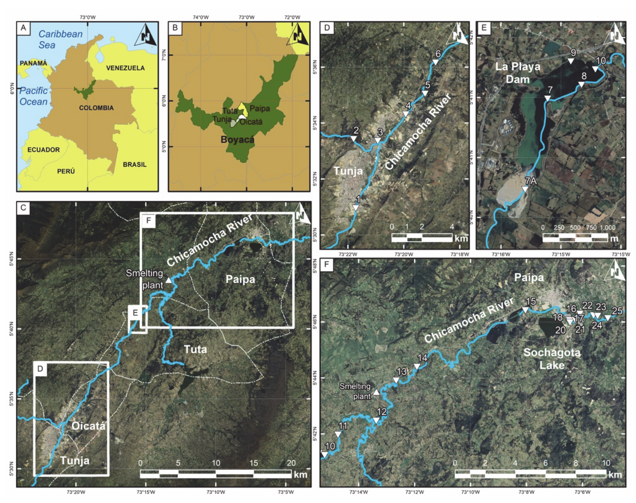
3.1. Materials
3.2. Mineralogical and Geochemical Analysis
4. Results
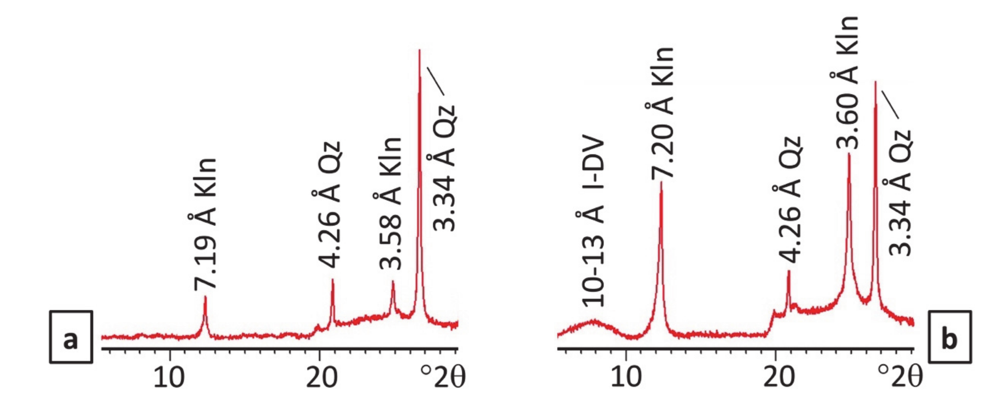
4.1. XRD Results
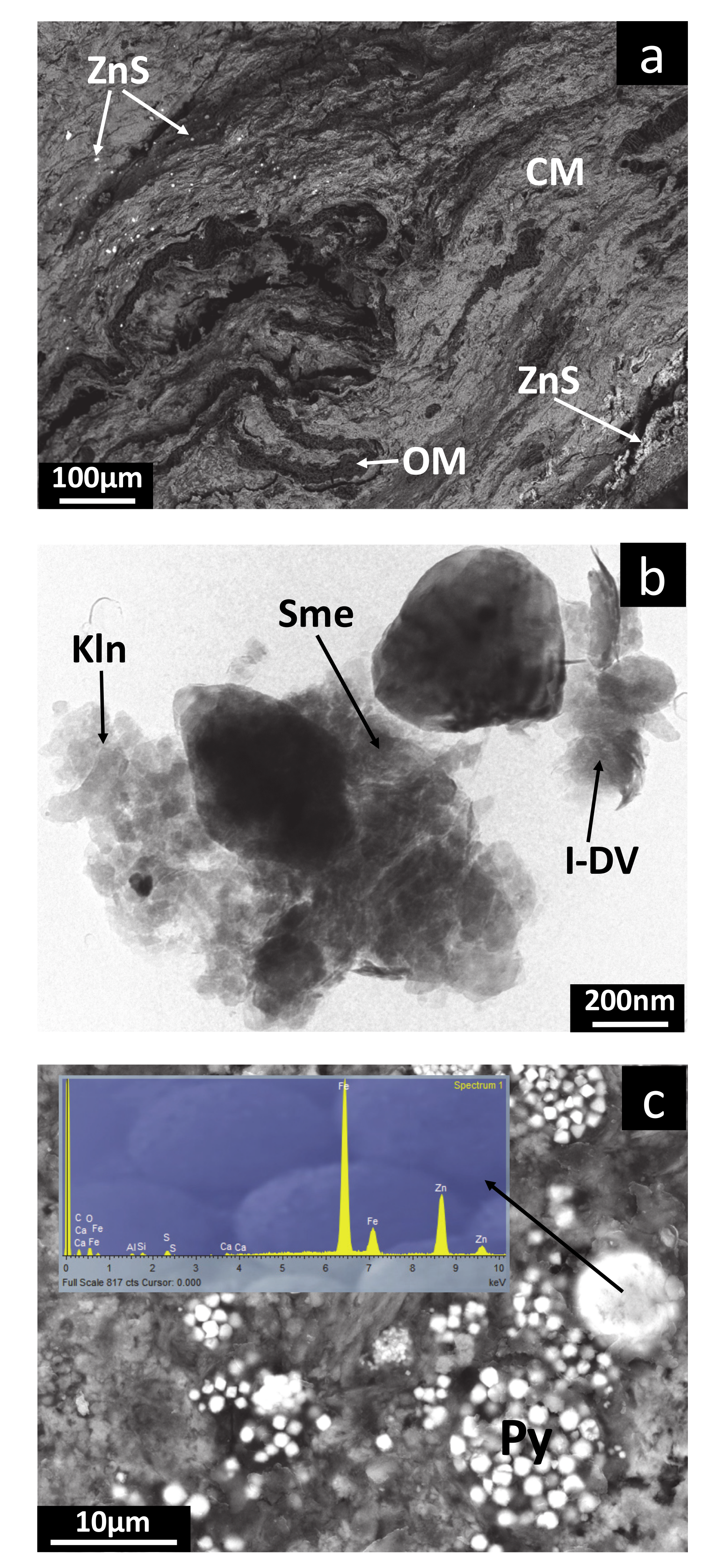
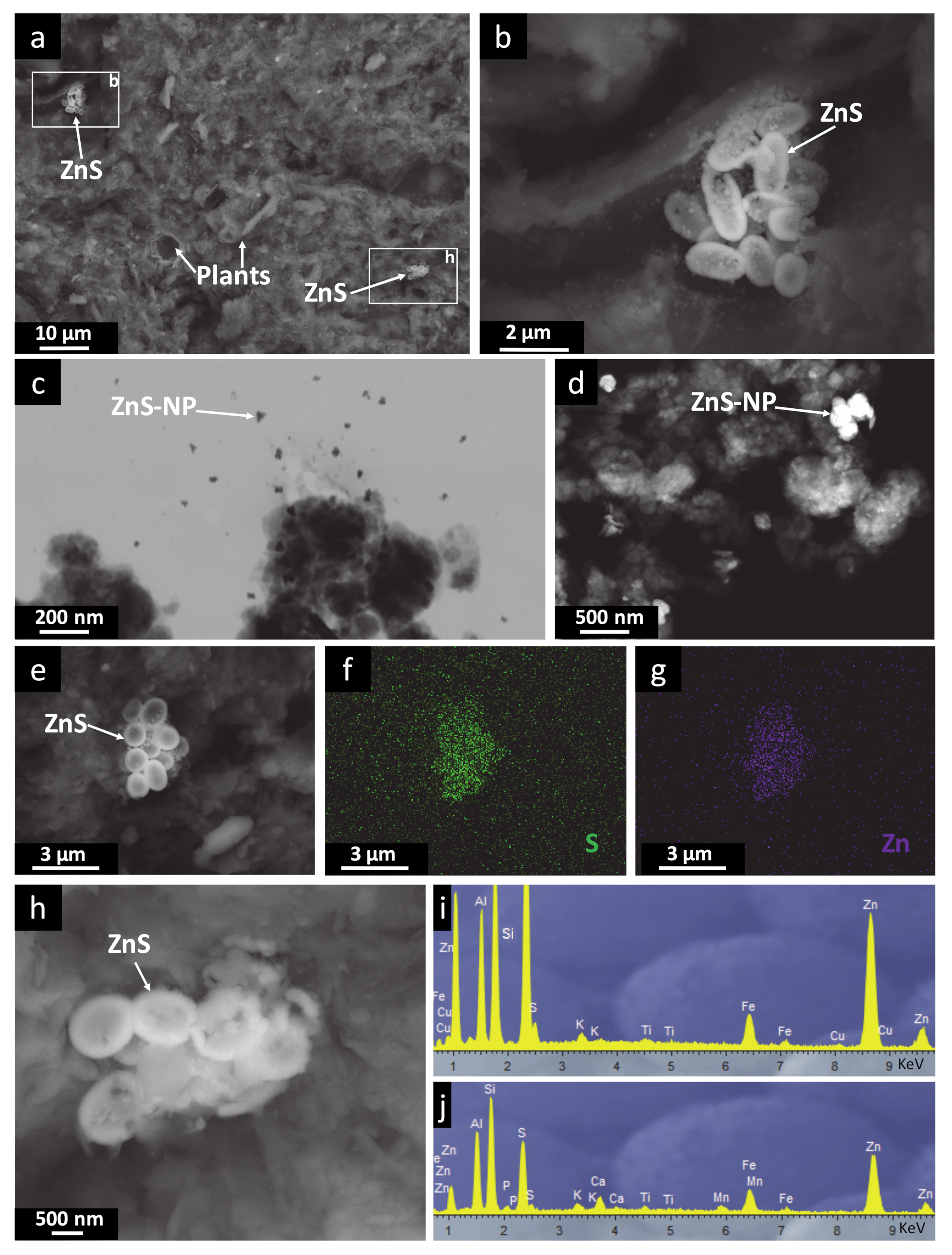
4.2. Electron Microscopy Results
4.3. Chemical Composition and Physicochemical Properties of the Sediments
5. Discussion
5.1. Flow Regime and Flooding of the Chicamocha River and Sediment Conditions
5.2. Heavy Metal Distribution in Sediments and Pollution Sources
5.3. The ZnS Formation
6. Conclusions
Supplementary Materials
Author Contributions
Funding
Acknowledgments
Conflicts of Interest
References
- Dietrich, D.; Huling, J.; Krekeler, M.P.S. Metal pollution investigation of Goldman Park, Middletown Ohio: Evidence for steel and coal pollution in a high child use setting. Sci. Total Environ. 2018, 618, 1350–1362. [Google Scholar] [CrossRef]
- Yun, S.W.; Baveye, P.C.; Kim, D.H.; Kang, D.H.; Lee, S.Y.; Kong, M.J.; Park, C.G.; Kim, H.D.; Son, J.; Yu, C. Analysis of metal(loid)s contamination and their continuous input in soils around a zinc smelter: Development of methodology and a case study in South Korea. Environ. Poll. 2018, 238, 140–149. [Google Scholar] [CrossRef]
- Marrugo-Negrete, J.; Pinedo, J.; Díez, S. Assessment of heavy metal pollution, spatial distribution and origin in agricultural soils along the Sinú River Basin, Colombia. Environ. Res. 2017, 154, 380–388. [Google Scholar] [CrossRef]
- Poot, A.; Gillissen, F.; Koelmans, A. Effects of Flow Regime and Flooding on Heavy Metal Availability in Sediment and Soil of Dynamic River System. Environ. Poll. 2007, 148, 779–787. [Google Scholar] [CrossRef]
- Derkach, T.; Khomenko, V. Essential and Toxic Microelements in the Medicinal Remedy Hyperichi herba by Different Producers. Res. J. Pharm. Technol. 2018, 11, 466–474. [Google Scholar] [CrossRef] [Green Version]
- Chen, H.; Teng, Y.; Lu, S.; Wang, Y.; Wang, J. Contamination features and health risk of soil heavy metals in China. Sci. Total Environ. 2015, 512, 143–153. [Google Scholar] [CrossRef] [PubMed]
- Shen, F.; Liao, R.; Ali, A.; Mahar, A.; Guo, D.; Li, R.; Sun, X.; Awasthi, M.K.; Wang, Q.; Zhang, Z. Spatial distribution and risk assessment of heavy metals in soil near a Pb/Zn smelter in Feng County, China. Ecotoxicol. Environ. Saf. 2017, 139, 254–262. [Google Scholar] [CrossRef]
- Kabata-Pendias, A.; Mukherjee, A.B. Trace Elements from Soil to Human; Springer: Berlin/Heidelberg, Germany, 2007. [Google Scholar]
- Yang, B.; Ren, J.; Wang, M.; Luo, H.; Cao, Y. Concentrations and chemical fractions of Cu, Zn, Cd, and Pb at ten metallurgical sites in China. Environ. Sci. Poll. Res. 2019, 26, 3603–3611. [Google Scholar] [CrossRef] [PubMed]
- Nolan, A.L.; Mclaughlin, M.J.; Mason, S.D. Chemical speciation of Zn, Cd, Cu, and Pb in pore waters of agricultural and contaminated soils using Donnan dialysis. Environ. Sci. Technol. 2003, 37, 90–98. [Google Scholar] [CrossRef]
- Yang, Y.; Nan, Z.; Zhao, Z.; Wang, S.; Wang, Z.; Wang, X. Chemical fractionations and bioavailability of cadmium and zinc to cole (Brassica campestris L.) grown in the multi-metals contaminated oasis soil, northwest of China. J. Environ. Sci. China 2011, 23, 275–281. [Google Scholar] [CrossRef]
- Foster, R.L.; Lott, P.F. X-ray diffractometry examination of air filters for compounds emitted by lead smelting operations. Environ. Sci. Technol. 1980, 14, 1240–1244. [Google Scholar] [CrossRef]
- Han, F.X.; Banin, A.; Triplett, G.B. Redistribution of heavy metals in arid-zone soils under a wetting-drying soil moisture regime. Soil Sci. 2001, 166, 18–28. [Google Scholar] [CrossRef]
- Castillo, J.; Pérez-López, R.; Caraballo, M.A.; Nieto, J.M.; Martins, M.; Costa, M.C.; Olías, M.; Cerón, J.C.; Tucoulou, R. Biologically-induced precipitation of sphalerite-wurtzite nanoparticles by sulfate-reducing bacteria: Implications for acid mine drainage treatment. Sci. Total Environ. 2012, 423, 176–184. [Google Scholar] [CrossRef] [PubMed]
- Labrenz, M.; Druschel, G.; Thomsen-Ebert, T.; Gilbert, B.; Welch, S.; Kemner, K.; Logan, G.; Summons, R.; Gilbert, P.; Bond, P.; et al. Formation of Sphalerite (ZnS) Deposits in Natural Biofilms of Sulfate-Reducing Bacteria. Science 2001, 290, 1744–1747. [Google Scholar] [CrossRef] [PubMed] [Green Version]
- Kosolapov, D.B.; Kuschk, P.; Vainshtein, M.B.; Vatsourina, A.V.; Wiessner, A.; Kästner, M.; Müller, R.A. Microbial processes of heavy metal removal from carbon-deficient effluents in constructed wetlands. Eng. Life Sci. 2004, 4, 403–411. [Google Scholar] [CrossRef]
- Gadd, G.M.; White, C. Microbial treatment of metal pollution—A working biotechnology. Trends Biotechnol. 1993, 11, 353–359. [Google Scholar] [CrossRef]
- Moreau, J.W.; Webb, R.I.; Banfield, J.F. Ultrastructure, aggregation state, and crystal growth of biogenic nanocrystalline sphalerite and wurtzite. Am. Mineral. 2004, 89, 950–960. [Google Scholar] [CrossRef]
- Rodríguez-Zambrano, A.; Aranguren-Riaño, N. Comunidad planctónica de un embalse con alta tensión ambiental: La Playa, cuenca alta del río Chicamocha (Tuta, Boyacá), Colombia. Biota Colomb. 2014, 15, 95–110. [Google Scholar]
- Silva-Rodríguez, J.D. Diseño de una red de logística inversa: Caso de estudio Usochicamocha—Boyacá. Ing. Cienc. 2017, 13, 91–113. [Google Scholar] [CrossRef]
- Parra, L.; Sánchez, D. Análisis de la Valorización de Escorias Negras Como Material Agregado Para Concreto en el Marco de la Gestión Ambiental de la Siderúrgica Diaco municipio de Tuta Boyacá; Universidad de la Salle: Bogotá, Colombia, 2010. [Google Scholar]
- Corpoboyacá—Plan de ordenamiento del Recurso Hídrico de la Cuenca Alta y Media del Rio Chicamocha; Corpoboyacá: Tunja, Colombia, 2015.
- Rodríguez, A.; Solano, O. Mapa Geológico del Departamento de Boyacá. In Instituto de Investigaciones en Geociencias, Minería y Química—Ingeominas; Ministerio de Minas y Energía de Colombia: Bogotá, Colombia, 2000. [Google Scholar]
- Corpoboyacá-Plan de Ordenación y Manejo Ambiental de la Cuenca Alta del Río Chicamocha; Corpoboyacá—Corporación Autónoma Regional de Boyacá, Universidad Nacional de Colombia: Tunja, Boyacá, Colombia, 2006.
- Márquez, G.; Guillot, G. Ecología y efecto Ambiental de Embalses—Aproximación a Casos Colombianos; Universidad Nacional de Colombia: Bogotá, Colombia, 2001; Volume 218. [Google Scholar]
- Gazulla, M.F.; Vicente, S.; Orduña, M.; Ventura, M.J. Chemical analysis of very small-sized samples by wavelength-dispersive X-ray fluorescence. X-Ray Spectrom. 2012, 41, 176–185. [Google Scholar] [CrossRef]
- Quevedo, C.P.; Jiménez-Millán, J.; Cifuentes, G.R.; Jiménez-Espinosa, R. Clay mineral transformations in anthropic organic matter-rich sediments under saline water environment. Effect on the detrital mineral assemblages in the upper Chicamocha river basin, Colombia. Appl. Clay Sci. 2020, 196, 105576. [Google Scholar] [CrossRef]
- Azam, H.M.; Finneran, K.T. Fe(III) reductionmediated phosphate removal as vivianite (Fe3(PO4)2·8H2O) in septic system wastewater. Chemosphere 2014, 97, 1–9. [Google Scholar] [CrossRef]
- Kabata-Pendias, A. Trace Elements in Soils and Plants, 4th ed.; CRC Press: Boca Ratón, FL, USA, 2011. [Google Scholar]
- Pérez-Sirvent, C.; Martínez-Sánchez, M.J.; García-Lorenzo, M.L.; Molina, J.; Tudela, M.L. Geochemical background levels of zinc, cadmium and mercury in anthropically influenced soils located in a semi-arid zone (SE, Spain). Geoderma 2009, 148, 307–317. [Google Scholar] [CrossRef]
- De Almeida Júnior, A.B.; do Nascimento, C.W.A.; Biondi, C.M.; de Souza, A.P.; Barros, F.M.; do Rêgo Barros, F.M. Background and reference values of metals in soils from Paraíba state, Brazil. Rev. Bras. Cienc. Sol. 2016, 40, 1–13. [Google Scholar] [CrossRef] [Green Version]
- Rueda-Saá, G.; Rodríguez-Victoria, J.A.; Madriñan-Molina, R. Methods for establishing baseline values for heavy metals in agricultural soils: Prospects for Colombia. AcAg 2011, 60, 203–218. [Google Scholar]
- Martinez-Mera, E.; Torregroza, A.; Crissien-Borrero, C.J.; Marrugo, J.; González-Márquez, L. Evaluation of contaminants in agricultural soils in an Irrigation District in Colombia. Heliyon 2019, 5, e02217-10. [Google Scholar]
- Gawlik, M.; Bidoglio, G. Background values in European soils and sewage sludges. Results of a JRC-coordinated study on background values. Conclus. Comments Recomm. 2006, 22265. [Google Scholar]
- Manceau, A.; Lanson, B.; Schlegel, M.L.; Harge, J.C.; Musso, M.; Eybert-Berard, L.; Hazemann, J.L.; Chateigner, D.; Lamble, G.M. Quantitative Zn speciation in smelter-contaminated soils by EXAFS spectroscopy. Am. J. Sci. 2000, 300, 289–343. [Google Scholar] [CrossRef]
- Li, Z.; Feng, X.; Li, G.; Bi, X.; Sun, G.; Zhu, J.; Qin, H.; Wang, J. Mercury and other metal and metalloid soil contamination near a Pb/Zn smelter in east Hunan province, China. Appl. Geochem. 2011, 26, 160–166. [Google Scholar] [CrossRef]
- Sajn, R.; Aliu, M.; Stafilov, T.; Alijagic, J. Heavy metal contamination of topsoil around a lead and zinc smelter in Kosovska Mitrovica/Mitrovice, Kosovo/Kosove. J. Geochem. Explor. 2013, l34, 1e16. [Google Scholar]
- Ghayoraneh, M.; Qishlaqi, A. Concentration, distribution and speciation of toxic metals in soils along a transect around a Zn/Pb smelter in the northwest of Iran. J. Geochem. Explor. 2017, 180, 1–14. [Google Scholar] [CrossRef]
- Li, P.; Lin, C.; Cheng, H.; Duan, X.; Lei, K. Contamination and health risks of soil heavy metals around a lead/zinc smelter in southwestern China. Ecotoxicol. Environ. Saf. 2015, 113, 391–399. [Google Scholar] [CrossRef] [PubMed]
- Silva, S.M.; Correa, F.J. Análisis de la contaminación del suelo: Revisión de la normativa y posibilidades de regulación económica. Semest. Econ. 2009, 12, 13–34. [Google Scholar]
- Yoon, S.; Yañez, C.; Bruns, M.A.; Martinez-Villegas, N.; Martinez, C.E. Natural zinc enrichment in peatlands: Biogeochemistry of ZnS formation. Geochim. Cosmochim. Acta 2012, 84, 165–176. [Google Scholar] [CrossRef]
- Cabała, J.; Smieja-Król, B.; Jablonska, M.; Chrost, L. Mineral components in a peat deposit: Looking for signs of early mining and smelting activities in Silesia-Cracow region (Southern Poland). Environ. Earth Sci. 2013, 69, 2559–2568. [Google Scholar] [CrossRef] [Green Version]
- Smieja-Król, B.; Janeczek, J.; Bauerek, A.; Thorseth, I.H. The role of authigenic sulfides in immobilization of potentially toxic metals in the Bagno Bory wetland, southern Poland. Environ. Sci. Poll. Res. 2015, 22, 15495–15505. [Google Scholar] [CrossRef] [Green Version]
- Xu, J.; Murayama, M.; Roco, C.M.; Veeramani, H.; Michel, F.M.; Rimstidt, J.D.; Winkler, C.; Hochella, M.F., Jr. Highly-defective nanocrystals of ZnS formed via dissimilatory bacterial sulfate reduction: A comparative study with their abiogenic analogues. Geochim. Cosmochim. Acta 2016, 180, 1–14. [Google Scholar] [CrossRef] [Green Version]
- Ferris, F.G.; Fyfe, W.S.; Beveridge, T.J. Bacteria as nucleation sites for authigenic minerals in a metal-contaminated lake sediment. Chem. Geol. 1987, 63, 225–232. [Google Scholar] [CrossRef]
- Picard, A.; Gartman, A.; Clarke, D.R.; Girguis, P.R. Sulfate-reducing bacteria influence the nucleation and growth of mackinawite and greigite. Geochim. Cosmochim. Acta 2018, 220, 367–384. [Google Scholar] [CrossRef]
- Cho, K.S.; Talapin, D.V.; Gaschler, W.; Murray, C.B. Designing PbSe nanowires and nanorings through oriented attachment of nanoparticles. J. Am. Chem. Soc. 2005, 127, 7140–7147. [Google Scholar] [CrossRef]
- Dey, A.; Bomans, P.H.; Müller, F.A.; Will, J.; Frederik, P.M.; de With, G.; Sommerdijk, N.A. The role of prenucleation clusters in surface-induced calcium phosphate crystallization. Nat. Mater. 2010, 9, 1010–1014. [Google Scholar] [CrossRef] [PubMed]
- De Yoreo, J.J.; Gilbert, P.U.P.A.; Sommerdijk, N.A.J.M.; Penn, R.L.; Whitelam, S.; Joester, D.; Zhang, H.; Rimer, J.D.; Navrotsky, A.; Banfield, J.F.; et al. Crystallization by particle attachment in synthetic, biogenic, and geologic environments. Science 2015, 349, 6247. [Google Scholar] [CrossRef] [PubMed]
- Falk, N.; Chaganti, S.R.; Weisener, C.G. Evaluating the microbial community and gene regulation involved in crystallization kinetics of ZnS formation in reduced environments. Geochim. Cosmochim. Acta 2018, 220, 201–216. [Google Scholar] [CrossRef] [Green Version]
- Becker, A.; Klöck, W.; Friese, K.; Schreck, P.; Treutler, H.C.; Spettel, B.; Duff, M. Lake Süßer See as a natural sink for heavy metals from copper mining. J. Geochem. Explor. 2001, 74, 205–217. [Google Scholar] [CrossRef]
- Giudici, G.; Pusceddu, C.; Medas, D.; Meneghini, C.; Gianoncelli, A.; Rimondi, V.; Podda, F.; Cidu, R.; Lattanzi, P.; Wanty, R.; et al. The role of natural biogeochemical barriers in limiting metal loading to a stream affected by mine drainage. Appl. Geochem. 2016, 76. [Google Scholar] [CrossRef] [Green Version]
- Kaya, A.; Oren, A.H. Adsorption of zinc from aqueous solutions to bentonite. J. Hazard. Mater. 2005, 125, 183–189. [Google Scholar] [CrossRef]
- Malamis, S.; Katsou, E. A review on zinc and nickel adsorption on natural and modified zeolite, bentonite and vermiculite: Examination of process parameters, kinetics and isotherms. J. Hazard. Mater. 2013, 252, 428–461. [Google Scholar] [CrossRef]
- Lahori, A.H.; Vu, N.H.; Du, J.; Quang, T.D.; Saif, U.R.; Zobia, N.; Muneer, A.; Zengqiang, Z. Stabilization of toxic metals in three contaminated soils by residual impact of lime integrated with biochar and clays. J. Soils Sedim. 2020, 20, 734–744. [Google Scholar] [CrossRef]


| Values | SiO2 | Al2O3 | FeO | MnO | MgO | CaO | Na2O | K2O | TiO2 | P2O5 | LOI | TOC |
|---|---|---|---|---|---|---|---|---|---|---|---|---|
| SW segment. Upstream La Playa dam (n = 6) | ||||||||||||
| Mean | 79.84 | 8.72 | 3.69 | 0.02 | 0.25 | 0.39 | 0.09 | 0.36 | 0.59 | 0.24 | 5.57 | 1.84 |
| Median | 81.18 | 7.60 | 3.07 | 0.02 | 0.25 | 0.33 | 0.10 | 0.35 | 0.57 | 0.32 | 5.70 | 1.90 |
| SD | 4.00 | 2.58 | 1.10 | 0.01 | 0.02 | 0.18 | 0.01 | 0.05 | 0.12 | 0.13 | 1.43 | 0.46 |
| Max | 84.01 | 13.32 | 5.40 | 0.04 | 0.27 | 0.61 | 0.11 | 0.46 | 0.80 | 0.33 | 7.48 | 2.49 |
| Min | 73.60 | 6.79 | 2.89 | 0.01 | 0.22 | 0.18 | 0.07 | 0.31 | 0.48 | 0.07 | 3.66 | 1.22 |
| Central segment. La Playa reservoir (n = 4) | ||||||||||||
| Mean | 61.24 | 17.17 | 4.05 | 0.01 | 0.14 | 0.39 | 0.06 | 1.12 | 0.42 | 0.58 | 14.37 | 10.72 |
| Median | 60.76 | 17.22 | 4.00 | 0.01 | 0.13 | 0.39 | 0.07 | 1.11 | 0.44 | 0.59 | 15.38 | 12.40 |
| SD | 1.44 | 0.92 | 0.26 | 0.00 | 0.02 | 0.23 | 0.04 | 0.20 | 0.07 | 0.04 | 2.61 | 4.49 |
| Max | 63.24 | 18.24 | 4.38 | 0.01 | 0.17 | 0.61 | 0.10 | 1.35 | 0.48 | 0.61 | 16.22 | 13.80 |
| Min | 60.18 | 15.99 | 3.81 | 0.01 | 0.12 | 0.18 | 0.02 | 0.91 | 0.31 | 0.51 | 10.51 | 4.29 |
| NE segment. Downstream La Playa dam (n = 10) | ||||||||||||
| Mean | 87.28 | 4.15 | 1.88 | 0.03 | 0.15 | 0.20 | 0.12 | 0.41 | 0.34 | 0.14 | 3.75 | 1.19 |
| Median | 89.60 | 2.16 | 1.39 | 0.01 | 0.12 | 0.16 | 0.10 | 0.29 | 0.25 | 0.09 | 2.03 | 0.68 |
| SD | 8.41 | 3.66 | 1.35 | 0.04 | 0.10 | 0.15 | 0.04 | 0.29 | 0.26 | 0.12 | 3.25 | 1.10 |
| Max | 96.25 | 11.57 | 5.01 | 0.13 | 0.41 | 0.55 | 0.20 | 1.23 | 0.87 | 0.36 | 10.83 | 3.61 |
| Min | 69.62 | 1.27 | 0.57 | 0.01 | 0.06 | 0.06 | 0.08 | 0.20 | 0.09 | 0.03 | 0.72 | 0.15 |
| Unpolluted sediment (n = 15) | ||||||||||||
| Mean | 91.74 | 5.66 | 2.46 | 0.02 | 0.17 | 0.25 | 0.12 | 0.36 | 0.42 | 0.16 | 4.00 | 1.28 |
| Sample | Cr | Ni | Cu | Zn | Pb |
|---|---|---|---|---|---|
| SW segment. Upstream La Playa dam (n = 6) | |||||
| Mean | 62 | 58 | 76 | 96 | 20 |
| Median | 48 | 41 | 64 | 71 | 20 |
| SD | 38 | 39 | 35 | 83 | 10 |
| Max | 133 | 130 | 142 | 264 | 35 |
| Min | 28 | 22 | 47 | 43 | 5 |
| Central segment. La Playa reservoir (n = 4) | |||||
| Mean | 184 | 144 | 262 | 423 | 40 |
| Median | 184 | 138 | 204 | 415 | 38 |
| SD | 8 | 33 | 133 | 86 | 10 |
| Max | 192 | 190 | 459 | 534 | 54 |
| Min | 175 | 112 | 180 | 328 | 29 |
| NE segment. Downstream La Playa dam (n = 10) | |||||
| Mean | 30 | 22 | 34 | 43 | 10 |
| Median | 15 | 12 | 17 | 24 | 7 |
| SD | 45 | 26 | 35 | 53 | 8 |
| Max | 161 | 81 | 110 | 191 | 28 |
| Min | 5 | 5 | 6 | 5 | 5 |
| Unpolluted sediment (n = 15) | |||||
| Mean | 29 | 26 | 41 | 42 | 12 |
| Sample | P | Cr | Ni | Cu | Zn | Pb |
|---|---|---|---|---|---|---|
| SW segment. Upstream La Playa dam (n = 6) | ||||||
| Mean | 419 | 1 | 4 | 8 | 43 | 1 |
| Median | 410 | 1 | 3 | 7 | 35 | 1 |
| SD | 246 | 0 | 1 | 2 | 20 | 0 |
| Max | 680 | 1 | 5 | 10 | 78 | 2 |
| Min | 30 | 0 | 2 | 6 | 28 | 0 |
| Central segment. La Playa reservoir (n = 4) | ||||||
| Mean | 7280 | 3 | 7 | 23 | 268 | 4 |
| Median | 7098 | 4 | 7 | 23 | 249 | 4 |
| SD | 556 | 2 | 1 | 9 | 146 | 2 |
| Max | 8078 | 6 | 8 | 33 | 459 | 5 |
| Min | 6847 | 1 | 5 | 12 | 115 | 2 |
| NE segment. Downstream La Playa dam (n = 10) | ||||||
| Mean | 221 | 0 | 2 | 4 | 27 | 1 |
| Median | 183 | 0 | 1 | 3 | 24 | 1 |
| SD | 172 | 0 | 1 | 3 | 14 | 1 |
| Max | 675 | 1 | 4 | 10 | 61 | 3 |
| Min | 14 | 0 | 1 | 1 | 14 | 0 |
| Variable | Cr | Ni | Cu | Zn | Pb | TOC | LOI | P2O5 |
|---|---|---|---|---|---|---|---|---|
| Cr | 1 | 0.960 | 0.854 | 0.942 | 0.919 | 0.828 | 0.932 | 0.871 |
| Ni | 1 | 0.848 | 0.899 | 0.898 | 0.732 | 0.857 | 0.855 | |
| Cu | 1 | 0.935 | 0.792 | 0.860 | 0.851 | 0.816 | ||
| Zn | 1 | 0.888 | 0.942 | 0.936 | 0.883 | |||
| Pb | 1 | 0.942 | 0.936 | 0.883 | ||||
| TOC | 1 | 0.922 | 0.832 | |||||
| LOI | 1 | 0.751 | ||||||
| P2O5 | 1 |
| Segments of the UCRB | pH | R.P. (mV) | E.C. (µS/cm) |
|---|---|---|---|
| SW segment. Upstream La Playa dam (n = 6) | 7.3 | 62 | 241 |
| Central segment. La Playa reservoir (n = 4) | 7.1 | −233 | 2625 |
| NE segment. Downstream La Playa dam (n = 10) | 7.4 | 89 | 189 |
© 2020 by the authors. Licensee MDPI, Basel, Switzerland. This article is an open access article distributed under the terms and conditions of the Creative Commons Attribution (CC BY) license (http://creativecommons.org/licenses/by/4.0/).
Share and Cite
Quevedo, C.P.; Jiménez-Millán, J.; Cifuentes, G.R.; Jiménez-Espinosa, R. Electron Microscopy Evidence of Zn Bioauthigenic Sulfides Formation in Polluted Organic Matter-Rich Sediments from the Chicamocha River (Boyacá-Colombia). Minerals 2020, 10, 673. https://doi.org/10.3390/min10080673
Quevedo CP, Jiménez-Millán J, Cifuentes GR, Jiménez-Espinosa R. Electron Microscopy Evidence of Zn Bioauthigenic Sulfides Formation in Polluted Organic Matter-Rich Sediments from the Chicamocha River (Boyacá-Colombia). Minerals. 2020; 10(8):673. https://doi.org/10.3390/min10080673
Chicago/Turabian StyleQuevedo, Claudia Patricia, Juan Jiménez-Millán, Gabriel Ricardo Cifuentes, and Rosario Jiménez-Espinosa. 2020. "Electron Microscopy Evidence of Zn Bioauthigenic Sulfides Formation in Polluted Organic Matter-Rich Sediments from the Chicamocha River (Boyacá-Colombia)" Minerals 10, no. 8: 673. https://doi.org/10.3390/min10080673