

Abstract
Microsphere based super-resolution imaging is a promising approach to achieve enhanced optical imaging with an ordinary optical microscope. In this study, we propose a new approach to enhance magnification factor in microsphere based super-resolution imaging. In the method, two microspheres are stacked in serial along the axial direction to implement the primary and the secondary magnifications, which is termed as stacked dual microscope (SDM) imaging. An analytical model is developed for SDM imaging, followed by the experimental investigation of several different impact factors on magnification factors M, such as microsphere diameters and separation distance between the two microspheres. The results show that the magnification factors as well as the field of view is significantly enlarged in SDM imaging, compared to the ordinary single microsphere super-resolution imaging.
Export citation and abstract BibTeX RIS
1. Introduction
Imaging with a conventional optical microscope remains as a primary approach for the observation of sample surfaces at micro\nanoscale [1–5]. However, we suffer from the limited imaging resolution for centuries because of the Abbe diffraction limit. As a result, one could not characterize sub-wavelength features of around 200 nm with an optical microscope [6]. Many methods have been proposed to achieve super-resolution optical imaging, such as stimulated emission depletion microscopy [7], photo activated localization microscopy [8], and stochastic optical reconstruction microscopy [9]. In these methods, target proteins are generally labelled by fluorescent materials, which could be potentially harmful to the proteins. Moreover, fluorescence molecules are required to be delicately selected and pretreated for optimized imaging. As a result, an easy approach is demanded for super-resolution imaging.
It has been demonstrated that dielectric microlenses placed in between an objective lens and a sample surface in an optical microscope can significantly enhance the imaging resolution [10–12]. This provides a promising approach to achieve real-time and non-invasive super-resolution imaging. In such a configuration, the microlenses play two important roles. First, they generate photonic nanojets [13, 14], which enhance backscattering of visible light by several orders of magnitude [13]. Second, they transform the evanescent waves that carry the high spatial-frequency information into far-field propagating waves, creating a magnified far-field image [11, 15]. The sub-diffraction-limit frequency components collected by the microlens dramatically reduces as the distance between the sample surface and the microsphere increases [16].
Many types of microlenses, such as metamaterial superlenses [17], nanoscale solid immersion lenses [10, 18–20], microdroplets [21, 22], and dielectric microspheres [11, 12, 15, 23–28] are applied for the super-resolution imaging. Among them, microspheres can provide a higher magnification factor [25]. For example, by using a silica microsphere with a diameter of 2.37 μm, an imaging resolution of 50 nm was achieved [11]. As a result, much effort has been put on microsphere-assisted super-resolution imaging to study imaging mechanism [12, 14, 29], optimize imaging quality [15, 23, 24, 27, 30–34], and develop imaging systems [16, 35–38].
Imaging resolution and magnification are two key aspects in super-resolution imaging. It is challenging to have the two parameters optimized at the same time. It is reported that microspheres can achieve super-resolution imaging only when the sphere-sample distance is within one wavelength [14, 16]. In this range, the imaging resolution increases while the magnification factor decreases with decreasing sphere-sample distance [16, 39]. To obtain an increased magnification factor, smaller microspheres are preferable. However, this will significantly reduce the field of view (FOV) of imaging [23, 27, 29]. As a result, the improved imaging resolution is often accompanied with a scarified magnification factor.
In this paper, a novel approach is proposed to achieve super-resolution imaging with an enhanced magnification factor and FOV. By stacking two silica microspheres in serial along the axial direction, which is referred to as stacked dual microspheres (SDM) in this study, a much improved magnification factor can be achieved in super-resolution imaging. A mathematical model of SDM super-resolution imaging is developed, followed by the systematic investigation of the major impact factors.
2. Experimental
The super-resolution imaging experiments were conducted with a standard inverted optical microscope (Model: IX70, Olympus, Japan). The experimental setup is illustrated in figure 1. In the setup, a piece of DVD disk with a 400 nm width stripe separated by 340 nm width grooves is taken as the sample for imaging. Two silica microspheres (MO-Sci Corporation) with the refractive index n = 1.46 are employed. The upper spheres (primary spheres) are directly deposited on the sample surface. The lower (secondary) ones are attached to the end of atomic force microscope (AFM) cantilevers using epoxy (Araldite Bostik, Coubert), as shown in the inset image. The cantilevers are mounted on a XYZ three degree of freedom (DOF) piezoelectric stage (Model: P-545.3C7, PI, Germany) to implement super-resolution imaging at specific positions over the sample surfaces.

Figure 1. Schematic diagram of the experimental setup for super-resolution imaging. The setup is integrated in a standard inverted optical microscope. It includes an upper (primary) microsphere, a lower (secondary) microsphere which is attached to the end of an AFM cantilever, and a XYZ three DOF piezoelectric stage. The scale bar in the inset image equals 20.0 μm.
Download figure:
Standard image High-resolution imageAll optical images are captured by a high speed camera (MC3010, Mikrotron) with a ×60 objective lens (LCPlanFl, NA = 0.7, Olympus, Japan) using a halogen lamp in the transmission mode. The acquired images are transmitted to a host computer (Tower7910, dual-Xeon-E5, HP) via a frame grabber (OR-X8C0-XPF00, DALSA). Moreover, an AFM (Resolve, Bruker, U.S.A.) is used to characterize the sample surface at nanoscale. During imaging, a lens nanopositioner (E725.1CD, Phsik Instrument, Germany) with motion control resolution of 0.65 nm is used to realize the accurate positioning of the objective lens along the axial direction. We take the position which gives the sharpest image as the focal plane. We repeated the operation to capture multiple images for each combination of the dual microspheres. From the multiple images, the measurement errors were estimated.
The numerical simulation was conducted using the finite-difference time-domain (FDTD) method. In the simulation, plane waves and perfectly matched layer (PML) are chosen as light source and boundaries, respectively. The wavelength of the plane wave is set to be 600 nm, which is equal to the peak value of the halogen lamp used in the experiment.
3. Results and discussion
3.1. Theoretical modeling of SDM super-resolution imaging
The enhanced super-resolution imaging by using SDMs is first experimentally demonstrated. Figure 2(a) shows an image taken with the inverted optical microscopy without any microsphere. No features can be observed in the image. That is because the stripes and grooves in the sample surface is smaller than the imaging resolution of 0.61λ/NA ≈ 523 nm [40] with the applied 60× objective. After a primary microsphere with a diameter of 20.0 μm was used, the DVD tracks can be clearly observed (figure 2(b)). Furthermore, the secondary microsphere with a diameter of 72.0 μm was placed under the primary one. A further magnified image with well-recognized DVD tracks was obtained (figure 2(c)). This indicates that the SDMs can enhance super-resolution imaging.

Figure 2. Demonstration and the mechanism of the enhanced magnification factor M in SDM imaging. (a)–(c) Optical microscope images without any microsphere (a), with a single microsphere (b), and with the stack dual microspheres (SDMs) (c). SDM imaging provides an enhanced magnification factor. (scale bar: 10 μm) (d) Ray tracking diagram showing the mechanism of SDM imaging. (e) Result of FDTD simulation for a single sphere imaging (D = 20 μm), from which, the focal length f for microspheres can be obtained. (f) The f–D dependence determined by FDTD simulation and geometric optics analysis. Geometric optics analysis provides a reduced f compared to that of FDTD simulation.
Download figure:
Standard image High-resolution imageWhat is the mechanism of the enhanced magnification factor in SDM imaging? A ray tracing diagram of the SDM imaging is illustrated in figure 2(d). From the diagram, one can see that the primary microsphere first forms a virtual image, which corresponds to the image obtained in figure 2(b). This is consistent with previous single sphere super-resolution imaging [11, 15, 16]. Once the secondary sphere is applied, a real image can be obtained.
According to the Guassian lens equation [41], we have the following two equations valid:
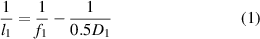
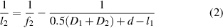
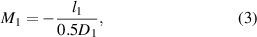
where l1 and l2 are the image distances, f1 and f2 the focal lengths, D1 and D2 the diameters of the primary and secondary microspheres, respectively, and d is the separation distance between two microspheres. Moreover, the magnification factor M for the primary and secondary microspheres can be given as:
and
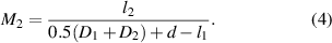
The overall magnification factor of the SDM imaging can then be given as

In equations (3)–(5), the focal lengths f1 and f2 are needed. When the size of an object is much smaller than that of microsphere radius, paraxial approximation [42] can be applied and the focal length f can be estimated as f ≈ nD/4(n−1), where n is the relative refractive index of the microsphere. However, in microsphere-assisted super-resolution imaging, the focal length of microspheres is supposed to be shorter because of the coupling of evanescent waves [43]. Figure 2(e) shows the light intensity distribution of a photonic nanojet. The position at which the light intensity reaches its maximum value is taken as the focal point. The distance between the center of the microsphere and the focal point can then be taken as the focal distance f in the FDTD simulation. A series of simulation was conducted for silica microshpheres with different diameters. The obtained focal length is shown in figure 2(f). One can see that f obtained in the FDTD simulation is smaller than that obtained through paraxial approximation. By linearly fitting the obtained simulation results with respect to D, f as a function of D of silica microspheres can be estimated as:

The above obtained relationship is applied to determine the focal lengths for the silica microspheres.
3.2. Magnification analysis
According to equations (1)–(6), one can see that M in SDM imaging is directly related to D1, D2, and d. In this section, the influence of the three impact factors on M in SDM imaging is investigated. First, primary microspheres with three different diameters of 28 μm, 49 μm, and 71 μm were used for the same secondary microsphere of D2= 112 μm. Three sample images are shown in figures 3(a)–(c). It clearly shows that the magnification factor decreases with increasing D1 (figure 3(d)). Similarly, experiments were conducted with a fixed primary microsphere of D1= 39 μm and changing D2. The results are shown in figures 3(e)–(g) for D2= 51 μm, 71 μm, and 121 μm, respectively. The magnification factor M increases with increasing D2. Moreover, the obtained values of M in these two series of experiments are compared with the theoretically calculated values, as shown in figures 3(d) and (h). In the figures, the experimentally measured values agree well with the theoretically predicted ones, as shown in the red solid curves.

Figure 3. Impact of microsphere diameters D1 and D2 on magnification factor M in SDM imaging when d = 0. (a)–(c) Images with different D1 of 28 μm (a), 49 μm (b), and 71 μm (c) when D2 = 112 μm. (e)–(g) Images with different D2 of 51 μm (e), 71 μm (f), and 121 μm (g) when D1 = 39 μm. (scale bars: 10 μm). (d), (h) The comparison of the theoretically and experimentally obtained M. The two values agree well with each other.
Download figure:
Standard image High-resolution imageFurthermore, the impact of the separation distance d between the two microspheres on magnification factor is investigated. In the experiment, the diameters of the primary and the secondary microspheres are 16.0 μm and 51.0 μm, respectively. The magnified images are shown in figures 4(a)–(d) for increasing separation distance d of 0 μm, 5 μm, 10 μm, and 15 μm, respectively. It clearly shows that M decreases with increasing d. Once again, the experimentally obtained M agrees well with that of the theoretically predicted results (figure 4(e)).

Figure 4. Impact of the separation distance d between the two microspheres on the magnification factor M in SDM imaging. (a)–(d) Images acquired with different d: 0 μm (a), 5 μm (b), 10 μm (c), and 15 μm (d) when D1 = 16 μm and D2 = 51 μm in SDM imaging (scale bars: 5 μm). (e) Comparison of the theoretically and experimentally obtained M with changing d. Both results agree well with each other.
Download figure:
Standard image High-resolution imageIn SDM imaging, whether or not the secondary microsphere helps to enhance the overall magnification M compared to the single microsphere imaging is strongly related to the combination of D1 and D2. Here the secondary magnification factor with different combinations of D1 and D2 is determined. To do so, the magnification factor M1 in single microsphere imaging with different values of D1 was first obtained, as shown in figure 5(a). One can see that the experimentally measured value M1 decreases with increasing D1, as predicted by equation (3) (red solid curve in figure 5(a)).

Figure 5. Dependence of the secondary magnification factor M2 on D1 and D2. (a) Magnification factor M1 in single microsphere imaging. (b) The secondary magnification factor M2 as a function of D1 and D2 when the separation distance d= 0. The bars are experimental measured M2, while the meshed surface is the theoretical predication of M2 from equation (4).
Download figure:
Standard image High-resolution imageFor each single primary microsphere, the colloidal probes with different sizes of microspheres were applied to implement SDM imaging. With the acquired images, M was obtained for each combination of D1 and D2. The secondary magnification factor M2 can be achieved as M2 = M/M1. The obtained M2 as a function of D1 and D2 is shown in figure 5(b). The transparent surface in the figure corresponds to the theoretically estimated M2 as a function of D1 and D2 when d = 0μm. From the figure, one can see that M2 increases with decreasing D1 and increasing D2. Therefore, a combination of a smaller primary microsphere and a larger secondary microsphere is favorable to achieve an enhanced M. Otherwise, a decreased M may be obtained. Moreover, one can see that when the secondary magnification factor M2 is larger than 1, one can obtain the enhanced overall magnification compared to that of the single microsphere imaging, regardless of the magnification factor of the applied objective lenses in optical microscopes. If an objective lens with a higher magnification factor is applied, a further improved overall imaging magnification will be achieved.
3.3. FOV analysis
The size of FOV is also an import factor in super-resolution imaging. The FOV for microsphere based super-resolution imaging is defined as the diameter of the circular area in which distinguishable features with no clear distortions can be obtained in the acquired images. The size of FOV in single microsphere and SDM imaging are presented in figure 6(a). In the figure, the green star points are measured FOV in single microsphere imaging. The data points of all other symbols correspond to the measured FOV in SDM imaging with different values of D2. One can see that the FOV in single sphere imaging increases with D1, which is consistent with that reported by others [23, 44]. Assuming super-resolution imaging can only be achieved when the distance between sample surface and the glass sphere is within one wavelength, the area in which the vertical distance in between the sample surface to the microspheres is less than one wavelength increases with microsphere diameters. As a result, FOV increases with D1.

Figure 6. Field of view (FOV) in stacked dual microsphere (SDM) super-resolution imaging. (a) Comparison of the FOV in single microsphere imaging and SDM imaging. Enlarged FOV is obtained in SDM imaging compared to single microsphere imaging. (b) The dependence of FOV on the magnification factor M in SDM imaging with different D2. All solid curves are obtained by fitting the measurement data with quadratic polynomial equations.
Download figure:
Standard image High-resolution image
From the above result, one can see that the SDM imaging enlarges FOV, especially for smaller D1. Moreover, a larger value of D2 exhibits an increased FOV for a given magnitude M, as shown in figure 6(b). For example, when M is equal to 2, the FOV is about 6 μm for D2= 51 μm. However, FOV is doubled when D2 increases to 112 μm.
Although a much enhanced M has been demonstrated in SDM imaging, readers have to note that the image quality, as well as imaging resolution for images presented in this paper is not optimized due to the limited experimental resources in our laboratory. We compared the imaging resolution of SDM with that of single microsphere imaging (data not shown) and found that there the resolution has not been significantly improved. As a result, this paper we mostly focus on the magnification factor and FOV. The image quality and imaging resolution can be optimized through the following two approaches. First, an upright optical microscope can be used for SDM imaging. Here we used an inverted optical microscope. Since the DVD sample is partially transparent, the sample cannot be well illuminated. This causes reduced image quality. Second, microspheres with higher refractive index can be used in SDM imaging, which will present improved imaging resolution, as studied somewhere else [25, 31].
4. Conclusions
To summarize, a novel approach is proposed to enhance magnification factors in microsphere based super-resolution imaging in an inverted optical microscopy. In the method, two microspheres, a primary one and a secondary one, are stacked in serial along the axial direction. The primary sphere directly contacts sample surface to implement single microsphere super-resolution imaging, while the secondary one further magnifies images for enhanced imaging. The influence of the primary microsphere diameter D1, the secondary microsphere diameter D2, as well the separation distance d between the two microspheres on the imaging magnification factor M was systematically investigated. The results show that M increases with decreasing D1 and d, and increasing D2. This shows great agreement with that of theoretical prediction obtained with a developed analytical model for SDM imaging. Moreover, experimental results demonstrate that the size of field of view in SDM imaging is larger than that of single microsphere imaging. We believe that the proposed SDM method can greatly improve the imaging performance and has strong bearings on various applications of topography characterization at micro/nanoscale.
Acknowledgments
Authors acknowledge the financial support from the Beijing Natural Science Foundation (Grant No. 3182022) and National Natural Science Foundation of China (Grant No. 51775028).