Review

Overview of Folic Acid Supplementation Alone or in Combination with Vitamin B12 in Dairy Cattle during Periparturient Period

1
Key Laboratory of Animal Genetics, Breeding, and Reproduction, Ministry of Agriculture & National Engineering Laboratory for Animal Breeding, College of Animal Science and Technology, China Agricultural University, Beijing 100193, China
2
State Key Laboratory of Animal Nutrition, Beijing Engineering Technology Research, Center of Raw Milk Quality and Safety Control, College of Animal Science and Technology, China Agricultural University, Beijing 100193, China
3
Genome Analysis Laboratory of the Ministry of Agriculture, Agricultural Genomics Institute at Shenzhen, Chinese Academy of Agricultural Sciences, Shenzhen 518120, China
*
Author to whom correspondence should be addressed.
These authors contributed equally to this work.
Metabolites 2020, 10(6), 263; https://doi.org/10.3390/metabo10060263
Submission received: 20 March 2020 / Revised: 20 May 2020 / Accepted: 26 May 2020 / Published: 25 June 2020
(This article belongs to the Special Issue Folate Absorption and Metabolism)

Abstract

:
The periparturient period is the period from three weeks before calving to three weeks post-calving. This period is important in terms of health, productivity and profitability, and is fundamental to successful lactation. During this period, the animal experiences stress because of hormonal changes due to pregnancy and the significant rise in milk production. In addition, a negative energy balance usually occurs, because the demand for nutrients to sustain milk production increases by more than the nutrient supply during the periparturient period. The immunity of dairy cattle is suppressed around parturition, which increases their susceptibility to infections. Special care regarding nutrition can reduce the risks of metabolism and immunity depression, which dairy cattle face during the periparturient span. Folic acid is relevant in this regard because of its critical role in the metabolism to maintain lactational performance and to improve health. Being a donor of one-carbon units, folic acid has a vital role in DNA and RNA biosynthesis. Generally, the folic acid requirements of dairy cattle can be met by the microbial synthesis in the rumen; however, in special cases, such as during the periparturient period, the requirement for this vitamin strictly increases. Vitamin B12 also has a critical role in the metabolism as a coenzyme of the enzyme methionine synthase for the transfer of a methyl group from folic acid to homocysteine for the regeneration of methionine. In the current review, we highlight the issues facing periparturient dairy cattle, and relevant knowledge and practices, and point out future research directions for utilization of the associated vitamins in ruminants, especially during the periparturient period.

1. Introduction

The periparturient period is the time span from 3 weeks prepartum until 3 weeks postpartum [1], and is crucial for the reproductive and productive competency of dairy cattle [2]. The peripartum period is characterized by a negative energy balance, immune suppression, and increased susceptibility to diseases in dairy cows [3]. The negative energy balance in periparturient cattle mainly develops due to a rapid increase in milk production during early lactation and dry matter intake depression [4,5]. Pregnancy burden is also one of the reasons for the low feed intake around parturition, resulting in low energy levels. Although the feed intake increases soon after calving, due to the high demand for milk production, the animal continues to be affected by a shortage of energy, which also leads to a negative energy balance [6,7]. Periparturient dairy cattle experience an alteration in the hormonal, digestive, and immune system, which intervenes in the immune function and causes immunity suppression [8,9,10]. Due to immunity depression, the periparturient period is considered an infection-prone period for dairy cattle [11].

1.1. Immunity and Periparturient Period

During the periparturient period, immune depression in dairy cows is mainly aggravated by physiological changes and negative energy balance [12,13,14]. It is well established that increases in the cortisol level near parturition results in humoral and cell-mediated immunity depression [15]. Many studies have reported the elevated level of non-esterified fatty acids (NEFA) and beta-hydroxybutyrate (BHBA) during the periparturient period, which causes depression of bovine peripheral blood mononuclear cells (PBMCs) [16], inhibits the interferon-γ production [17], and impairs the function of polymorphonuclear neutrophils (PMNLs) [18]. Additionally, the bactericidal efficiency of phagocyte and blood polymorphonuclear leukocytes (PMNs) is severely decreased during the first week of lactation [19]. This decrease in PMNL level exposes dairy cattle to mammary and uterine infections [20]. The process of parturition alters the gene expression profile in neutrophils, which influence the immune system of dairy cattle [21]. Moreover, during the transition period in dairy cattle, the cells involved in the phagocytosis and bacteria encounter activity [18], and immunoglobulin production by B-cells are affected [22]. Studies have shown that the occurrence of metabolic and production-related diseases, such as mastitis, metritis, milk fever, and ketosis, rises in dairy cattle during the periparturient period [23,24].
To cope with this situation, proper nutrition is essential for the normal performance of dairy cows, particularly during the periparturient period [25], because nutrition works as a backbone for the maintenance of good health [26]. Many food supplements have been provided during the periparturient period to avoid stressors, such as immunity depression, negative energy balance, and low dry matter intake due to pregnancy, and to maintain the normal metabolism and productive efficiency of dairy cows. In this regard, folic acid, alone or in combination with vitamin B12, has received considerable attention in ruminant research.

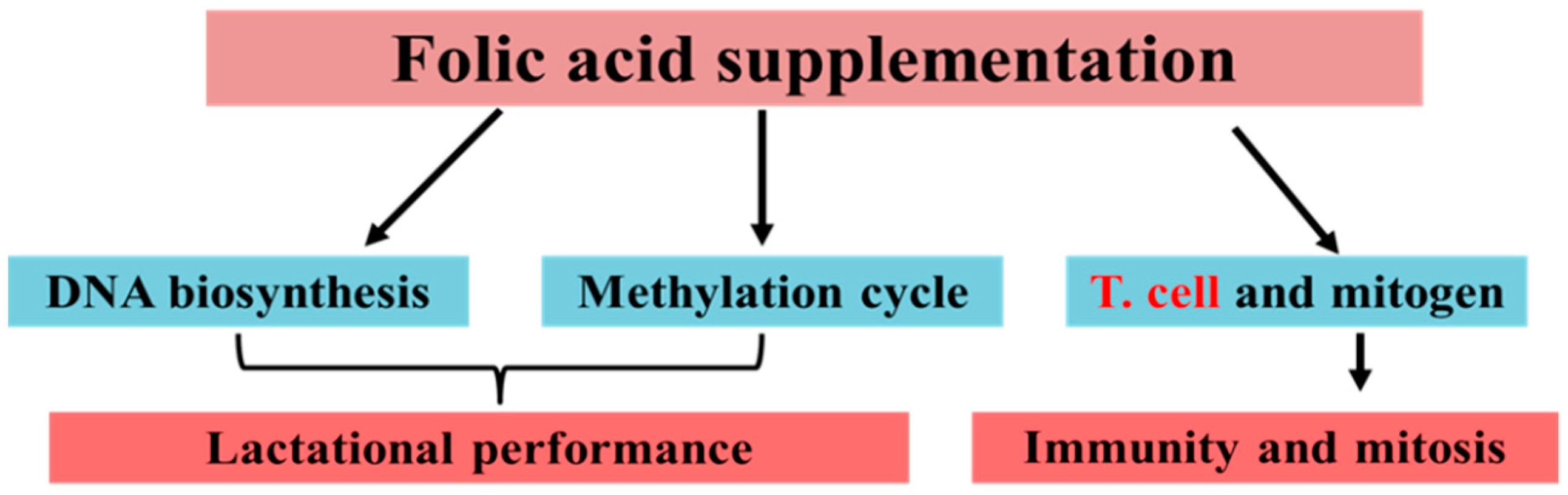
1.2. Folic Acid and Vitamin B12 Functions

Folic acid is a B-complex vitamin for which the primary biochemical function in animals is to provide the one-carbon unit [27]. Folic acid has multiple roles and has received considerable attention in animal studies, particularly during pregnancy. Folic acid has multiple functions, including neurotransmission regulation and gene expression [28,29], and has a protective role in the immune system [30,31], while its deficiency leads to anemia, granulocytopenia, and lymphocytopenia [32,33,34]. Hollingsworth reported that the utero exposure to the methyl donor might affect the expression of the critical gene that plays a central role in immunity [35]. In addition, folic acid in combination with B12 improves the efficiency of the energy metabolism in dairy cattle [36,37]. Vitamin B12 works as an essential coenzyme for the methionine synthase, which is necessary for the transfer of a methyl group from 5-methyl-tetrahydrofolate to homocysteine for the regeneration of methionine. The methionine, in the next step, changes to a major methyl group donor called S-adenosyl-methionine (SAM) [38]. Thus, the deficiency of B12 may lead to a reduction in methionine supply and de novo synthesis of methyl groups. It has been reported that a deficiency of folic acid can lead to a decrease in the levels of SAM [39,40]. Folic acid is also involved in T cell and mitogen regulation, which is essential for immunity and growth. Moreover, folic acid influences the methylation cycle, and DNA and RNA biosynthesis, which is essential for lactational performance (Figure 1). In addition, dairy cows have a high demand for methyl groups during early lactation [41]. Due to its central role in accepting and releasing one-carbon units in mammals, folic acid is essential for the de novo synthesis of methyl groups for the formation of methylated molecules [42]. Thus, the adequate supply of folic acid is highly desirable for optimal metabolic pathways and milk production during the periparturient period [43].
Changes in folic acid can alter the activity of methylenetetrahydrofolate reductase (MTHFR) and methionine synthase reductase (MTRR) genes, which have an essential role in chromosomal stability. Although the bacteria in the rumen can synthesize folic acid [44], under special conditions, such as during the periparturient period, the folate synthesized in the rumen by bacteria is not sufficient to fulfill the requirements of dairy cows. Furthermore, it was noted that folic acid transfers to the fetus in the uterus after absorption from the small intestine of pig [45], and the storing capacity of folic acid increases in the fetus during the late gestation period. The serum folate decreases by up to 40%, two months before calving in dairy cattle [46]. Similarly, a low concentration of B12 was also noted in dairy cattle during early lactation [47]. Thus, the current review aims to summarize the role of folic acid alone or in combination with vitamin B12 to regulate the metabolism, milk production, and immune system in periparturient dairy cattle. In addition, the review highlights the limitations and future research directions for folic acid as a supplement to the ruminant’s diet, specifically around parturition.

2. The Research Progress of Folic Acid and Vitamin B12 in Dairy Cattle

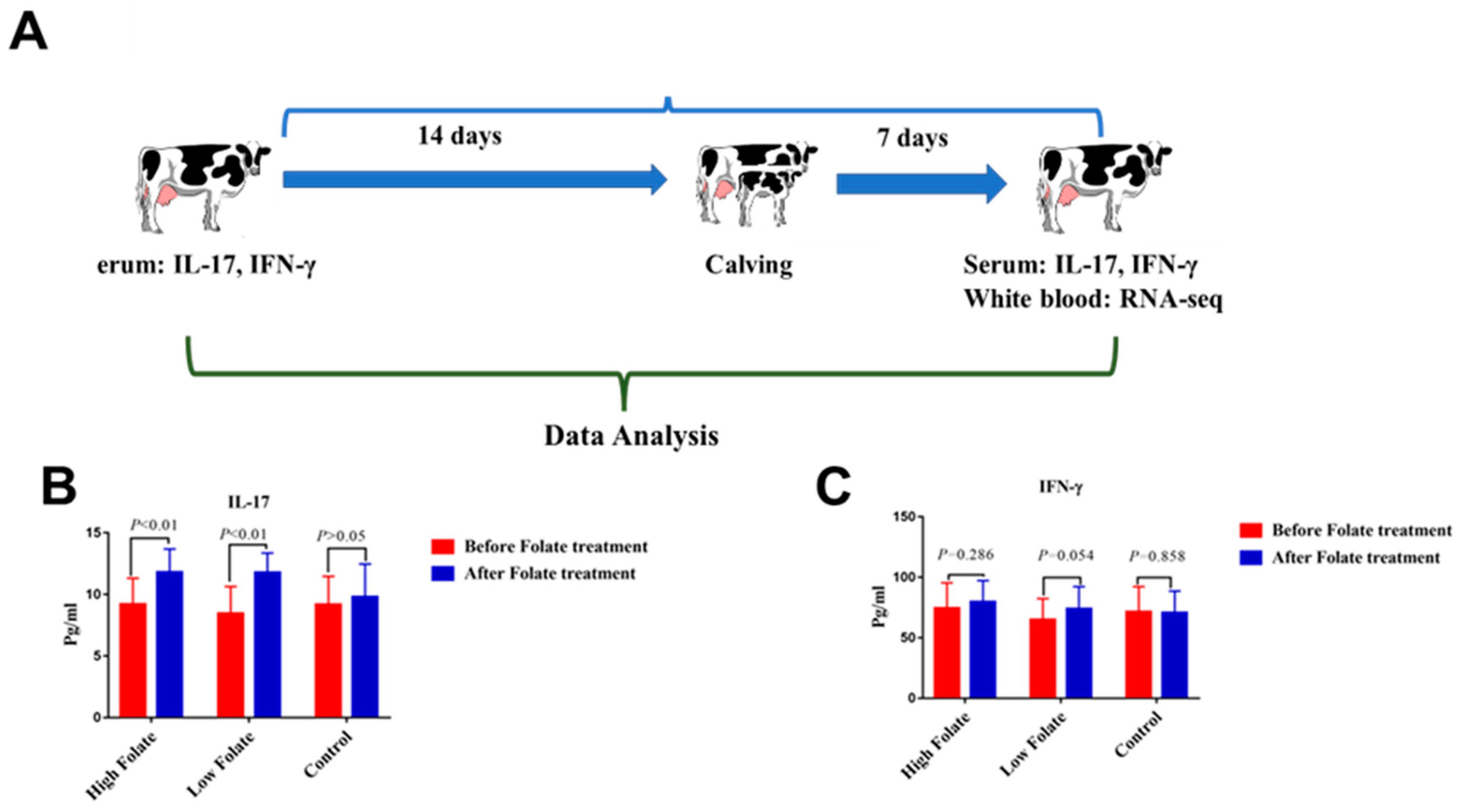
2.1. Folic Acid Significantly Regulates Immunity in Periparturient Dairy Cattle

Interferon-gamma (IFN-γ) is involved in antibacterial activity. It has been noted that, during the periparturient period, the level of IFN-γ is significantly decreased [48]. Recently, our research team observed that folic acid treatment increases the level of IFN-γ and interleukin 17 (IL-17) [49], as shown in Figure 2.
The transcriptomic profiling of peripheral blood lymphocytes in Chinese Holstein showed that folic acid treatment regulated several differentially expressed genes (DEGs), such as C1QB, ANXA1, CSF1, JSP.1, CCL5, CCL8, CCL16, CXCR5, HCK, MYD88, NFKBIA, MMP9, C1QB, SERPING1, TNFSF13, FOS, NFKBIA, NFKBIE, and IL9R, which have a strong association with immunity and anti-inflammation [49]. In addition, folic-acid-supplementation-mediated CD4, CD59, C8G, C8B, C8A, FOS, and NFKBIA in periparturient dairy cattle, which are the key genes of the immunity-associated pathways, e.g., T cell signaling, Staphylococcus aureus infection, and complement and coagulation cascades [49]. Furthermore, it was found that folic acid supplementation causes the down-regulation of NFKB inhibitor alpha (NFKBIA) and TNF alpha-induced protein 3 (TNFAIP3), which directed the negative mediation of NF-kappaB transcription factor activity. The suppressor of cytokine signaling 3 (SOCS3) was also down-regulated, which is involved in the negative mediation of cytokines that signal through the JAK/STAT pathway. The myeloid differentiation primary response 88 (MYD88), nucleotide-binding oligomerization domain containing 2 (NOD2), and mitogen-activated protein kinase 13 (MAPK13) are the positive mediators of interleukin 6 (IL-6), and were up-regulated by folic acid supplementation in periparturient dairy cattle [49]. The study conducted by Ouattara and colleagues in 2016 attempted to elucidate the effects of folic acid and vitamin B12, given separately or combined in two kinds of tissues (liver and mammary gland) of periparturient cows, to regulate their metabolism and milk production, and establish the molecular mechanism under which these changes take place. Moreover, some important genes, such as DLK1, LOXL4, SAA3, LOC100126815, MGC126945, and IGLL1, were reported to be mainly engaged in B-cell-mediated immunity, macrophage activation, and the apoptotic process [50]. The distribution of each lymphocyte and granulocyte subset is essential for the immune responses [51], while it has been shown that a folate-deficient diet in rats is associated with a decline in the total number of circulatory lymphocytes and granulocytes [33]. Recently, a study illustrated that folic acid regulates the immunity linked genes (IL2RG, TLR2, IRF1, IRF7, IRF8, CD40, DQB, RSAD2, ICOSLG, MX1, and MX2) and their pathways (immune response signaling, Toll-like receptor signaling pathway, cytokine–cytokine receptor interaction, and the NF-kappa B signaling pathway) in Hu sheep offspring [52]. From the above-mentioned published studies, it has been concluded that folic acid regulates immunity and could be the best choice to overcome immunity suppression, which is the primary concern in periparturient dairy cattle. Additionally, very limited development of the association of folic acid/vitamin B12 with immunity regulation in periparturient dairy cattle has been undertaken. Thus, it is suggested that trials be extended to provide more in-depth evidence.

2.2. Folic Acid Regulates Mastitis Resistance and Down-Regulates the Genes Involved in Mastitis Development

In previous research, we documented some important immunity and anti-inflammatory-associated genes in response to folic acid treatment [49]. The selected genes from previously published data [49] and recent Staphylococcus aureus infected mastitis cattle studies [53,54] are shown in Table 1.
Genes such as NFKBIA, SOCS3, and PIM1, which play a key role in the development of mastitis, were noted to be up-regulated in the S. aureus-infected mammary gland tissue of dairy cattle [53]. NFKBIA and TNFAIP3 play a key role in the negative mediation of NF-kappaB transcription factor activity. SOCS3 negatively regulates IL-6 [56], which is the key player in immune regulation and has an important role in mastitis resistance. SOCS3 is also responsible for the negative regulation of cytokines that signal through the JAK/STAT pathway [57]. These two pathways play an essential role in immunity and anti-inflammation. However, the mentioned genes were significantly down-regulated by folic acid administration in periparturient dairy cows [49], as shown in Table 1. Huang and his team concluded that maintaining a low expression of SOCS3 is also needed for milk synthesis well [57]. ARG1 plays a critical position in the identification of bacterial sources, while PTX3 is helpful in the apoptosis process. Additionally, our research team documented some essential signaling, such as antigen presentation, processing signaling, Staphylococcus aureus infection, and Toll-like receptor pathways, in response to folic acid supplementation in dairy cattle [49], which were potentially associated with mastitis resistance [53,54]. Moreover, the down-regulation of KIT and LPL genes negatively affects immunity and also interferes in the metabolic process [53]. In contrast, our research team noted the up-regulation of the mentioned genes [49]. By comparing the gene expression status in the above-published studies (Table 1), we expect that the role of folic acid as a key regulator of many immunity-associated genes might be target for mastitis control research in the future.

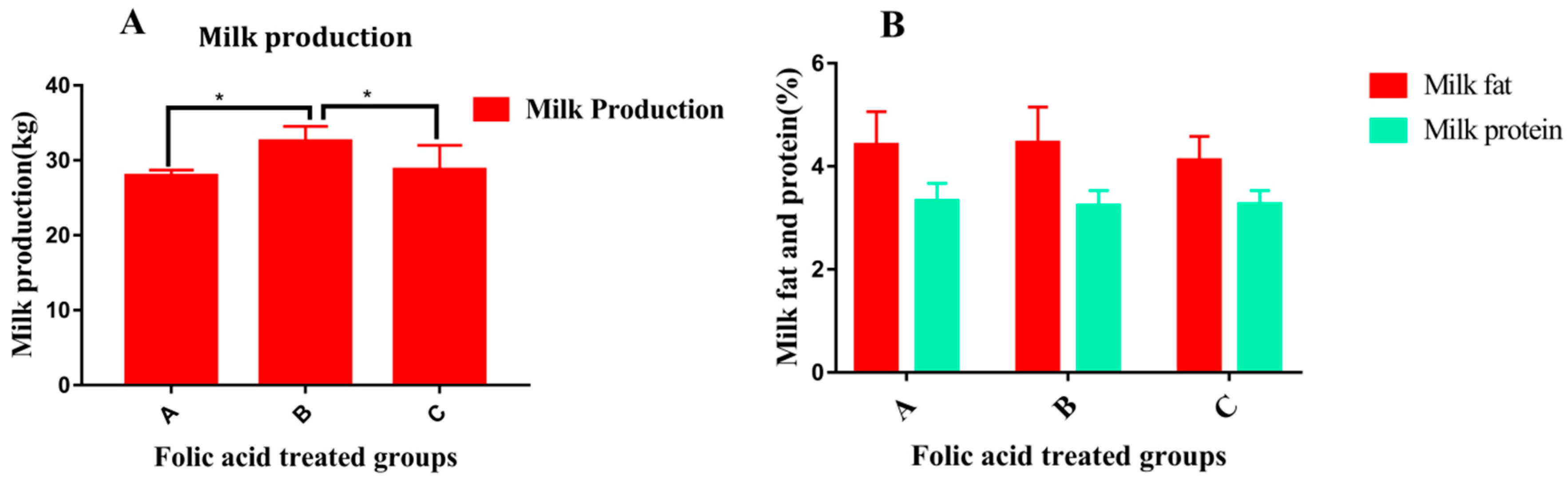
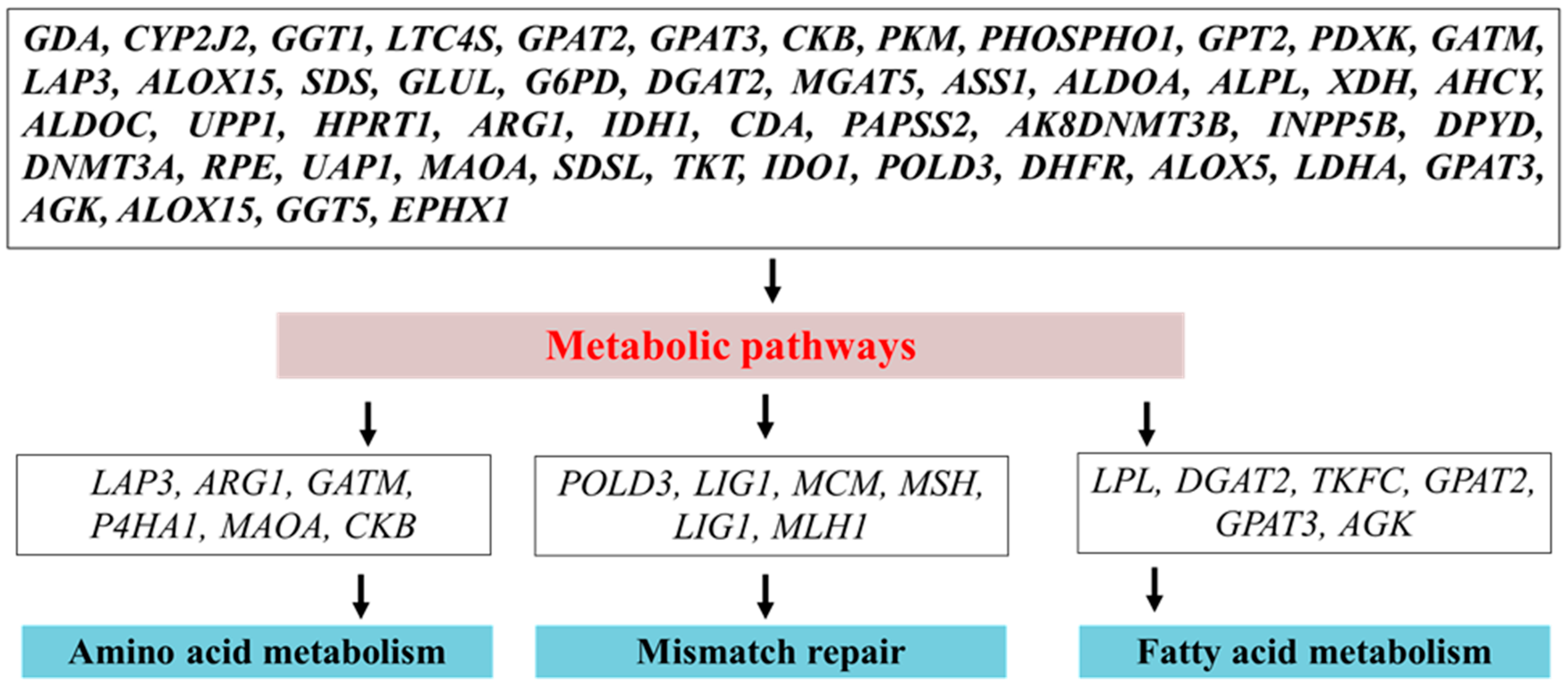
2.3. Folic Acid Alone or in Combination with Vitamin B12 Improves Milk Production and Metabolism in Periparturient Dairy Cattle

Recently, our research team investigated the association of folic acid supplementation alone with milk production and milk variables [58], as shown in Figure 3. Furthermore, it was also observed that the quantity of folic acid plays a role in lactational performance [58].
Note: Overall, Figure 3 shows that treatment in group B was effective for the improvement in milk production. In addition, folic acid treatment in all groups (A, B, C) did not show any effect on milk variables (milk fat and protein).
Folic acid supplements increase milk yield and milk protein in dairy cattle [43,59,60,61,62,63]. However, some studies did not note any effect of folic acid on lactational performance, which might be due to the low concentration of vitamin B12 in the plasma of dairy cattle [64,65]. Folate metabolism seems to have a vital influence on milk protein synthesis in mammary epithelial cells [66]. Recently, our published study noted that oral supplementation of folic acid significantly regulates milk production in periparturient dairy cows [58]. The effects of folic acid on milk production performance may be due to its correlation with DNA synthesis and the methylation cycle. The above studies showed that folic acid plays an essential role in milk yield improvement and should be supplied to periparturient dairy cattle.
A recent study [58] performed transcriptomic profiling of blood lymphocytes of folic acid-treated periparturient dairy cattle. The findings of the analysis illustrated that folic acid treatment mediated key DEGs (LAP3, SOCS3, BMX, ZC3H12A, UBE2C, MRPL57, BRCA1, IGF1R, LPL, HK3, PKM, VLDLR, XDH, SLC25A25, CTSB, DGAT2, UBE2R2, BRCA1, GGT, CALCRL, SDSL, ALOX5, SPP1, LYZ, and PPARD) that were notably associated with metabolism, milk fat, protein, milk production, and mammary gland development [58]. Cui and colleagues recorded several genes, such as RPL23A, SLC25A38, BMX, and ZC3H14, with a strong correlation with milk fat and protein yield [67]. Similarly, DGAT2, UBE2R2, BRCA1, VLDLR, and CTSB genes show an association with milk production [68]. The calcitonin receptor-like receptor (CALCRL) serves as a receptor for the further action of calcitonin genes. Using RNA-Seq, Seo and co-workers demonstrated that calcitonin genes are extensively linked to milk fat yield [69]. Two genes, namely, SPP1 and LYZ, were predominantly involved in the growth of mammary tissue and milk yield in Rhesus macaques [70]. Similarly, GGT, SDSL, PPRAD, and ALOX5 regulate pathways associated with mammary gland development and fatty acid metabolism, and are also involved in milk production [71].
From the above-mentioned published data, it has been shown that the response of mRNA to folic acid in periparturient dairy cows is close to that observed for the phenotypic milk production outcome. Furthermore, the reported genes associated with milk production in the above studies in response to folic acid might be targets as milk improvement markers in dairy cattle. The high production of milk raises the energy demand, resulting in a negative energy balance occurring in the body [7]. Moreover, the studies regarding folic acid and vitamin B12 treatment during the periparturient period in dairy cattle have revealed that proper metabolism is a crucial requirement to maintain milk production.
Folic acid is essential for the growth of ruminal bacteria and boosts the population of the cellulolytic bacterial population and the digestion of cellulose [72]. Intake of dry matter will normally decline during the peripartum period. However, the rumen-protected treatment of folic acid substantially promotes the intake of dry matter and also promotes the metabolism and energy balance [73]. Although the major absorption site of folic acid is the proximal duodenum, however, some portions of folic acid are also absorbed in the rumen [74]. Folic acid supplementation promotes metabolic effectiveness, dry matter intake, and overall fiber degradability in dairy cattle [72,73]. It was revealed, through a molecular-based mechanism, that folic acid and vitamin B12 regulate several genes that are associated with metabolism and milk yield. Metabolic pathways engage in a chain of chemical reactions that take place in the cell. These metabolic pathways are associated with either synthesizing molecules by utilizing energy or breaking down molecules, or releasing energy in the process known as the catabolic pathway [75]. Pathways are important for sustaining homeostasis within an organism, and the flow of metabolites through a pathway is controlled based on the cell’s needs and the substrate’s availability. We also noted in our previously published study that the majority of genes demonstrated their importance for the mediation of metabolic signaling, which further speeds up various biological processes related to metabolism [58]. Folic acid acts in the acceptance and release of one-carbon units [27], which are vital for the biosynthesis of purine, pyrimidine, and DNA, and also offers the methyl group for the formation of S-adenosylmethionine. Methyl-tetrahydrofolate is the cofactor providing a methyl group for remethylation of homocysteine into methionine, and this reaction is irreversible. Methionine works as a substrate for SAM, a cofactor and methyl group donor for many methylation reactions, such as methylation of DNA, RNA, proteins, and phospholipids [76]. The inadequate supply of folic acid may lead to a DNA mismatch repair and abnormal cell growth because of the essential role in the synthesis of DNA. Similarly, in a recently published study, many essential genes, such as ALDOA, PKM, SDS, SDSL, ASS1, POLD3, XDH, and ARG1, in response to folic acid treatment, were documented as being significantly associated with the biosynthesis of amino acid signaling [58]. Moreover, MCM2, MCM3, POLD3, and LIG1 were documented to have an involvement in DNA replication and mismatch repair, while PHOSPHO1, LPCAT2, GPAT2, and GPAT3 were found to control the glycerophospholipid metabolism. For milk lactose synthesis in dairy cattle, glucose is essential, and is provided by the processes of glucogenesis and glycogenolysis [77]. The combined supplement of folic acid and vitamin B12 promote gluconeogenesis in periparturient dairy cattle [65], while a recently published study has also shown the association of folic acid linkages to the regulation of glycolysis/gluconeogenesis and related genes, i.e., LDHA, ALDOC, and ALDH3B1 [58]. The metabolic-associated genes further facilitate the biological function processes (Figure 4).
Additionally, HERC6, PPP1R3B, PYCR1, ID1, MYOM1, CDK5R1, DLK1, HP, LOXL4, FUT5, HERC6, LOC515676 ISG15, PYCR1, and CACNA2D1 were noted to be associated with metabolic processes, while MEP1B, DLK1, MYOM1, SFRP1, MEP1B, IGLL1, and SPP1 genes were linked to developmental functions [58]. The above-mentioned published research revealed that folic acid, in combination with vitamin B12, may be the best option for maximizing the milk production and metabolic efficiency of periparturient dairy cows.

3. Future Direction for Research of Folic Acid and Vitamin B12 in Transition Dairy Cattle

Based on the published studies on folic acid and vitamin B12, we note that, although folic acid has a crucial role in disease control, as shown in human and mouse studies, to date, no study has established the association of folic acid with disease prevention in dairy cattle. Thus, we suggest that further research is needed in dairy cattle to examine the influence of folate supplementation, alone or in combination with vitamin B12, on disease control. As our recently published study documented the association of folic acid supplementation with mastitis-linked genes, it would be of interest to extend the study in further mastitis research. To date, the exact requirement of folic acid supplementation has not yet been reported in periparturient dairy cattle research. Thus, it is an important topic for future research, because both low and high levels of folic acid may have serious consequences. In addition, it has been proved that high doses of folic acid are toxic and reduced the natural killer cells (NK) cytotoxicity responsible for the regulation of the innate immunity system in mice [78].
This review showed that controversy remains regarding the influence of folic acid and vitamin B12 on milk production, and the effect of feed intake on the availability of folic acid in dairy cattle. These issues thus need to be addressed in future research. Additionally, the microorganisms in rumens should be characterized for folic acid needs and synthesis during the transition period. In vitro study is also recommended to examine the individual and combined influence on milk production, metabolism, and immunity. Considering folic acid is a methyl donor, we strongly recommend an epigenomic study to investigate the mechanism by which folic acid influences the epigenetic modifications of different beneficial genes responsible for disease resistance, metabolism, and milk production.

4. Conclusions

Based on the cited literature, we concluded that the periparturient period is vital in dairy cattle; special managemental care, including balanced intake of folic acid and vitamin B12, is needed. The review suggests that folic acid and vitamin B12 are essential nutrients and, as such, should be supplemented in dairy cattle during the periparturient period to overcome metabolic and immune stress. Milk production can be improved with the supplementation of folic acid and vitamin B12, which are key concerns for animal scientists. Folic acid quantity should be considered during supplementation because an excessive intake of folic acid suppressed NK-cells. Folic acid supplementation regulates immunity and mastitis-linked genes. Thus, the targeting of folic acid in mastitis control studies in periparturient dairy cattle is recommended. Genes regulated by folic acid treatments might be a useful addition to the marker selection for milk production and metabolic regulation in peripartum cattle.

Author Contributions

Conceptualization, M.Z.K and Y.Y.; writing—original draft preparation, M.Z.K and A.K. writing—review and editing, M.Z.K., L.L., J.D., J.X., and Y.Y. All authors have read and agreed to the published version of the manuscript.

Funding

This research was financially supported by the NSFC-PSF Joint Project (31961143009), Beijing Natural Science Foundation (6182021), National Natural Science Foundation of China (31272420) and Modern Agro-industry Technology Research System (CARS-36).

Acknowledgments

We are thankful to the Chinese Government Scholarship Council and China Agricultural University, Beijing, China, for providing us an environment of learning. Without this platform the completion of this work would not have been an easy task.

Conflicts of Interest

The authors declare no conflict of interest.

References

  1. Grummer, R.R.; Wiltbank, M.C.; Fricke, P.M.; Watters, R.D.; Silva-Del-Rio, N. Management of dry and transition cows to improve energy balance and reproduction. J. Reprod. Dev. 2010, 56, S22–S28. [Google Scholar] [CrossRef] [Green Version]
  2. Esposito, G.; Irons, P.C.; Webb, E.C.; Chapwanya, A. Interactions between negative energy balance, metabolic diseases, uterine health and immune response in transition dairy cows. Anim. Reprod. Sci. 2014, 144, 60–71. [Google Scholar] [CrossRef] [Green Version]
  3. Lacasse, P.; Vanacker, N.; Ollier, S.; Ster, C. Innovative dairy cow management to improve resistance to metabolic and infectious diseases during the transition period. Res. Vet. Sci. 2018, 116, 40–46. [Google Scholar] [CrossRef]
  4. Bell, A.W. Regulation of organic nutrient metabolism during transition from late pregnancy to early lactation. J. Anim. Sci. 1995, 73, 2804–2819. [Google Scholar] [CrossRef]
  5. Janovick, N.A.; Drackley, J.K. Prepartum dietary management of energy intake affects postpartum intake and lactation performance by primiparous and multiparous Holstein cows. J. Dairy Sci. 2010, 93, 3086–3102. [Google Scholar] [CrossRef]
  6. Roche, J.R.; Bell, A.W.; Overton, T.R.; Loor, J.J. Nutritional management of the transition cow in the 21st century-a paradigm shift in thinking. Anim. Prod. Sci. 2013, 53, 1000–1023. [Google Scholar] [CrossRef]
  7. Grummer, R.R. Impact of changes in organic nutrient metabolism on feeding the transition dairy cow. J. Anim. Sci. 1995, 73, 2820–2833. [Google Scholar] [CrossRef]
  8. Gonzalez, M.; Yabuta, A.K.; Galindo, F. Behaviour and adrenal activity of first parturition and multiparous cows under a competitive situation. Appl. Anim. Behav. Sci. 2003, 83, 259–266. [Google Scholar] [CrossRef]
  9. Meglia, G.E.; Johannisson, A.; Agenas, S.; Holtenius, K.; Waller, K.P. Effects of feeding intensity during the dry period on leukocyte and lymphocyte sub-populations, neutrophil function and health in periparturient dairy cows. Vet. J. 2005, 169, 376–384. [Google Scholar] [CrossRef]
  10. Kulberg, S.; Storset, A.K.; Heringstad, B.; Larsen, H.J.S. Reduced levels of total leukocytes and neutrophils in Norwegian cattle selected for decreased mastitis incidence. J. Dairy Sci. 2002, 85, 3470–3475. [Google Scholar] [CrossRef] [Green Version]
  11. Mulligan, F.J.; Doherty, M.L. Production diseases of the transition cow. Vet. J. 2008, 176, 3–9. [Google Scholar] [CrossRef]
  12. Mallard, B.A.; Dekkers, J.C.; Ireland, M.J.; Leslie, K.E.; Sharif, S.; Vankampen, C.L.; Wagter, L.; Wilkie, B.N. Alteration in Immune Responsiveness during the Peripartum Period and Its Ramification on Dairy Cow and Calf Health. J. Anim. Sci. 1998, 81, 585–595. [Google Scholar] [CrossRef]
  13. Dänicke, S.; Meyer, U.; Kersten, S.; Frahm, J. Animal models to study the impact of nutrition on the immune system of the transition cow. Res. Vet. Sci. 2018, 116, 15–27. [Google Scholar] [CrossRef]
  14. Trevisi, E.; Minuti, A. Assessment of the innate immune response in the periparturient cow. Res. Vet. Sci. 2018, 116, 47–54. [Google Scholar] [CrossRef]
  15. Jacob, S.K.; Ramnath, V.; Philomina, P.T.; Raghunandhanan, K.V.; Kannan, A. Assessment of physiological stress in periparturient cows and neonatal calves. Indian J. Physiol. Pharmacol. 2001, 45, 233–238. [Google Scholar]
  16. Lacetera, N.; Franci, O.; Scalia, D.; Bernabucci, U.; Ronchi, B.; Nardone, A. Effects of nonesterified fatty acids and β-hydroxybutyrate on functions of mononuclear cells obtained from ewes. Am. J. Vet. Res. 2002, 63, 414–418. [Google Scholar] [CrossRef]
  17. Bai, H.; Talukder, M.S.; Hiroki, K.; Tsukino, I.; Manabu, K.; Masashi, T. Evaluation of the immune status of peripheral blood monocytes from dairy cows during the periparturition period. J. Reprod. Dev. 2019, 65, 313–318. [Google Scholar] [CrossRef] [Green Version]
  18. Hammon, D.S.; Evjen, I.M.; Dhiman, T.R.; Goff, J.P.; Walters, J.L. Neutrophil function and energy status in Holstein cows with uterine health disorders. Vet. Immunol. Immunopathol. 2006, 113, 21–29. [Google Scholar] [CrossRef]
  19. Kehrli, M.E.; Nonnecke, B.J.; Roth, J.A. Alterations in bovine neutrophil function during the periparturient period. Am. J. Vet. Res. 1989, 50, 207–214. [Google Scholar]
  20. Sheldon, I.M.; Cronin, J.; Goetze, L.; Donofrio, G.; Schuberth, H.J. Defining postpartum uterine disease and the mechanisms of infection and immunity in the female reproductive tract in cattle. Biol. Reprod. 2009, 81, 1025–1032. [Google Scholar] [CrossRef]
  21. Crookenden, M.A.; Heiser, A.; Murray, A.; Dukkipati, V.S.R.; Kay, J.K.; Loor, J.J.; Meier, S.; Mitchell, M.D.; Moyes, K.M.; Walker, C.G.; et al. Parturition in dairy cows temporarily alters the expression of genes in circulating neutrophils. J. Dairy Sci. 2016, 99, 6470–6483. [Google Scholar] [CrossRef] [Green Version]
  22. Nonnecke, B.J.; Kimura, K.; Goff, J.P.; Kehrli, M.E. Effects of the mammary gland on functional capacities of blood mononuclear leukocyte populations from periparturient cows 1. J. Dairy Sci. 2003, 86, 2359–2368. [Google Scholar] [CrossRef]
  23. Itle, A.J.; Huzzey, J.M.; Weary, D.M.; Von Keyserlingk, M.A.G. Clinical ketosis and standing behavior in transition cows. J. Dairy Sci. 2015, 98, 128–134. [Google Scholar] [CrossRef] [Green Version]
  24. Bertoni, G.; Trevisi, E.; Han, X.; Bionaz, M. Effects of inflammatory conditions on liver activity in puerperium period and consequences for performance in dairy cows. J. Dairy Sci. 2008, 91, 3300–3310. [Google Scholar] [CrossRef] [Green Version]
  25. McArt, J.A.A.; Nydam, D.V.; Oetzel, G.R. Epidemiology of subclinical ketosis in early lactation dairy cattle. J. Dairy Sci. 2012, 95, 5056–5066. [Google Scholar] [CrossRef] [Green Version]
  26. Ribeiro, E.S.; Lima, F.S.; Greco, L.F.; Bisinotto, R.S.; Monteiro, A.P.A.; Favoreto, M.; Ayres, H.; Marsola, R.S.; Martinez, N.; Thatcher, W.W.; et al. Prevalence of periparturient diseases and effects on fertility of seasonally calving grazing dairy cows supplemented with concentrates. J. Dairy Sci. 2013, 96, 5682–5697. [Google Scholar] [CrossRef]
  27. Choi, S.-W.; Mason, J.B. Folate and carcinogenesis: An integrated scheme. J. Nutr. 2000, 130, 129–132. [Google Scholar] [CrossRef] [Green Version]
  28. Ghoshal, K.; Li, X.; Datta, J.; Bai, S.; Pogribny, I.; Pogribny, M.; Huang, Y.; Young, D.; Jacob, S.T. A folate- and methyl-deficient diet alters the expression of DNA methyltransferases and methyl CpG binding proteins involved in epigenetic gene silencing in livers of F344 rats. J. Nutr. 2006, 136, 1522–1527. [Google Scholar] [CrossRef]
  29. Pogribny, I.P.; Karpf, A.R.; James, S.R.; Melnyk, S.; Han, T.; Tryndyak, V.P. Epigenetic alterations in the brains of Fisher 344 rats induced by long-term administration of folate/methyl-deficient diet. Brain Res. 2008, 1237, 25–34. [Google Scholar] [CrossRef]
  30. Mikkelsen, K.; Apostolopoulos, V. Vitamin B12, Folic Acid, and the Immune System. In Nutrition and Immunity; Springer: Cham, Switzerland, 2019; pp. 103–114. [Google Scholar]
  31. Kunisawa, J.; Eri, H.; Izumi, I.; Hiroshi, K.A. Pivotal role of vitamin B9 in the maintenance of regulatory T cells in vitro and in vivo. PLoS ONE 2012, 7, e32094. [Google Scholar] [CrossRef] [Green Version]
  32. Abe, I.; Shirato, K.; Hashizume, Y.; Mitsuhashi, R.; Kobayashi, A.; Shiono, C.; Sato, S.; Tachiyashiki, K.; Imaizumi, K. Folate-deficiency induced cell-specific changes in the distribution of lymphocytes and granulocytes in rats. Environ. Health Prev. Med. 2013, 18, 78–84. [Google Scholar] [CrossRef] [PubMed] [Green Version]
  33. Metz, J. A high prevalence of biochemical evidence of vitamin B12 or folate deficiency does not translate into a comparable prevalence of anemia. Food Nutr. Bull. 2008, 29, S74–S85. [Google Scholar] [CrossRef] [PubMed]
  34. Antony, A.C. In utero physiology: Role of folic acid in nutrient delivery and fetal development. Am. J. Clin. Nutr. 2007, 85, 598S–603S. [Google Scholar] [CrossRef] [Green Version]
  35. Hollingsworth, J.W.; Maruoka, S.; Boon, K.; Garantziotis, S.; Li, Z.; Tomfohr, J.; Bailey, N.; Potts, E.N.; Whitehead, G.; Brass, D.M.; et al. In utero supplementation with methyl donors enhances allergic airway disease in mice. J. Clin. Investig. 2008, 118, 3462–3469. [Google Scholar] [CrossRef] [Green Version]
  36. Preynat, A.; Lapierre, H.; Thivierge, C.M.; Palin, F.M.; Cardinault, N.; Matte, J.J.; Desrochers, A.; Girard, L.C. Effects of supplementary folic acid and vitamin B12 on hepatic metabolism of dairy cows according to methionine supply. J. Dairy Sci. 2010, 93, 2130–2142. [Google Scholar] [CrossRef] [Green Version]
  37. Duplessis, M.; Girard, C.L.; Santschi, D.E.; Lefebvre, D.M.; Pellerin, D. Folic acid and vitamin B12 supplement enhances energy metabolism of dairy cows in early lactation. J. Dairy Sci. 2012, 95, 118. [Google Scholar]
  38. Froese, S.D.; Brian, F.; Matthias, R.B. Vitamin B12, folate, and the methionine remethylation cycle—Biochemistry, pathways, and regulation. J. Inherit. Metab. Dis. 2019, 42, 1–13. [Google Scholar] [CrossRef] [Green Version]
  39. Miller, J.W.; Nadea, M.R.; Smith, J.; Smith, D.; Selhub, J. Folate-deficiency-induced homocysteinaemia in rats: Disruption of S-adenosylmethionine’s co-ordinate regulation of homocysteine metabolism. Biochem. J. 1994, 298, 415–419. [Google Scholar] [CrossRef] [Green Version]
  40. James, S.J.; Miller, B.J.; Mcgarrity, L.J.; Morris, S.M. The effect of folic-acid and/or methionine deficiency on deoxyribonucleotide pools and cell cycle cell cycle distribution in mitogen-stimulated rat lymphocytes. Cell Prolif. 1994, 27, 395–406. [Google Scholar] [CrossRef]
  41. Girard, C.L.; Matte, J.J. Folic acid and vitamin B12 requirements of dairy cows: A concept to be revised. Livest. Prod. Sci. 2005, 98, 123–133. [Google Scholar] [CrossRef]
  42. Bailey, L.B.; Gregory, J.F. Folate metabolism and requirements. J. Nutr. 1999, 129, 779–782. [Google Scholar] [CrossRef] [Green Version]
  43. Girard, C.L.; Matte, J.J. İmpact of B-vitamin supply on major metabolic pathways of lactating dairy cows. Can. J. Anim. Sci. 2006, 86, 213–220. [Google Scholar] [CrossRef] [Green Version]
  44. Bechdel, S.I.; Honeywell, H.E.; Dutcher, R.A.; Knutsen, M.H. Synthesis of vitamin B in the rumen of the cow. J. Biol. Chem. 1928, 80, 231–238. [Google Scholar]
  45. Matte, J.J.; Girard, C.L.; Tremblay, G.F. Effect of long-term addition of folic acid on folate status, growth performance, puberty attainment, and reproductive capacity of gilts. J. Anim. Sci. 1993, 71, 151–157. [Google Scholar] [CrossRef]
  46. Girard, C.L.; Matte, J.J.; Tremblay, G.F. Serum folates in gestating and lactating dairy cows. J. Dairy. Sci. 1989, 72, 3240–3246. [Google Scholar] [CrossRef]
  47. Girard, C.L.; Lapierre, H.; Matte, J.J.; Lobley, G.E. Effects of dietary supplements of folic acid and rumen-protected methionine on lactational performance and folate metabolism of dairy cows. J. Dairy Sci. 2005, 88, 660–670. [Google Scholar] [CrossRef]
  48. Ster, C.; Loiselle, M.C.; Lacasse, P. Effect of postcalving serum nonesterified fatty acids concentration on the functionality of bovine immune cells. J. Dairy Sci. 2012, 95, 708–717. [Google Scholar] [CrossRef]
  49. Khan, M.Z.; Zhang, Z.; Liu, L.; Wang, D.; Mi, S.; Liu, X.; Liu, G.; Guo, G.; Li, X.; Wang, Y.; et al. Folic acid supplementation regulates key immunity-associated genes and pathways during the periparturient period in dairy cows. Asian-Australas. J. Anim. Sci. 2020, 00, 1–13. [Google Scholar] [CrossRef] [Green Version]
  50. Ouattara, B.; Bissonnette, N.; Duplessis, M.; Girard, C.L. Supplements of vitamins B9 and B12 affect hepatic and mammary gland gene expression profiles in lactating dairy cows. BMC. Genom. 2016, 17, 640. [Google Scholar] [CrossRef] [Green Version]
  51. Shirato, K.; Motohashi, N.; Tanihata, J.; Tachiyashiki, K.; Tomoda, A.; Imaizumi, K. Effects of two types of inactivity on the number of white blood cells in rats. Eur. J. Appl. Physiol. 2006, 98, 590–600. [Google Scholar] [CrossRef]
  52. Wang, B.; Li, H.; Zhen, L.; Jian, L.; Gao, Y.; Qu, Y.; Liu, C.; Xu, C.; Li, Y.; Diao, Z.; et al. Folic acid supplementation modulates the growth performance, muscle development and immunity of Hu sheep offspring of different litter size. J. Nutr. Biochem. 2019, 70, 194–201. [Google Scholar] [CrossRef]
  53. Fang, L.; Hou, Y.; An, J.; Li, B.; Song, M.; Wang, X.; Sørensen, P.; Dong, Y.; Liu, C.; Wang, Y.; et al. Genome-wide transcriptional and post-transcriptional regulation of innate immune and defense responses of bovine mammary gland to Staphylococcus aureus. Front. Cell. Infect. Microbiol. 2016, 6. [Google Scholar] [CrossRef] [Green Version]
  54. Kosciuczuk, E.M.; Lisowski, P.; Jarczak, J.; Majewska, A.; Rzewuska, M.; Zwierzchowski, L.; Bagnicka, E. Transcriptome profiling of Staphylococci-infected cow mammary gland parenchyma. BMC. Vet. Res. 2017, 13, 161. [Google Scholar] [CrossRef]
  55. He, Y.S.; Song, M.; Zhang, Y.; Li, X.; Song, J.; Zhang, Y.; Yu, Y. Whole-genome regulation analysis of histone H3 lysin 27 trimethylation in subclinical mastitis cows infected by Staphylococcus aureus. BMC Genom. 2016, 17, 565. [Google Scholar] [CrossRef] [Green Version]
  56. Croker, A.B.; Danielle, L.K.; Zhang, J.; Sam, W.; Tracy, A.W.; Edouard, G.S.; Lorraine, R.; Christopher, J.G.; Förster, I.; Clausen, B.E.; et al. SOCS3 negatively regulates IL-6 signaling In Vivo. Nat. Immunol. 2003, 4, 540–545. [Google Scholar] [CrossRef]
  57. Huang, Y.; Feng, Z.; Luo, C.; Zhang, X.; Yu, S.; Sun, Z.; Zhang, L.; Li, Q.; Xue-Jun, G. SOCS3-Mediated Blockade Reveals Major Contribution of JAK2/STAT5 Signaling Pathway to Lactation and Proliferation of Dairy Cow Mammary Epithelial Cells In Vitro. Molecules 2013, 18, 12987–13002. [Google Scholar] [CrossRef] [Green Version]
  58. Khan, Z.M.; Lei, L.; Zhichao, Z.; Adnan, K.; Di, W.; Siyuan, M.; Tahir, U.; Gang, L.; Gang, G.; Xizhi, L.; et al. Folic acid supplementation regulates milk production variables, metabolic associated genes and pathways in perinatal holsteins. J. Anim. Physiol. Anim. Nutr. 2020, 104, 438–492. [Google Scholar] [CrossRef]
  59. Graulet, B.; Matte, J.J.; Desrochers, A.; Doepel, L.; Palin, M.F.; Girard, C.L. Effects of dietary supplements of folic acid and vitamin B12 on metabolism of dairy cows in early lactation. J. Dairy Sci. 2007, 90, 3442–3455. [Google Scholar] [CrossRef] [Green Version]
  60. Girard, C.L.; Matte, J.J. Dietary Supplements of Folic Acid during Lactation: Effects on the Performance of Dairy Cows. J. Dairy Sci. 1998, 81, 1412–1419. [Google Scholar] [CrossRef]
  61. Preynat, A.; Lapierre, H.; Thivierge, M.C.; Palin, M.F.; Matte, J.J.; Desrochers, A.; Girard, C.L. Influence of methionine supply on the response of lactational performance of dairy cows to supplementary folic acid and vitamin B12. J. Dairy Sci. 2009, 92, 1685–1695. [Google Scholar] [CrossRef]
  62. Li, H.Q.; Liu, Q.; Wang, C.; Yang, Z.M.; Guo, G.; Huo, W.J.; Pei, C.X.; Zhang, Y.L.; Zhang, S.L.; Wang, H.; et al. Effects of dietary supplements of rumen-protected folic acid on lactation performance, energy balance, blood parameters and reproductive performance in dairy cows. Anim. Feed Sci. Technol. 2016, 213, 55–63. [Google Scholar] [CrossRef]
  63. Girard, C.L.; Matte, J.J. Effects of intramuscular injections of vitamin B12 on lactation performance of dairy cows fed dietary supplements of folic acid and rumen-protected methionine. J. Dairy Sci. 2005, 98, 671–676. [Google Scholar] [CrossRef]
  64. Girard, C.L.; Benchaar, C.; Chiquette, J.; Desrochers, A. Net flux of nutrients across the rumen wall of lactating dairy cows as influenced by dietary supplements of folic acid. J. Dairy Sci. 2009, 92, 6116–6122. [Google Scholar] [CrossRef] [Green Version]
  65. Preynat, A.; Lapierre, H.; Thivierge, M.C.; Palin, M.F.; Matte, J.J.; Desrochers, A.; Girard, C.L. Effects of supplements of folic acid, vitamin B12, and rumen-protected methionine on whole body metabolism of methionine and glucose in lactating dairy cows. J. Dairy Sci. 2009, 92, 677–689. [Google Scholar] [CrossRef]
  66. Menzies, K.K.; Lefèvre, C.; Sharp, J.A.; Macmillan, K.L.; Sheehy, P.A.; Nicholas, K.R. A novel approach identified the FOLR1 gene, a putative regulator of milk protein synthesis. Mamm. Genome 2009, 20, 498–503. [Google Scholar] [CrossRef]
  67. Cui, X.; Yali, H.; Shaohua, Y.; Yan, X.; Shengli, Z.; Yuan, Z.; Qin, Z.; Xuemei, L.; George, E.L.; Sun, D. Transcriptional profiling of mammary gland in Holstein cows with extremely different milk protein and fat percentage using RNA sequencing. BMC Genom. 2014, 15, 226. [Google Scholar] [CrossRef] [Green Version]
  68. Wickramasinghe, S.; Rincon, G.; Islas-Trejo, A.; Medrano, J.F. Transcriptional profiling of bovine milk using RNA sequencing. BMC. Genom. 2012, 13, 45. [Google Scholar] [CrossRef] [Green Version]
  69. Seo, M.; Lee, H.J.; Kim, K.; Caetano-Anolles, K.; Jeong, J.Y.; Park, S.; Oh, Y.K.; Cho, S.; Kim, H. Characterizing milk production related genes in Holstein using RNA-seq. Asian-Australas. J. Anim. Sci. 2016, 29, 343–351. [Google Scholar] [CrossRef] [Green Version]
  70. Lemay, D.G.; Hovey, R.C.; Hartono, S.R.; Hinde, K.; Smilowitz, J.T.; Ventimiglia, F.; Schmidt, K.A.; Lee, J.W.S.; Islas-Trejo, A.; Silva, P.I.; et al. Sequencing the transcriptome of milk production: Milk trumps mammary tissue. BMC Genom. 2013, 14, 872. [Google Scholar] [CrossRef] [Green Version]
  71. Yang, J.; Jiang, J.; Liu, X.; Wang, H.; Guo, G.; Zhang, Q.; Jiang, L. Differential expression of genes in milk of dairy cattle during lactation. Anim. Genet. 2016, 47, 174–180. [Google Scholar] [CrossRef] [Green Version]
  72. Wang, C.; Liu, Q.; Guo, G.; Huo, W.J.; Liang, Y.; Pei, C.X.; Zhang, S.L.; Yang, W.Z.; Wang, H. Effects of different dietary protein levels and rumen-protected folic acid on ruminal fermentation, degradability, bacterial populations and urinary excretion of purine derivatives in beef steers. J. Agric. Sci. 2017, 155, 1477–1486. [Google Scholar] [CrossRef]
  73. Wang, C.; Liu, Q.; Guo, G.; Huo, W.J.; Ma, L.; Zhang, Y.L.; Pei, C.X.; Zhang, S.L.; Wang, H. Effects of dietary supplementation of rumen-protected folic acid on rumen fermentation, degradability and excretion of urinary purine derivatives in growing steers. Arch. Anim. Nutr. 2016, 70, 441–454. [Google Scholar] [CrossRef] [PubMed]
  74. Ragaller, V.; Peter, L.; Wiebke, B.; Karl-Heinz, S.; Liane, H.; Gerhard, F. Effects of folic acid supplementation to rations differing in the concentrate to roughage ratio on ruminal fermentation, nutrient flow at the duodenum, and on serum and milk variables of dairy cows. Arch. Anim. Nutr. 2010, 64, 484–503. [Google Scholar] [CrossRef] [PubMed]
  75. Reece, J.B. Campbell Biology, 9th ed.; Benjamin Cummings: Boston, MA, USA, 2011; p. 143. ISBN 978-0-321-55823-7. [Google Scholar]
  76. Wagner, C. Biochemical role of folate in cellular metabolism. Clin. Res. Regul. Aff. 2001, 18, 161–180. [Google Scholar] [CrossRef]
  77. Nafikov, R.A.; Beitz, D.C. Carbohydrate and Lipid Metabolism in Farm Animals. J. Nutr. 2007, 137, 702–705. [Google Scholar] [CrossRef] [Green Version]
  78. Sawaengsri, H.; Wang, J.; Christina, R.; Josiane, S.; Wu, D.; Jacob, S.; Ligi, P. High folic acid intake reduces natural killer cell cytotoxicity in aged mice. J. Nutr. Biochem. 2016, 30, 102–107. [Google Scholar] [CrossRef] [Green Version]
Figure 1. Folic acid regulates the remethylation cycle of methionine, DNA biosynthesis, and T cells.
Metabolites 10 00263 g001
Figure 2. The influence of folic acid supplementation on serum cytokines of periparturient dairy cows [49]. (A) Experimental procedures of the entire study. (B) The level of IL-17 and (C) IFN-γ (pg/mL) in Holstein dairy cattle before and after folic acid supplementation. High folate (240 mg/500 kg), low (120 mg/500 kg body weight).
Metabolites 10 00263 g002
Figure 3. The effect of folic acid quantity on milk production variables in periparturient dairy cows [58]. (A) Comparison of the average milk production status over three months in group B (120 mg/500 kg body weight/cow), group A (240 mg/500 kg body weight/cow), and group C (control). (B) Comparison of average milk fat and protein percentage in three folic acid-treated dairy cattle groups (A, B, and C). The * denotes that the difference is significant and the p-value < 0.05.
Metabolites 10 00263 g003
Figure 4. Folic acid supplementation mediates the essential genes linked to metabolic pathways (amino acid and fatty acid) and mismatch repair. For ease, the genes mainly involved in metabolic pathways, mismatch repair, fatty acid metabolism, and protein synthesis are summarized.
Metabolites 10 00263 g004
Table 1. Comparison of gene regulation status in folic acid-treated and S. aureus-infected mastitis cows.
Gene S. aureus TreatmentFolic Acid-Treated Group [49]
PIM1, SOCS3, ATP12A, NFKBIA, DUSP4, ZC3H12, ESPNL, TNFAIP3Up-regulated [53]Down-regulated
CX3CR1, ALOX5, KIT, LPLDown-regulated [53]Up-regulated
C1QA, C1QB, CCL5, MMP9, VNN, BLA-DQBUp-regulated [54]Up-regulated
ICAM1, CXCL10Up-regulated [54]Down-regulated
PPARD, ARG1, PTX3, CD4Up-regulated [55]Up-regulated

