Abstract
In Uganda, the role of ticks in zoonotic disease transmission is not well described, partly, due to limited available information on tick diversity. This study aimed to identify the tick species that infest cattle. Between September and November 2017, ticks (n = 4362) were collected from 5 districts across Uganda (Kasese, Hoima, Gulu, Soroti, and Moroto) and identified morphologically at Uganda Virus Research Institute. Morphological and genetic validation was performed in Germany on representative identified specimens and on all unidentified ticks. Ticks were belonging to 15 species: 8 Rhipicephalus species (Rhipicephalus appendiculatus, Rhipicephalus evertsi evertsi, Rhipicephalus microplus, Rhipicephalus decoloratus, Rhipicephalus afranicus, Rhipicephalus pulchellus, Rhipicephalus simus, and Rhipicephalus sanguineus tropical lineage); 5 Amblyomma species (Amblyomma lepidum, Amblyomma variegatum, Amblyomma cohaerens, Amblyomma gemma, and Amblyomma paulopunctatum); and 2 Hyalomma species (Hyalomma rufipes and Hyalomma truncatum). The most common species were R. appendiculatus (51.8%), A. lepidum (21.0%), A. variegatum (14.3%), R. evertsi evertsi (8.2%), and R. decoloratus (2.4%). R. afranicus is a new species recently described in South Africa and we report its presence in Uganda for the first time. The sequences of R. afranicus were 2.4% divergent from those obtained in Southern Africa. We confirm the presence of the invasive R. microplus in two districts (Soroti and Gulu). Species diversity was highest in Moroto district (p = 0.004) and geographical predominance by specific ticks was observed (p = 0.001). The study expands the knowledge on tick fauna in Uganda and demonstrates that multiple tick species with potential to transmit several tick-borne diseases including zoonotic pathogens are infesting cattle.
Introduction
Ticks are associated with significant medical and veterinary health problems globally (Brites-Neto et al. 2015). Ticks are obligate hematophagous ectoparasites, which during feeding on their vertebrate hosts, can cause various clinical manifestations including tissue injury, body paralysis, and sometimes anemia during massive infestations (Giraldo-Ríos and Betancur 2018). Since the turn of the nineteenth century when the first description of a tick-transmitted infection was made (Smith and Kilborne 1893), many tick species are now known reservoirs and vectors of a multitude of pathogens that cause significant morbidity and mortality in both humans and animals. Some of the diseases that have since been described such as East Coast fever and Crimean-Congo hemorrhagic fever are challenging public health, veterinary, and socio-economic threats due to their increasing occurrence, pathogenicity, and economic impact (Adams et al. 2016; Kuehn 2019; Wesołowski et al. 2014).
In Uganda, the overall threat of ticks and tick-borne diseases to public health is not well known, partly due to limited knowledge on the natural diversity of ticks across the country. In fact, the most detailed and nationally representative surveys of tick species in Uganda that involved a variety of animal species were done in the 1970s, or earlier (Matthysee and Colbo 1987; Tukei et al. 1970), while the most recent studies have focused mainly on either specific geographical areas or veterinary aspects (Byaruhanga et al. 2015; Magona et al. 2011; Rubaire-Akiiki et al. 2006; Socolovschi et al. 2007). According to Walker et al. (2014), there are approximately 27 species of ticks infesting domestic animals in Uganda that are of socio-economic, veterinary, and human health importance. With the increasing reports of geographical expansion of many tick species (Gasmi et al. 2018; Leger et al. 2013; Nyangiwe et al. 2017; Raghavan et al. 2019; Sonenshine 2018), it is important that regular tick surveys are undertaken for inventory revisions. In this study, we aimed to identify the species of ticks currently infesting cattle across various agroecological zones of Uganda, as well as to provide a baseline investigation to a larger study on ticks and tick-borne diseases in Uganda (Malmberg and Hayer 2019). In order to achieve a more precise taxonomic classification of ticks in our study, we complemented the traditional morphotaxonomic approach with molecular techniques as recently suggested and applied in some studies (Brahma et al. 2014; Ernieenor et al. 2017; Estrada-Peña et al. 2017; Estrada-Peña et al. 2013). Molecular analyses were also done in order to provide sequence information for those tick species in Uganda that were not yet available in GenBank. We used cattle as sentinels because they can be infested with a variety of tick species (Rehman et al. 2017). In Uganda, particularly, intensity of tick infestation on cattle is high and tick-borne diseases are a major problem to cattle keepers (Ocaido et al. 2009). According to the Uganda Bureau of Statistics (2017), cattle is the most socially and economically important type of livestock in the country. Therefore, contact with cattle and/or their products is potentially among the most important routes through which many people come in direct, or indirect, contact with tick-borne zoonoses in Uganda.
Material and methods
Study areas
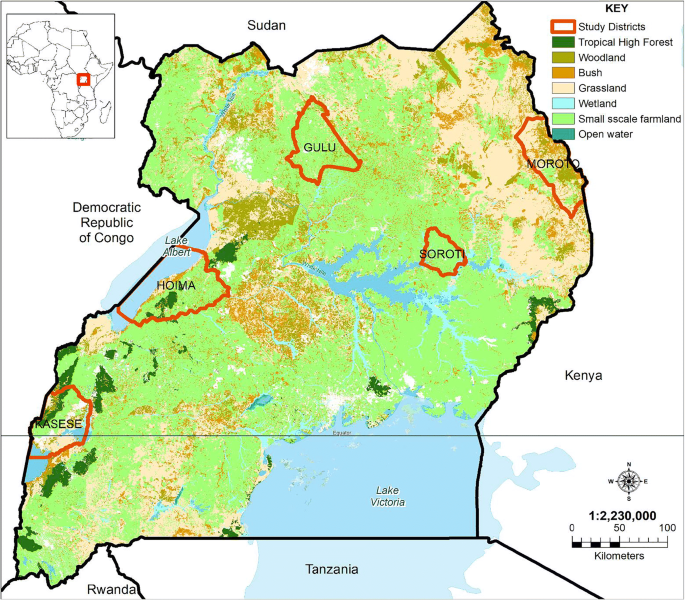
This study was conducted in the five districts of Kasese, Hoima, Gulu, Soroti, and Moroto in Uganda. As shown in Fig. 1, and based on previous studies by Wortmann and Eledu (1999) and Drichi (2003), these districts represent different agroecological zones of Uganda. Briefly, Kasese and Moroto districts have a semi-arid climatic environment and represent the extreme ends of the Ugandan livestock farming borderlines. Soroti and Gulu lie within a semi-moist zone with scattered subsistence mixed agricultural practices, amidst large swathes of open bushland. These districts are also equidistant to the expansive low-lying swampy areas of Lake Kyoga. On the other hand, Hoima district represented areas with low to medium altitudes that also practice extensive and commercialized agricultural and livestock farming. Additionally, Kasese and Gulu districts border with two major wildlife conservation areas, and therefore are ideal study sites for characterizing ticks at the livestock-wildlife interface. Moroto district represented areas with extensive transboundary migrations of livestock between multiple countries mainly Uganda, Kenya, and South Sudan.
Study design
This was a cross-sectional study, in which all tick samples were collected between September and November, 2017. To identify animals for sampling, a multistage approach was applied such that in each district, 2 sub-counties were purposively selected based on environmental diversity, and differences in animal management practices. Thereafter, a random selection of one parish from each sub-county was made based on the sampling frame provided by the local administrators. From each parish, 5 villages were identified based on geographical spread. And from each village, 5 households with cattle were selected based on convenience and willingness of the farmer to participate in the study. For tick collection, from each household, two animals were selected from the herd based on the farmer’s choice and/or those with visible ticks on them. Totally, ticks were collected from 100 cattle from each district.
Tick collection and identification
Ticks were handpicked from one side of the animal’s body, with attention to predilection sites, for approximately 5–10 min per animal. Ticks from each animal were placed separately into 50-ml tubes in which the lid had been perforated with pinholes to allow continuous circulation of fresh air. We also placed 3–4 pieces of fresh grass into each tick-containing tube in an effort to mimic the ticks’ natural environment. All tick-containing tubes were transported in a cool box to Uganda Virus Research Institute (UVRI), Entebbe, Uganda, within 5 days of collection. At UVRI, ticks were identified to species level using morphological characters under a stereomicroscope (Stereo Discovery V12, Zeiss, Birkerød, Denmark) and a Keyence VHX-900 microscope (Itasca, IL, USA) as previously described (Apanaskevich and Horak 2008a; Apanaskevich and Horak 2008b; Voltzit and Keirans 2003; Walker et al. 2014). Representative ticks from each of the identified species and ticks that could not be fully identified at UVRI, were shipped to Bundeswehr Institute of Microbiology, Munich, Germany, to confirm the morphological identification, and where necessary, validate it genetically. For genetic validation, DNA was extracted from individual ticks using a commercially available kit (QIAamp Mini Kit, Qiagen GmbH, Hilden, Germany) according to the manufacturer’s instructions. The 16S rDNA gene was amplified using the polymerase chain reaction protocol as described by Mangold et al. (1998). Thereafter, all obtained sequence data from this study, as well as additional data from GenBank, were compiled into a dataset of 71 sequences. Sequences from GenBank were chosen to encompass the range of Rhipicephalus and Amblyomma species that occur in Uganda, as well as closely related species. Validity of species identification for these sequences follows from recent studies that include large-scale taxonomic investigations to verify species identity by phylogenetic analysis and correlated morphology (Bakkes et al. 2020; Black and Piesman 1994; Chitimia-Dobler et al. 2017; Dantas-Torres et al. 2013; Nava et al. 2018). The prevalence of misidentified tick species among sequence data in GenBank is a growing problem that can only be addressed by large-scale taxonomic studies. Sequence data were aligned using MAFFT (Q-INS-i, 200PAM/k = 2; Gap opening penalty, 1.53) (Katoh et al. 2002). The optimal nucleotide substitution model was selected using BIC calculations in W-IQ-TREE (Trifinopoulos et al. 2016) and was determined as TPM2+F+G4. Maximum likelihood analysis was performed in MEGA v7.0.14 (Kumar et al. 2016) with 1000 bootstraps, as well as calculation of pairwise p-distances. Average p-distances between conspecific sequences from GenBank and collected samples were calculated to determine species identification validity according to the generally accepted threshold of 5% or greater sequence divergence between species (Bakkes et al. 2020; Bakkes et al. 2018; Chitimia-Dobler et al. 2017; Lado et al. 2018; Li et al. 2018; Mans et al. 2019).
Statistical analysis
All statistical data analyses were performed in STATA v14.2 software (College Station, TX). Chi-square or Fisher’s exact tests were used as appropriate to compare the differences between tick frequencies obtained from study districts and/or identified species. For all comparisons, a p value < 0.05 was statistically significant.
Results
Five hundred cattle were examined for ticks and only nine (1.8%) were found with no visible tick infestation. Overall, a total of 4362 ticks were collected from cattle in the five studied districts with no significant difference between the total number of ticks collected in each district (χ2 = 4.0; p = 0.40). Altogether, 15 tick species from three genera (Rhipicephalus, Amblyomma, and Hyalomma) were identified. As shown in Table 1, the most dominant tick species collected in this survey were R. appendiculatus (n = 2259; 51.79%), A. lepidum (n = 916; 21.00%), A. variegatum (n = 625; 14.33%), R. evertsi evertsi (n = 359; 8.23%), and R. decoloratus (n = 104; 2.38%). Moreover, 4 species including R. appendiculatus, R. evertsi evertsi, R. decoloratus, and A. variegatum were found in all study districts, albeit with significant variations in their respective levels of abundance (p = 0.001). On the other hand, the least abundant species were R. simus and A. paulopunctatum—each of them had only a single tick collected from the entire survey.
Moroto district had a significantly higher number of tick species (n = 13; p = 0.004) including all ticks belonging to R. sanguineus tropical lineage, R. pulchellus, R. simus, A. gemma, A. paulopunctatum, and H. truncatum. Importantly, a recently described tick species, R. afranicus (formerly R. turanicus, see Bakkes et al. (2020)), was also found only in Moroto district. Additionally, 99.23% of all A. lepidum in the study was found in Moroto district. On the other hand, Kasese district had 6 species (R. appendiculatus, R. evertsi evertsi, R. decoloratus, A. variegatum, A. lepidum, and A. cohaerens); Hoima district had 6 species (R. appendiculatus, R. evertsi evertsi, R. decoloratus, A. variegatum, A. cohaerens, and H. rufipes), while Gulu and Soroti districts had a uniform distribution of 5 tick species (R. appendiculatus, R. evertsi evertsi, R. decoloratus, R. microplus, and A. variegatum). Our study highlights identification of R. microplus in Gulu and Soroti districts as possible recent expansion and colonization into the area.
Amblyomma variegatum specimens (7 females and 6 males) which had been morphologically classified as Amblyomma pomposum in Uganda due to their color pattern (especially males), were confirmed genetically as A. variegatum with 16S rDNA gene sequencing. Additionally, two Rhipicephalus specimens morphologically identified as R. sanguineus, were confirmed genetically as R. afranicus (male) and R. sanguineus (female) tropical lineage. Average pairwise p-distances between conspecific sequences from GenBank versus collected samples were below 5% divergence and supported morphological identification (R. sanguineus tropical lineage, 0.7%; R. afranicus, 2.4%; R. appendiculatus, 0.4%; A. variegatum, 2.5%). In summary, from our study, we have generated sequence information for 14 ticks including two sequences belonging to R. afranicus that we have deposited in GenBank (Accession numbers: MN994300-MN994317) as shown in Fig. 2.
Maximum likelihood phylogenetic analysis of 16S rDNA sequences obtained from ticks infesting cattle in Uganda, 2017, using a TPM2+F+G4 nucleotide substitution model. Indicated are species/lineage and sample names as well as GenBank accession numbers and bootstrap support values. Bold samples refer to sequences generated in this study
Discussion
The main purpose of this study was to identify the species of ticks that infest cattle in Uganda. In total, 15 tick species were identified. Rhipicephalus species were the most abundant, among which the commonest species was R. appendiculatus. Together with R. evertsi evertsi and R. decoloratus, they were found across all the study areas. This was followed by Amblyomma species, of which only A. variegatum was distributed across all study areas. These aforementioned species, together with A. lepidum which formed the largest collection from Moroto district, were the major species of ticks found feeding on cattle in Uganda during the study period. The finding of these species in diverse ecological environments has been reported elsewhere (Bazarusanga et al. 2007; Kalume et al. 2013; Sungirai et al. 2015). In fact, the richness of Rhipicephalus and Amblyomma species in continental Africa is reportedly high (Guglielmone and Nava 2014; Voltzit and Keirans 2003). According to Walker et al. (2014), R. appendiculatus covers a more eastern and central African distribution, ranging from South Sudan to the northern parts of South Africa, while R. evertsi evertsi is more widespread including parts of West Africa. Similarly, R. decoloratus is widely distributed in most areas south of the Sahara, typically within grasslands and wooded areas used as pasture for cattle (Walker et al. 2014). Rhipicephalus microplus, an invasive tick species of Asian origin and considered one of the most widespread ectoparasites of livestock, was identified from ticks collected from Soroti and Gulu districts. This is an interesting finding because there have not been any reports of this tick species in Uganda, other than the recent report by Muhanguzi et al. (2020) who morphologically and genetically confirmed its presence in one subcounty of Serere district, south-eastern Uganda. Therefore, taking into account its high dispersal rate as reported in Southern Africa (Nyangiwe et al. 2017), the finding of R. microplus in our study, collectively with the findings of Muhanguzi et al. (2020), warrants further investigation about its distribution as a major component of the tick fauna in Uganda. In many countries, so far, the economic costs associated with the control of R. microplus is already high (Grisi et al. 2014; Rodriguez Vivas et al. 2017).
The above rhipicephaline distribution in our study was almost mirrored by Amblyomma species, with A. variegatum, as the most widespread member of this genus as previously reported (Matthysee and Colbo 1987). According to Voltzit and Keirans (2003), and by Walker et al. (2014), in most of the tropical and subtropical Africa, A. variegatum has a northern borderline that stretches from Senegal to Ethiopia, and a southern borderline that covers parts of Namibia, through Zambia, northern Zimbabwe, Botswana, and northern Mozambique.
In this study, we noted significant differences in the levels of abundance among the tick species obtained from the different study districts, perhaps depicting the differences in the geoclimatic conditions between the areas. Our study was performed from September to November, which generally in Uganda is rainy, and humid in many parts of the western, central, and eastern regions, and dry in the north-eastern Karamoja region where Moroto district is located (Funk et al. 2012). In particular, R. appendiculatus and A. variegatum were less abundant in the drier Moroto district, while appearing commonly in the moist and humid district of Soroti in the eastern region. Using a GIS-based model that was supplemented by actual specimen collection, Lynen et al. (2007) observed that R. appendiculatus and A. variegatum share the same ecological range in Tanzania, being more abundant around the humid lake regions and largely absent in dry areas. This could be associated with their relatively short three-host life cycle that tends to avoid desiccation and long diapause situations (Randolph 1993; Solomon and Kaaya 1998). Conversely, R. evertsi evertsi and A. lepidum were most abundant in Moroto district, with A. lepidum almost exclusively found in this district. Both species are known to have a preference for arid conditions as recently observed in South Africa (Yawa et al. 2018). On the other hand, R. decoloratus was almost uniformly distributed across all the study areas reflecting its wide distribution in most of Africa (Walker et al. 2014), with capability to survive at various elevations during wet and dry conditions throughout the year (Abera et al. 2010). However, unlike in the recent findings of Muhanguzi et al. (2020) who concluded that R. decoloratus has been displaced by R. microplus in Serere district, we found both tick species in sympatry in the neighboring districts of Soroti and Gulu. Although more investigations are necessary to further understand the ecological relationship between these two tick species in Uganda, we think that the displacement process of one species by another in a natural setting is gradual, hence the finding of both species in the same habitat at one point in time. Other tick species, such as R. simus, R. pulchellus, A. gemma, and both Hyalomma spp., were less frequent, mainly restricted to Moroto district as similarly described in a previous study (Matthysee and Colbo 1987).
We used molecular tools to correct any morphological misidentifications, as well as to elucidate on the biosystematics of some tick species in Uganda. Herein, we confirm that A. pomposum, previously not described in eastern Africa, was not identified in our study, contrary to what was considered from the morphological identification. According to Cumming (1999) and Walker et al. (2014), A. pomposum is restricted to parts of Southern-Central African region including Angola, Zambia, and western Democratic Republic of Congo (DRC). We attempted to expound on the biosystematics of R. sanguineus in Uganda. As previously reported, the R. sanguineus complex includes species with very similar morphology which can easily be misidentified (Chitimia-Dobler et al. 2017; Dantas-Torres et al. 2013; Nava et al. 2012). Consequently, there are wide-ranging nomenclatural and identification ambiguities in this group of ticks (Nava et al. 2015), and a description by their divergent genetic lineages, rather than by the assigned species’ names, has been proposed (Chitimia-Dobler et al. 2017; Nava et al. 2015). So far, at least three lineages, namely, tropical, temperate, and south-eastern lineages, have been identified (Chitimia-Dobler et al. 2017; Nava et al. 2012), but their geographical spread around the world is not well known. Moreover, major differences in the ecology, vector competence, crossbreeding, and other biological attributes of these lineages have also been observed (Eremeeva et al. 2011; Labruna et al. 2017; Levin et al. 2012; Zemtsova et al. 2016). Therefore, a well-documented distribution of R. sanguineus lineages is needed. From our study, we confirm that some ticks of the R. sanguineus complex in Uganda belong to the tropical lineage. This lineage also includes ticks from South America, Sub-Saharan African, and parts of Southern Asia (Dantas-Torres et al. 2013). Furthermore, we expand on the recently resolved biosystematics of African R. turanicus for which a new name, R. afranicus, has been proposed (Bakkes et al. 2020). This taxon was recently described as a distinct species that was previously confounded with the name R. turanicus in Afrotropical regions (Bakkes et al. 2020). Sequence data for the 16S rDNA gene corroborate separate species status between Africa and the Palearctic (Fig. 2). Within Africa, Ugandan R. afranicus showed an average of 2.4% sequence divergence from Southern African samples (Fig. 2), indicating that two distinct populations of this species may exist between Southern and East Africa.
Overall, our findings are similar to what has been observed in recent tick surveys in Uganda (Byaruhanga et al. 2015; Magona et al. 2011; Muhanguzi et al. 2020; Rubaire-Akiiki et al. 2006), as well as in nearby Rwanda (Bazarusanga et al. 2007), Tanzania (Lynen et al. 2007), DRC (Kalume et al. 2013), and Zimbabwe (Sungirai et al. 2015). Interestingly, Byaruhanga et al. (2015) obtained similar frequencies in Uganda for A. variegatum, R. appendiculatus, A. gemma, and R. pulchellus as in our study, an indication of their possible endemic stability in the country.
However, our study was limited by the cross-sectional nature of its design as the density of many tick species can vary considerably depending on the prevailing bioclimatic factors (Estrada-Peña et al. 2013). Nevertheless, it demonstrates the high diversity and abundance of multiple tick species infesting cattle in Uganda, thereby raising the potential for the existence of numerous tick-borne zoonoses, perhaps, beyond those that are already known in the country. In fact, in several recent reviews (Brites-Neto et al. 2015; Oguntomole et al. 2018; Shi et al. 2018; Vandegrift and Kapoor 2019), many tick species identified in this study, such as A. variegatum, H. rufipes, H. truncatum, R. sanguineus, R. afranicus, R. decoloratus, and R. microplus, are cited as known vectors of a multitude of tick-borne infections in various places around the world, a majority of which are known zoonoses, or suspected to be of zoonotic potential. However, the actual prevalence of these disease agents needs to be determined in order to establish proper public health actions in Uganda.
Data availability
Sequence information for the 16S rDNA gene for 14 ticks sequenced in this study have been deposited in GenBank (Accession numbers: MN994300-MN994317). Selected ticks from this study representing the different species have been deposited at Uganda Virus Research Institute Tick Museum, Entebbe, Uganda.
References
Abera M, Mohammed T, Abebe R, Aragaw K, Bekele J (2010) Survey of ixodid ticks in domestic ruminants in Bedelle district, southwestern Ethiopia. Trop Anim Health Prod 42(8):1677–1683. https://doi.org/10.1007/s11250-010-9620-4
Adams D et al (2016) Nationally Notifiable Infectious Conditions Group. Summary of notifiable infectious diseases and conditions—United States, 2014. MMWR Morb Mortal Wkly Rep 63:1–152. https://doi.org/10.15585/mmwr.mm6354a1
Apanaskevich D, Horak I (2008a) The genus Hyalomma Koch, 1844.V. Re-evaluation of the taxonomic rank of taxa comprising the H. (Euhyalomma) marginatum Koch complex of species (Acari: Ixodidae) with redescription of all parasitic stages and notes on biology. Int J Acarol 34:13–42. https://doi.org/10.1080/01647950808683704
Apanaskevich DA, Horak IG (2008b) The genus Hyalomma. VI. Systematics of H. (Euhyalomma) truncatum and the closely related species, H. (E.) albiparmatum and H. (E.) nitidum (Acari: Ixodidae). Exp Appl Acarol 44(2):115–136. https://doi.org/10.1007/s10493-008-9136-z
Bakkes DK, De Klerk D, Latif AA, Mans BJ (2018) Integrative taxonomy of Afrotropical Ornithodoros (Ornithodoros) (Acari: Ixodida: Argasidae). Ticks Tick Borne Dis 9(4):1006–1037. https://doi.org/10.1016/j.ttbdis.2018.03.024
Bakkes DK et al (2020) Integrative taxonomy and species delimitation of Rhipicephalus turanicus (Acari: Ixodida: Ixodidae). Int J Parasitol in press
Bazarusanga T, Geysen D, Vercruysse J, Madder M (2007) An update on the ecological distribution of Ixodid ticks infesting cattle in Rwanda: countrywide cross-sectional survey in the wet and the dry season. Exp Appl Acarol 43(4):279–291. https://doi.org/10.1007/s10493-007-9121-y
Black WC, Piesman J (1994) Phylogeny of hard- and soft-tick taxa (Acari: Ixodida) based on mitochondrial 16S rDNA sequences. Proc Natl Acad Sci U S A 91(21):10034–10038. https://doi.org/10.1073/pnas.91.21.10034
Brahma RK, Dixit V, Sangwan AK, Doley R (2014) Identification and characterization of Rhipicephalus (Boophilus) microplus and Haemaphysalis bispinosa ticks (Acari: Ixodidae) of northeast India by ITS2 and 16S rDNA sequences and morphological analysis. Exp Appl Acarol 62(2):253–265. https://doi.org/10.1007/s10493-013-9732-4
Brites-Neto J, Duarte KMR, Martins TF (2015) Tick-borne infections in human and animal population worldwide. Vet World 8(3):301–315. https://doi.org/10.14202/vetworld.2015.301-315
Byaruhanga C, Collins N, Knobel D, Kabasa W, Oosthuizen M (2015) Endemic status of tick-borne infections and tick species diversity among transhumant zebu cattle in Karamoja Region, Uganda: support for control approaches. Vet Parasitol Reg Stud Reports 1-2:21–30. https://doi.org/10.1016/j.vprsr.2015.11.001
Chitimia-Dobler L, Langguth J, Pfeffer M, Kattner S, Küpper T, Friese D, Dobler G, Guglielmone AA, Nava S (2017) Genetic analysis of Rhipicephalus sanguineus sensu lato ticks parasites of dogs in Africa north of the Sahara based on mitochondrial DNA sequences. Vet Parasitol 239:1–6. https://doi.org/10.1016/j.vetpar.2017.04.012
Cumming GS (1999) Host distributions do not limit the species ranges of most African ticks (Acari: Ixodida). Bull Entomol Res 89(4):303–327. https://doi.org/10.1017/S0007485399000450
Dantas-Torres F, Latrofa MS, Annoscia G, Giannelli A, Parisi A, Otranto D (2013) Morphological and genetic diversity of Rhipicephalus sanguineus sensu lato from the New and Old Worlds. Parasit Vectors 6:213. https://doi.org/10.1186/1756-3305-6-213
Drichi P (2003) National biomass study : technical report of 1996–2002. Forest Department, Kampala
Eremeeva M et al (2011) Rickettsia rickettsii in Rhipicephalus ticks, Mexicali, Mexico. J Med Entomol 48:418–421. https://doi.org/10.1603/ME10181
Ernieenor FCL, Ernna G, Mariana A (2017) Phenotypic and genotypic identification of hard ticks of the genus Haemaphysalis (Acari: Ixodidae) in Peninsular Malaysia. Exp Appl Acarol 71(4):387–400. https://doi.org/10.1007/s10493-017-0120-3
Estrada-Peña A, Gray JS, Kahl O, Lane RS, Nijhof AM (2013) Research on the ecology of ticks and tick-borne pathogens--methodological principles and caveats. Front Cell Infect Microbiol 3:29. https://doi.org/10.3389/fcimb.2013.00029
Estrada-Peña A, D’Amico G, Palomar AM, Dupraz M, Fonville M, Heylen D, Habela MA, Hornok S, Lempereur L, Madder M, Núncio MS, Otranto D, Pfaffle M, Plantard O, Santos-Silva MM, Sprong H, Vatansever Z, Vial L, Mihalca AD (2017) A comparative test of ixodid tick identification by a network of European researchers. Ticks Tick Borne Dis 8(4):540–546. https://doi.org/10.1016/j.ttbdis.2017.03.001
Funk C, Rowland J, Eilerts G, White L, Martin T, Maron J (2012) A climate trend analysis of Uganda. In: US Geological Survey Fact Sheet. https://pubs.usgs.gov/fs/2012/3062/FS2012-3062.pdf. Accessed 2 January 2020 2020
Gasmi S, Bouchard C, Ogden NH, Adam-Poupart A, Pelcat Y, Rees EE, Milord F, Leighton PA, Lindsay RL, Koffi JK, Thivierge K (2018) Evidence for increasing densities and geographic ranges of tick species of public health significance other than Ixodes scapularis in Québec, Canada. PLoS One 13(8):e0201924. https://doi.org/10.1371/journal.pone.0201924
Giraldo-Ríos C, Betancur O (2018) Economic and health impact of the ticks in production animals. https://doi.org/10.5772/intechopen.81167 Accessed 20 January 2020
Grisi L et al (2014) Reassessment of the potential economic impact of cattle parasites in Brazil. Rev Bras Parasitol Vet 23:150–156. https://doi.org/10.1590/S1984-29612014042
Guglielmone AA, Nava S (2014) Names for Ixodidae (Acari: Ixodoidea): valid, synonyms, incertae sedis, nomina dubia, nomina nuda, lapsus, incorrect and suppressed names--with notes on confusions and misidentifications. Zootaxa 3767:1–256. https://doi.org/10.11646/zootaxa.3767.1.1
Kalume MK, Saegerman C, Mbahikyavolo DK, Makumyaviri AM’P, Marcotty T, Madder M, Caron Y, Lempereur L, Losson B (2013) Identification of hard ticks (Acari: Ixodidae) and seroprevalence to Theileria parva in cattle raised in North Kivu Province, Democratic Republic of Congo. Parasitol Res 112(2):789–797. https://doi.org/10.1007/s00436-012-3200-7
Katoh K, Misawa K, Kuma K, Miyata T (2002) MAFFT: a novel method for rapid multiple sequence alignment based on fast Fourier transform. Nucleic Acids Res 30(14):3059–3066. https://doi.org/10.1093/nar/gkf436
Kuehn B (2019) Tickborne diseases increasing. JAMA 321(2):138. https://doi.org/10.1001/jama.2018.20464
Kumar S, Stecher G, Tamura K (2016) MEGA7: molecular evolutionary genetics analysis version 7.0 for bigger datasets. Mol Biol Evol 33(7):1870–1874. https://doi.org/10.1093/molbev/msw054
Labruna MB, Gerardi M, Krawczak FS, Moraes-Filho J (2017) Comparative biology of the tropical and temperate species of Rhipicephalus sanguineus sensu lato (Acari: Ixodidae) under different laboratory conditions. Ticks Tick Borne Dis 8(1):146–156. https://doi.org/10.1016/j.ttbdis.2016.10.011
Lado P, Nava S, Mendoza-Uribe L, Caceres AG, Delgado-de la Mora J, Licona-Enriquez JD, Delgado-de la Mora D, Labruna MB, Durden LA, Allerdice MEJ, Paddock CD, Szabó MPJ, Venzal JM, Guglielmone AA, Beati L (2018) The Amblyomma maculatum Koch, 1844 (Acari: Ixodidae) group of ticks: phenotypic plasticity or incipient speciation? Parasit Vectors 11(1):610. https://doi.org/10.1186/s13071-018-3186-9
Leger E, Vourc'h G, Vial L, Chevillon C, McCoy KD (2013) Changing distributions of ticks: causes and consequences. Exp Appl Acarol 59(1–2):219–244. https://doi.org/10.1007/s10493-012-9615-0
Levin ML, Studer E, Killmaster L, Zemtsova G, Mumcuoglu KY (2012) Crossbreeding between different geographical populations of the brown dog tick, Rhipicephalus sanguineus (Acari: Ixodidae). Exp Appl Acarol 58(1):51–68. https://doi.org/10.1007/s10493-012-9561-x
Li L-H, Zhang Y, Wang JZ, Li XS, Yin SQ, Zhu D, Xue JB, Li SG (2018) High genetic diversity in hard ticks from a China-Myanmar border county. Parasit Vectors 11(1):469. https://doi.org/10.1186/s13071-018-3048-5
Lynen G, Zeman P, Bakuname C, di Giulio G, Mtui P, Sanka P, Jongejan F (2007) Cattle ticks of the genera Rhipicephalus and Amblyomma of economic importance in Tanzania: distribution assessed with GIS based on an extensive field survey. Exp Appl Acarol 43:303–319. https://doi.org/10.1007/s10493-007-9123-9
Magona JW, Walubengo J, Kabi F (2011) Response of Nkedi Zebu and Ankole cattle to tick infestation and natural tick-borne, helminth and trypanosome infections in Uganda. Trop Anim Health Prod 43(5):1019–1033. https://doi.org/10.1007/s11250-011-9801-9
Malmberg M, Hayer J (2019) Ticks and tickborne diseases in Africa. https://ticksinafrica.org/. Accessed 27 October 2019
Mangold AJ, Bargues MD, Mas-Coma S (1998) Mitochondrial 16S rDNA sequences and phylogenetic relationships of species of Rhipicephalus and other tick genera among Metastriata (Acari: Ixodidae). Parasitol Res 84(6):478–484. https://doi.org/10.1007/s004360050433
Mans BJ, Featherston J, Kvas M, Pillay KA, de Klerk DG, Pienaar R, de Castro MH, Schwan TG, Lopez JE, Teel P, Pérez de León AA, Sonenshine DE, Egekwu NI, Bakkes DK, Heyne H, Kanduma EG, Nyangiwe N, Bouattour A, Latif AA (2019) Argasid and ixodid systematics: implications for soft tick evolution and systematics, with a new argasid species list. Ticks Tick Borne Dis 10(1):219–240. https://doi.org/10.1016/j.ttbdis.2018.09.010
Matthysee JG, Colbo MH (1987) The ixodid ticks of Uganda. Entomological Society of America, College Park
Muhanguzi D, Byaruhanga J, Amanyire W, Ndekezi C, Ochwo S, Nkamwesiga J, Mwiine FN, Tweyongyere R, Fourie J, Madder M, Schetters T, Horak I, Juleff N, Jongejan F (2020) Invasive cattle ticks in East Africa: morphological and molecular confirmation of the presence of Rhipicephalus microplus in south-eastern Uganda. Parasit Vectors 13(1):165. https://doi.org/10.1186/s13071-020-04043-z
Nava S, Mastropaolo M, Venzal JM, Mangold AJ, Guglielmone AA (2012) Mitochondrial DNA analysis of Rhipicephalus sanguineus sensu lato (Acari: Ixodidae) in the Southern Cone of South America. Vet Parasitol 190(3–4):547–555. https://doi.org/10.1016/j.vetpar.2012.06.032
Nava S, Estrada-Peña A, Petney T, Beati L, Labruna MB, Szabó MPJ, Venzal JM, Mastropaolo M, Mangold AJ, Guglielmone AA (2015) The taxonomic status of Rhipicephalus sanguineus (Latreille, 1806). Vet Parasitol 208(1–2):2–8. https://doi.org/10.1016/j.vetpar.2014.12.021
Nava S, Beati L, Venzal JM, Labruna MB, Szabó MPJ, Petney T, Saracho-Bottero MN, Tarragona EL, Dantas-Torres F, Silva MMS, Mangold AJ, Guglielmone AA, Estrada-Peña A (2018) Rhipicephalus sanguineus (Latreille, 1806): Neotype designation, morphological re-description of all parasitic stages and molecular characterization. Ticks Tick Borne Dis 9(6):1573–1585. https://doi.org/10.1016/j.ttbdis.2018.08.001
Nyangiwe N, Horak IG, van der Mescht L, Matthee S (2017) Range expansion of the economically important Asiatic blue tick, Rhipicephalus microplus, in South Africa. J S Afr Vet Assoc 88:1–7. https://doi.org/10.4102/jsava.v88i0.1482
Ocaido M, Muwazi RT, Opuda JA (2009) Economic impact of ticks and tick-borne diseases on cattle production systems around Lake Mburo National Park in South Western Uganda. Trop Anim Health Prod 41(5):731–739. https://doi.org/10.1007/s11250-008-9245-z
Oguntomole O, Nwaeze U, Eremeeva ME (2018) Tick-, flea-, and louse-borne diseases of public health and veterinary significance in Nigeria. Trop Med Infect Dis 3(1). https://doi.org/10.3390/tropicalmed3010003
Raghavan RK, Peterson AT, Cobos ME, Ganta R, Foley D (2019) Current and future distribution of the lone star tick, Amblyomma americanum (L.) (Acari: Ixodidae) in North America. PLoS One 14(1):e0209082. https://doi.org/10.1371/journal.pone.0209082
Randolph SE (1993) Climate, satellite imagery and the seasonal abundance of the tick Rhipicephalus appendiculatus in southern Africa: a new perspective. Med Vet Entomol 7(3):243–258. https://doi.org/10.1111/j.1365-2915.1993.tb00684.x
Rehman A, Nijhof AM, Sauter-Louis C, Schauer B, Staubach C, Conraths FJ (2017) Distribution of ticks infesting ruminants and risk factors associated with high tick prevalence in livestock farms in the semi-arid and arid agro-ecological zones of Pakistan. Parasit Vectors 10(1):190. https://doi.org/10.1186/s13071-017-2138-0
Rodriguez Vivas RI et al (2017) Potential economic impact assessment for cattle parasites in Mexico. Rev Mex Cienc Pecu 8:61–74. https://doi.org/10.22319/rmcp.v8i1.4305
Rubaire-Akiiki CM, Okello-Onen J, Musunga D, Kabagambe EK, Vaarst M, Okello D, Opolot C, Bisagaya A, Okori C, Bisagati C, Ongyera S, Mwayi MT (2006) Effect of agro-ecological zone and grazing system on incidence of East Coast fever in calves in Mbale and Sironko districts of eastern Uganda. Prev Vet Med 75(3–4):251–266. https://doi.org/10.1016/j.prevetmed.2006.04.015
Shi J, Hu Z, Deng F, Shen S (2018) Tick-borne viruses. Virol Sin 33(1):21–43. https://doi.org/10.1007/s12250-018-0019-0
Smith T, Kilborne FL (1893) Investigations into the nature, causation, and prevention of Texas or southern cattle fever, vol 1-5. U.S. Department of Agriculture, Bureau of Animal Industry, Washington, D.C.
Socolovschi C, Matsumoto K, Marie J-L, Davoust B, Raoult D, Parola P (2007) Identification of Rickettsiae, Uganda and Djibouti. Emerg Infect Dis 13(10):1508–1509. https://doi.org/10.3201/eid1310.070078
Solomon G, Kaaya GP (1998) Development, reproductive capacity and survival of Amblyomma variegatum and Boophilus decoloratus in relation to host resistance and climatic factors under field conditions. Vet Parasitol 75(2):241–253. https://doi.org/10.1016/S0304-4017(97)00184-2
Sonenshine DE (2018) Range expansion of tick disease vectors in North America: implications for spread of tick-borne disease. Int J Environ Res Public Health 15(3):478. https://doi.org/10.3390/ijerph15030478
Sungirai M, Madder M, Moyo DZ, De Clercq P, Abatih EN (2015) An update on the ecological distribution of the Ixodidae ticks in Zimbabwe. Exp Appl Acarol 66(2):269–280. https://doi.org/10.1007/s10493-015-9892-5
Trifinopoulos J, Nguyen LT, von Haeseler A, Minh BQ (2016) W-IQ-TREE: a fast online phylogenetic tool for maximum likelihood analysis. Nucleic Acids Res 44(W1):W232–W235. https://doi.org/10.1093/nar/gkw256
Tukei PM, Williams MC, Mukwaya LG, Henderson BE, Kafuko GW, McCrae AW (1970) Virus isolations from ixodid ticks in Uganda. I. Isolation and characterisation of ten strains of a virus not previously described from Eastern Africa. East Afr Med J 47(5):265–272
Uganda Bureau of Statistics: Statistical abstract (2017) https://www.ubos.org/wp-content/uploads/publications/03_20182017_Statistical_Abstract.pdf. Accessed 27 October 2019
Vandegrift JK, Kapoor A (2019) The ecology of new constituents of the tick virome and their relevance to public health. Viruses 11(6). https://doi.org/10.3390/v11060529
Voltzit OV, Keirans JE (2003) A review of African Amblyomma species (Acari, Ixodida, Ixodidae). Acarina 11(2):135–214
Walker A et al (2014) Ticks of domestic animals in Africa: a guide to identification of species. Bioscience Reports. http://www.alanrwalker.com/assets/PDF/tickguide-africa.pdf. Accessed 10 August, 2018
Wesołowski R, Woźniak A, Mila-Kierzenkowska C (2014) The importance of tick-borne diseases in public health. Med Biol Sci 28:51–55. https://doi.org/10.12775/MBS.2014.009
Wortmann CS, Eledu CS (1999) Uganda’s agroecological zones: a guide for planners and policy markers. Centro Internacional de Agricultura Tropical (CIAT), Kampala
Yawa M, Nyangiwe N, Muchenje V, Kadzere CT, Mpendulo TC, Marufu MC (2018) Ecological preferences and seasonal dynamics of ticks (Acari: Ixodidae) on and off bovine hosts in the eastern Cape Province, South Africa. Exp Appl Acarol 74(3):317–328. https://doi.org/10.1007/s10493-018-0234-2
Zemtsova GE, Apanaskevich DA, Reeves WK, Hahn M, Snellgrove A, Levin ML (2016) Phylogeography of Rhipicephalus sanguineus sensu lato and its relationships with climatic factors. Exp Appl Acarol 69(2):191–203. https://doi.org/10.1007/s10493-016-0035-4
Acknowledgments
We would like to thank Mr. Samuel Twongyeirwe, Mr. Ivan Odur, Mr. Joseph Mutyaba, and Dr. Luke Nyakarahuka for their assistance in field data collection and analysis. We appreciate the technical and administrative support that we received from Makerere University College of Veterinary Medicine, Animal Resources and Biosecurity, Uganda Virus Research Institute, and Conservation and Ecosystem Health Alliance (CEHA).
Funding
Open access funding provided by Swedish University of Agricultural Sciences. This study was funded by the Swedish Research Council (Grant 2016-05705) through the Swedish University of Agricultural Sciences, Uppsala, Sweden; College of Veterinary Medicine, Animal Resources & Biosecurity (COVAB), Makerere University, Kampala, Uganda, and Uganda Virus Research Institute, Entebbe, Uganda.
Author information
Authors and Affiliations
Corresponding author
Ethics declarations
Conflict of interest
The authors declare that they have no conflict of interest.
Ethical approval
This study was duly approved by the Animal Ethics Committee, School of Veterinary Medicine & Animal Resources (SVAR), Makerere University (Reference Number: SVARREC/03/2017) and the Uganda National Council of Science and Technology (UNCST) (Reference Number: UNCST A580). Additionally, a written consent was obtained from all animal owners or their representative following detailed explanation on the study purpose.
Code availability
Not applicable.
Additional information
Section Editor: Charlotte Oskam
Publisher’s note
Springer Nature remains neutral with regard to jurisdictional claims in published maps and institutional affiliations.
Rights and permissions
Open Access This article is licensed under a Creative Commons Attribution 4.0 International License, which permits use, sharing, adaptation, distribution and reproduction in any medium or format, as long as you give appropriate credit to the original author(s) and the source, provide a link to the Creative Commons licence, and indicate if changes were made. The images or other third party material in this article are included in the article's Creative Commons licence, unless indicated otherwise in a credit line to the material. If material is not included in the article's Creative Commons licence and your intended use is not permitted by statutory regulation or exceeds the permitted use, you will need to obtain permission directly from the copyright holder. To view a copy of this licence, visit http://creativecommons.org/licenses/by/4.0/.
About this article
Cite this article
Balinandi, S., Chitimia-Dobler, L., Grandi, G. et al. Morphological and molecular identification of ixodid tick species (Acari: Ixodidae) infesting cattle in Uganda. Parasitol Res 119, 2411–2420 (2020). https://doi.org/10.1007/s00436-020-06742-z
Received:
Accepted:
Published:
Issue Date:
DOI: https://doi.org/10.1007/s00436-020-06742-z

