New Directions in Exercise Prescription: Is There a Role for Brain-Derived Parameters Obtained by Functional Near-Infrared Spectroscopy?

 ,
,  ,
, 
 ,
, 

Abstract
:1. Introduction
2. Which Portable Neuroimaging Tools Can Be Used to Assess Brain Activation During Physical Exercises?
3. Neurophysiological Mechanisms and Physical Principles of fNIRS
- (i)
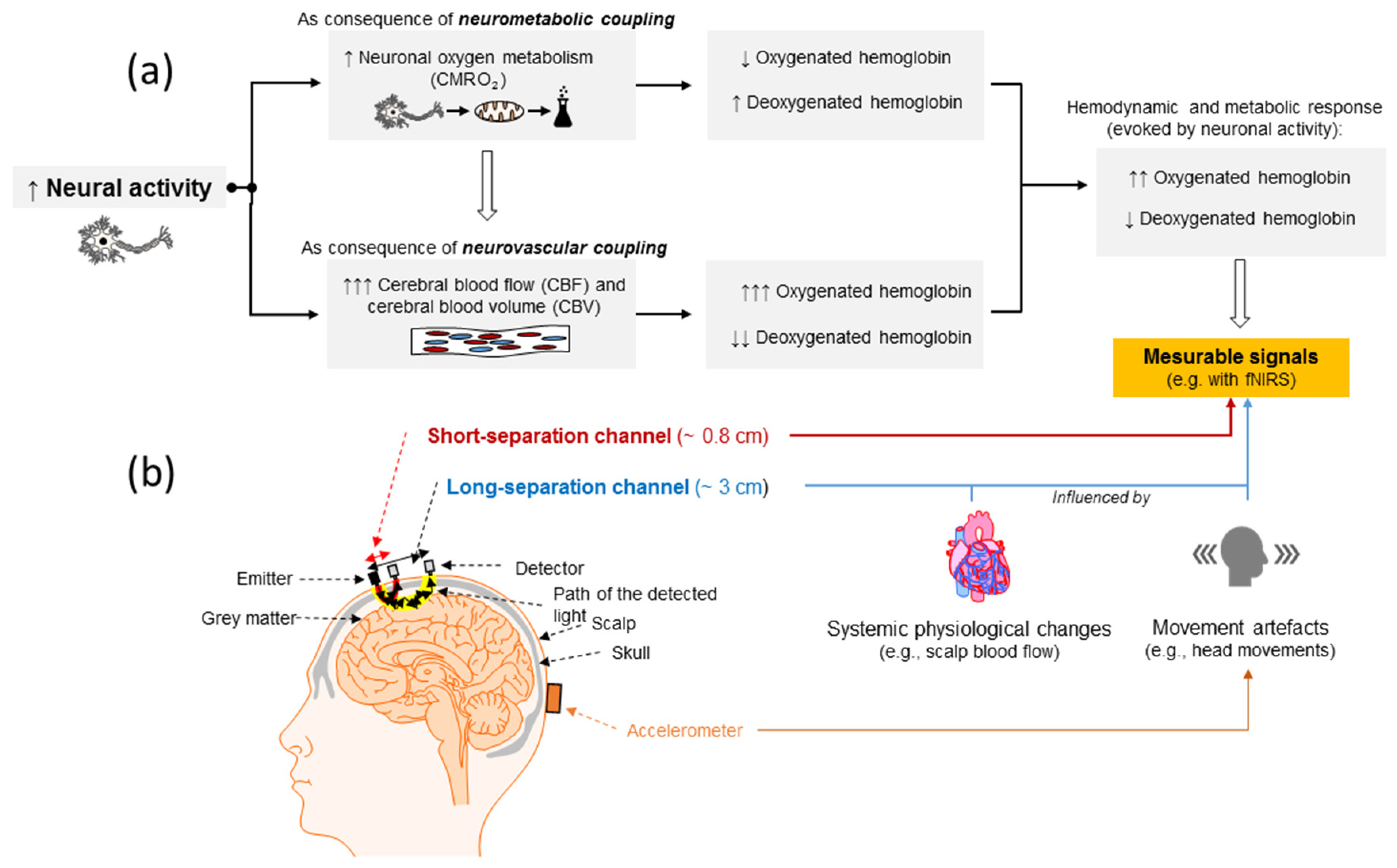
- In CW-NIRS devices, the changes in light intensity (i.e., attenuation) are used to calculate the relative concentration changes in chromophores (e.g., oxyHb and deoxyHb) [98,171,174,180,181]. Light with a distinct intensity is emitted into the tissue (e.g., brain and scalp tissue) via an emitter (e.g., placed on the scalp) and the non-absorbed light components leaving the tissue at a distinct point are measured via a detector, from which the intensity of outgoing light is obtained. By using CW-NIRS, changes in the attenuation coefficient can be calculated and used to determine the relative concentration changes in the chromophores (e.g., relative to baseline) [171,182].
- (ii)
- SRS-NIRS is a special type of CW-NIRS. In SRS-NIRS, at least two detectors (e.g., placed on the head surface) are used to measure the light which leaves the examined tissue after traveling through it (e.g., brain and scalp tissue) [183]. The information of the two detectors is used to determine the local gradients of light attenuation which, in turn, can be used to calculate the absolute concentration changes in chromophores (e.g., oxyHb and deoxyHb) and the tissue oxygenation index (TOI) [184,185]. TOI is the ratio of oxyHb to total hemoglobin (sum of oxy- and deoxyHb) and is also known as the tissue saturation index (TSI) and regional tissue oxygen saturation (StO2, and rSO2).
- (iii)
- In FD-NIRS, the source(s) (e.g., placed on the scalp) continuously emit(s) light with a distinct intensity into the tissue, whose amplitude is modulated at a specific frequency in the MHz range. A detector (e.g., placed on the head’s surface) measures the phase shift (delay) and light attenuation of the measured and non-absorbed light components, which, in turn, are used to determine the absorption and scattering properties of the specific tissue (e.g., brain tissue). Using the individual-specific information about the scattering and absorption properties of the distinct tissue allows the quantification of absolute concentration changes in the chromophores (e.g., oxyHb and deoxyHb) [98,171,174,180,181,186,187].
- (iv)
- In TD-NIRS, multiple sources emit extremely short light impulses into the tissue (e.g., brain and scalp tissue) and detectors, placed at a certain distance from the light emitting source, quantify the time of flight, the temporal distribution, and the shape of the temporal distribution of the non-absorbed photons, which leave the examined tissue (e.g., brain and scalp tissue). The information about the time of flight, temporal distribution, and shape of the temporal distribution of the non-absorbed photons are used to determine the scattering and absorption properties of the distinct tissue (e.g., brain tissue). In general, photons that travelled through the cerebral tissue are more delayed than photons that are only traveling through the scalp. The obtained information about scattering and absorption enables the calculation of absolute concentrations changes in chromophores (e.g., oxyHb and deoxyHb) [98,171,174,180,181,186,187].
4. Advantages of Brain-Derived Indicators of Internal Load
- (a)
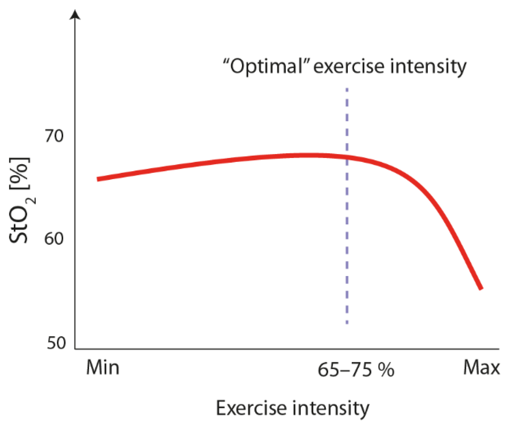
- cortical hemodynamics are sensitive to the level of physical load (e.g., exercise intensity) [125,126,128,139] and a decline in prefrontal oxygenation (i.e., oxyHb and StO2/TOI) at very high exercise intensities was observed [126,158,164,197,198]. The latter corroborates the notion of a “central governor” limiting maximal exercise performance [198,199,200,201,202,203,204,205],
- (b)
- (c)
- cortical hemodynamics during physical exercise are indicators of responsiveness, as levels of oxyHb in the right ventrolateral prefrontal cortex (PFC) during exercise are higher in individuals who show superior performance in a spatial memory task (e.g., being responders) after an acute bout of physical exercise [207],
- (d)
- the level of cortical hemodynamics during physical exercise might act as an indicator of the optimal brain state since lower levels of oxygenated hemoglobin in the PFC during physical exercise were associated with slower reaction times in an executive function task (i.e., Stroop task [208].
- (e)
- (f)
- (g)
- (h)
- (i)
5. Practical Implementation of Brain-Derived Parameters to Prescribe Physical Exercise
5.1. How Can We Prescribe Exercise Intensity by Using fNIRS-Derived Brain Parameters?
5.2. Which Cortical Brain Area Should Be Targeted?
- (a)
- Regarding the ventrolateral PFC; it was observed that young adults with superior performance in a spatial memory tasks in response to an acute bout of endurance exercise (i.e., responders) exhibited higher levels of oxyHb in the right ventrolateral PFC during the exercise [207].
- (b)
- (c)
- Regarding the FPA; it was observed that in young adults, higher levels of oxyHb in the left FPA (measured during a cognitive test after exercising) [227] were associated with exercise-induced behavioral changes in the performance of the Stroop test. Similar findings have been observed for older adults regarding the right FPA [229].
5.3. How Can We Minimize Confounders in Order to Successfully Apply fNIRS during Physical Exercise?
5.4. Which Additional Internal Load Indicators Should Be Recorded Alongside fNIRS?
6. Summary and Conclusion
Author Contributions
Funding
Acknowledgments
Conflicts of Interest
References
- Hillman, C.H.; Erickson, K.I.; Kramer, A.F. Be smart, exercise your heart: Exercise effects on brain and cognition. Nat. Rev. Neurosci. 2008, 9, 58–65. [Google Scholar] [CrossRef] [PubMed]
- Liu-Ambrose, T.; Barha, C.; Best, J.R. Physical activity for brain health in older adults. Appl. Physiol. Nutr. Metab. 2018, 43, 1105–1112. [Google Scholar] [CrossRef] [PubMed]
- Erickson, K.I.; Hillman, C.; Stillman, C.M.; Ballard, R.M.; Bloodgood, B.; Conroy, D.E.; Macko, R.; Marquez, D.X.; Petruzzello, S.J.; Powell, K.E.; et al. Physical Activity, Cognition, and Brain Outcomes. Med. Sci. Sports Exerc. 2019, 51, 1242–1251. [Google Scholar] [CrossRef] [PubMed]
- Jackson, P.; Pialoux, V.; Corbett, D.; Drogos, L.; Erickson, K.I.; Eskes, G.A.; Poulin, M.J. Promoting brain health through exercise and diet in older adults: A physiological perspective. J. Physiol. 2016, 594, 4485–4498. [Google Scholar] [CrossRef]
- Tyndall, A.V.; Clark, C.M.; Anderson, T.J.; Hogan, D.B.; Hill, M.D.; Longman, R.S.; Poulin, M.J. Protective Effects of Exercise on Cognition and Brain Health in Older Adults. Exerc. Sport Sci. Rev. 2018, 46, 215–223. [Google Scholar] [CrossRef]
- Liu-Ambrose, T.; Best, J.R. Exercise is Medicine for the Aging Brain. Kinesiol. Rev. 2017, 6, 22–29. [Google Scholar] [CrossRef]
- Stillman, C.M.; Esteban-Cornejo, I.; Brown, B.; Bender, C.M.; Erickson, K.I. Effects of Exercise on Brain and Cognition Across Age Groups and Health States. Trends Neurosci. 2020. [Google Scholar] [CrossRef] [PubMed]
- Esteban-Cornejo, I.; Tejero-González, C.M.; Sallis, J.F.; Veiga, O.L. Physical activity and cognition in adolescents: A systematic review. J. Sci. Med. Sport 2015, 18, 534–539. [Google Scholar] [CrossRef]
- Greeff, J.W.D.; Bosker, R.J.; Oosterlaan, J.; Visscher, C.; Hartman, E. Effects of physical activity on executive functions, attention and academic performance in preadolescent children: A meta-analysis. J. Sci. Med. Sport 2018, 21, 501–507. [Google Scholar] [CrossRef]
- Hillman, C.H.; Logan, N.E.; Shigeta, T.T. A Review of Acute Physical Activity Effects on Brain and Cognition in Children. Transl. J. Acsm 2019, 17, 132–136. [Google Scholar] [CrossRef]
- Ludyga, S.; Gerber, M.; Pühse, U.; Looser, V.N.; Kamijo, K. Systematic review and meta-analysis investigating moderators of long-term effects of exercise on cognition in healthy individuals. Nat. Hum. Behav. 2020, 1–10. [Google Scholar] [CrossRef] [PubMed]
- Stillman, C.M.; Cohen, J.; Lehman, M.E.; Erickson, K.I. Mediators of Physical Activity on Neurocognitive Function: A Review at Multiple Levels of Analysis. Front. Hum. Neurosci. 2016, 10, 626. [Google Scholar] [CrossRef] [Green Version]
- Stimpson, N.J.; Davison, G.; Javadi, A.-H. Joggin’ the Noggin: Towards a Physiological Understanding of Exercise-Induced Cognitive Benefits. Neurosci. Biobehav. Rev. 2018, 88, 177–186. [Google Scholar] [CrossRef] [PubMed]
- Herold, F.; Hamacher, D.; Schega, L.; Müller, N.G. Thinking While Moving or Moving While Thinking—Concepts of Motor-Cognitive Training for Cognitive Performance Enhancement. Front. Aging Neurosci. 2018, 10, 228. [Google Scholar] [CrossRef] [Green Version]
- Herold, F.; Müller, P.; Gronwald, T.; Müller, N.G. Dose-Response Matters!—A Perspective on the Exercise Prescription in Exercise-Cognition Research. Front. Psychol. 2019, 10, 2338. [Google Scholar] [CrossRef] [Green Version]
- Rolland, Y.; Van Kan, G.A.; Vellas, B. Healthy Brain Aging: Role of Exercise and Physical Activity. Clin. Geriatr. Med. 2010, 26, 75–87. [Google Scholar] [CrossRef]
- Bherer, L.; Erickson, K.I.; Liu-Ambrose, T. A Review of the Effects of Physical Activity and Exercise on Cognitive and Brain Functions in Older Adults. J. Aging Res. 2013, 2013, 1–8. [Google Scholar] [CrossRef] [Green Version]
- Voelcker-Rehage, C.; Niemann, C. Structural and functional brain changes related to different types of physical activity across the life span. Neurosci. Biobehav. Rev. 2013, 37, 2268–2295. [Google Scholar] [CrossRef]
- Lauenroth, A.; Ioannidis, A.E.; Teichmann, B. Influence of combined physical and cognitive training on cognition: A systematic review. BMC Geriatr. 2016, 16, 141. [Google Scholar] [CrossRef] [Green Version]
- Cai, Y.; Abrahamson, K. Does Exercise Impact Cognitive Performance Research Article Op in Community-dwelling Older Adults with Mild Cognitive Impairment? A Systematic Review. Qual. Prim. Care 2015, 23, 214–222. [Google Scholar]
- Tait, J.; Duckham, R.L.; Milte, C.M.; Main, L.C.; Daly, R.M. Influence of Sequential vs. Simultaneous Dual-Task Exercise Training on Cognitive Function in Older Adults. Front. Aging Neurosci. 2017, 9, 368. [Google Scholar] [CrossRef]
- Herold, F.; Törpel, A.; Schega, L.; Müller, N.G. Functional and/or structural brain changes in response to resistance exercises and resistance training lead to cognitive improvements—Asystematic review. Eur. Rev. Aging Phys. Act. 2019, 16, 10. [Google Scholar] [CrossRef]
- Soga, K.; Masaki, H.; Gerber, M.; Ludyga, S. Acute and Long-term Effects of Resistance Training on Executive Function. J. Cogn. Enhanc. 2018, 2, 200–207. [Google Scholar] [CrossRef]
- Wollesen, B.; Voelcker-Rehage, C. Training effects on motor–cognitive dual-task performance in older adults. Eur. Rev. Aging Phys. Act. 2013, 11, 5–24. [Google Scholar] [CrossRef] [Green Version]
- Gronwald, T.; Budde, H. Commentary: Physical Exercise as Personalized Medicine for Dementia Prevention? Front. Physiol. 2019, 10, 1358. [Google Scholar] [CrossRef] [PubMed] [Green Version]
- Hofmann, P.; Tschakert, G. Special Needs to Prescribe Exercise Intensity for Scientific Studies. Cardiol. Res. Pract. 2010, 2011, 1–10. [Google Scholar] [CrossRef] [PubMed] [Green Version]
- Mann, T.N.; Lamberts, R.P.; Lambert, M. Methods of Prescribing Relative Exercise Intensity: Physiological and Practical Considerations. Sports Med. 2013, 43, 613–625. [Google Scholar] [CrossRef] [PubMed]
- Gass, G.C.; McLellan, T.M.; Gass, E.M. Effects of prolonged exercise at a similar percentage of maximal oxygen consumption in trained and untrained subjects. Eur. J. Appl. Physiol. Occup. Physiol. 1991, 63, 430–435. [Google Scholar] [CrossRef]
- Katch, V.; Weltman, A.; Sady, S.; Freedson, P. Validity of the relative percent concept for equating training intensity. Eur. J. Appl. Physiol. Occup. Physiol. 1978, 39, 219–227. [Google Scholar] [CrossRef]
- Meyer, T.; Gabriel, H.H.; Kindermann, W. Is determination of exercise intensities as percentages of VO2max or HRmax adequate? Med. Sci. Sports Exerc. 1999, 31, 1342–1345. [Google Scholar] [CrossRef]
- Scharhag-Rosenberger, F.; Meyer, T.; Gäßler, N.; Faude, O.; Kindermann, W. Exercise at given percentages of VO2max: Heterogeneous metabolic responses between individuals. J. Sci. Med. Sport 2010, 13, 74–79. [Google Scholar] [CrossRef]
- Weltman, A.; Weltman, J.; Rutt, R.; Seip, R.; Levine, S.; Snead, D.; Kaiser, D.; Rogol, A. Percentages of Maximal Heart Rate, Heart Rate Reserve, and VO2peak for Determining Endurance Training Intensity in Sedentary Women*. Int. J. Sports Med. 1989, 10, 212–216. [Google Scholar] [CrossRef] [PubMed]
- Weltman, A.; Snead, D.; Seip, R.; Schurrer, R.; Weltman, J.; Rutt, R.; Rogol, A. Percentages of Maximal Heart Rate, Heart Rate Reserve and VO 2 max for Determining Endurance Training Intensity in Male Runners. Int. J. Sports Med. 1990, 11, 218–222. [Google Scholar] [CrossRef]
- Weatherwax, R.; Harris, N.; Kilding, A.E.; Dalleck, L. The incidence of training responsiveness to cardiorespiratory fitness and cardiometabolic measurements following individualized and standardized exercise prescription: Study protocol for a randomized controlled trial. Trials 2016, 17, 601. [Google Scholar] [CrossRef] [Green Version]
- Tschakert, G.; Hofmann, P. High-Intensity Intermittent Exercise: Methodological and Physiological Aspects. Int. J. Sports Physiol. Perform. 2013, 8, 600–610. [Google Scholar] [CrossRef] [PubMed] [Green Version]
- Schneider, J.; Schlüter, K.; Sprave, T.; Wiskemann, J.; Rosenberger, F. Exercise intensity prescription in cancer survivors: Ventilatory and lactate thresholds are useful submaximal alternatives to VO2peak. Support. Care Cancer 2020, 1–8. [Google Scholar] [CrossRef] [Green Version]
- Gronwald, T.; Velasques, B.; Ribeiro, P.; Machado, S.; Murillo-Rodríguez, E.; Ludyga, S.; Yamamoto, T.; Budde, H. Increasing exercise’s effect on mental health: Exercise intensity does matter. Proc. Natl. Acad. Sci. USA 2018, 115, E11890–E11891. [Google Scholar] [CrossRef] [Green Version]
- Suwabe, K.; Byun, K.; Hyodo, K.; Reagh, Z.M.; Roberts, J.M.; Matsushita, A.; Saotome, K.; Ochi, G.; Fukuie, T.; Suzuki, K.; et al. Reply to Gronwald et al.: Exercise intensity does indeed matter; maximal oxygen uptake is the gold-standard indicator. Proc. Natl. Acad. Sci. USA 2018, 115, E11892–E11893. [Google Scholar] [CrossRef] [Green Version]
- Gronwald, T.; Alves, A.C.D.B.; Murillo-Rodríguez, E.; Latini, A.; Schuette, J.; Budde, H. Standardization of exercise intensity and consideration of a dose-response is essential. Commentary on “Exercise-linked FNDC5/irisin rescues synaptic plasticity and memory defects in Alzheimer’s models”, by Lourenco et al., published 2019 in Nature Medicine. J. Sport Health Sci. 2019, 8, 353–354. [Google Scholar] [CrossRef]
- Burgess, D.J. The Research Doesn’t Always Apply: Practical Solutions to Evidence-Based Training-Load Monitoring in Elite Team Sports. Int. J. Sports Physiol. Perform. 2017, 12, S2136–S2141. [Google Scholar] [CrossRef]
- Bourdon, P.C.; Cardinale, M.; Murray, A.; Gastin, P.; Kellmann, M.; Varley, M.; Gabbett, T.J.; Coutts, A.J.; Burgess, D.J.; Gregson, W.; et al. Monitoring Athlete Training Loads: Consensus Statement. Int. J. Sports Physiol. Perform. 2017, 12, S2161–S2170. [Google Scholar] [CrossRef] [PubMed]
- McLaren, S.J.; MacPherson, T.; Coutts, A.J.; Hurst, C.; Spears, I.R.; Weston, M. The Relationships Between Internal and External Measures of Training Load and Intensity in Team Sports: A Meta-Analysis. Sports Med. 2017, 48, 641–658. [Google Scholar] [CrossRef] [Green Version]
- Vanrenterghem, J.; Nedergaard, N.J.; Robinson, M.A.; Drust, B. Training Load Monitoring in Team Sports: A Novel Framework Separating Physiological and Biomechanical Load-Adaptation Pathways. Sports Med. 2017, 47, 2135–2142. [Google Scholar] [CrossRef] [PubMed]
- Wallace, L.K.; Slattery, K.; Coutts, A.J. The Ecological Validity and Application of the Session-RPE Method for Quantifying Training Loads in Swimming. J. Strength Cond. Res. 2009, 23, 33–38. [Google Scholar] [CrossRef] [PubMed] [Green Version]
- Halson, S. Monitoring training load to understand fatigue in athletes. Sports Med. 2014, 44, S139–S147. [Google Scholar] [CrossRef] [Green Version]
- Impellizzeri, F.M.; Marcora, S.M.; Coutts, A.J. Internal and External Training Load: 15 Years On. Int. J. Sports Physiol. Perform. 2019, 14, 270–273. [Google Scholar] [CrossRef]
- Soligard, T.; Schwellnus, M.; Alonso, J.-M.; Bahr, R.; Clarsen, B.; Dijkstra, H.P.; Gabbett, T.; Gleeson, M.; Hägglund, M.; Hutchinson, M.R.; et al. How much is too much? (Part 1) International Olympic Committee consensus statement on load in sport and risk of injury. Br. J. Sports Med. 2016, 50, 1030–1041. [Google Scholar] [CrossRef] [Green Version]
- Maslov, P.Z.; Schulman, A.; Lavie, C.J.; Narula, J. Personalized exercise dose prescription. Eur. Heart J. 2017, 39, 2346–2355. [Google Scholar] [CrossRef] [Green Version]
- Pickering, C.; Kiely, J. Do Non-Responders to Exercise Exist—And If So, What Should We Do About Them? Sports Med. 2018, 49, 1–7. [Google Scholar] [CrossRef] [Green Version]
- Perrey, S.; Besson, P. Studying brain activity in sports performance: Contributions and issues. Prog. Brain Res. 2018, 240, 247–267. [Google Scholar] [CrossRef]
- Tan, S.J.; Kerr, G.; Sullivan, J.P.; Peake, J.M. A Brief Review of the Application of Neuroergonomics in Skilled Cognition During Expert Sports Performance. Front. Hum. Neurosci. 2019, 13, 278. [Google Scholar] [CrossRef] [PubMed]
- Wang, C.-H.; Moreau, D.; Kao, S.-C. From the Lab to the Field: Potential Applications of Dry EEG Systems to Understand the Brain-Behavior Relationship in Sports. Front. Mol. Neurosci. 2019, 13, 893. [Google Scholar] [CrossRef] [PubMed] [Green Version]
- Peake, J.M.; Kerr, G.; Sullivan, J.P. A Critical Review of Consumer Wearables, Mobile Applications, and Equipment for Providing Biofeedback, Monitoring Stress, and Sleep in Physically Active Populations. Front. Physiol. 2018, 9, 743. [Google Scholar] [CrossRef] [PubMed]
- Cohen, M.X. Where Does EEG Come From and What Does It Mean? Trends Neurosci. 2017, 40, 208–218. [Google Scholar] [CrossRef]
- Yucel, M.; Selb, J.J.; Huppert, T.J.; Franceschini, M.A.; Boas, D. Functional Near Infrared Spectroscopy: Enabling routine functional brain imaging. Curr. Opin. Biomed. Eng. 2017, 4, 78–86. [Google Scholar] [CrossRef]
- Rahman, M.; Karwowski, W.; Fafrowicz, M.; Hancock, P.A. Neuroergonomics Applications of Electroencephalography in Physical Activities: A Systematic Review. Front. Hum. Neurosci. 2019, 13, 182. [Google Scholar] [CrossRef] [Green Version]
- Hülsdünker, T.; Mierau, A.; Neeb, C.; Kleinöder, H.; Strüder, H. Cortical processes associated with continuous balance control as revealed by EEG spectral power. Neurosci. Lett. 2015, 592, 1–5. [Google Scholar] [CrossRef]
- Collado-Mateo, D.; Adsuar, J.C.; Olivares, P.R.; Cano-Plasencia, R.; Gusi, N. Using a dry electrode EEG device during balance tasks in healthy young-adult males: Test-retest reliability analysis. Somat. Mot. Res. 2015, 32, 219–226. [Google Scholar] [CrossRef]
- Slobounov, S.; Sebastianelli, W.; Hallett, M. Residual brain dysfunction observed one year post-mild traumatic brain injury: Combined EEG and balance study. Clin. Neurophysiol. 2012, 123, 1755–1761. [Google Scholar] [CrossRef] [Green Version]
- Choi, W.; Lee, S.; Park, J. EEG-biofeedback Intervention Improves balance in Stroke Survivor. Indian J. Sci. Technol. 2015, 8, 8. [Google Scholar] [CrossRef]
- Hülsdünker, T.; Mierau, A.; Strüder, H.K. Higher Balance Task Demands are Associated with an Increase in Individual Alpha Peak Frequency. Front. Hum. Neurosci. 2016, 9, 85. [Google Scholar] [CrossRef] [PubMed] [Green Version]
- Mierau, A.; Hülsdünker, T.; Strüder, H.K. Changes in cortical activity associated with adaptive behavior during repeated balance perturbation of unpredictable timing. Front. Behav. Neurosci. 2015, 9, 233. [Google Scholar] [CrossRef] [PubMed] [Green Version]
- Mierau, A.; Pester, B.; Hülsdünker, T.; Schiecke, K.; Strüder, H.K.; Witte, H. Cortical Correlates of Human Balance Control. Brain Topogr. 2017, 30, 434–446. [Google Scholar] [CrossRef] [PubMed] [Green Version]
- Beurskens, R.; Steinberg, F.; Antoniewicz, F.; Wolff, W.; Granacher, U. Neural Correlates of Dual-Task Walking: Effects of Cognitive versus Motor Interference in Young Adults. Neural Plast. 2016, 2016, 1–9. [Google Scholar] [CrossRef] [PubMed] [Green Version]
- Bruijn, S.; Van Dieën, J.; Daffertshofer, A. Beta activity in the premotor cortex is increased during stabilized as compared to normal walking. Front. Hum. Neurosci. 2015, 9, 321. [Google Scholar] [CrossRef] [PubMed] [Green Version]
- Castermans, T.; Duvinage, M.; Cheron, G.; Dutoit, T. About the cortical origin of the low-delta and high-gamma rhythms observed in EEG signals during treadmill walking. Neurosci. Lett. 2014, 561, 166–170. [Google Scholar] [CrossRef] [Green Version]
- Nordin, A.D.; Hairston, W.D.; Ferris, D.P. Human electrocortical dynamics while stepping over obstacles. Sci. Rep. 2019, 9, 4693. [Google Scholar] [CrossRef]
- Oliveira, A.S.; Schlink, B.R.; Hairston, W.D.; Konig, P.; Ferris, D.P. Restricted vision increases sensorimotor cortex involvement in human walking. J. Neurophysiol. 2017, 118, 1943–1951. [Google Scholar] [CrossRef]
- Peterson, S.M.; Ferris, D.P. Differentiation in Theta and Beta Electrocortical Activity between Visual and Physical Perturbations to Walking and Standing Balance. eNeuro 2018, 5, 207–218. [Google Scholar] [CrossRef] [Green Version]
- Flanagan, S.D.; Dunn-Lewis, C.; Comstock, B.A.; Maresh, C.M.; Volek, J.S.; Denegar, C.R.; Kraemer, W.J. Cortical Activity during a Highly-Trained Resistance Exercise Movement Emphasizing Force, Power or Volume. Brain Sci. 2012, 2, 649–666. [Google Scholar] [CrossRef]
- Falvo, M.J.; Sirevaag, E.J.; Rohrbaugh, J.W.; Earhart, G.M. Resistance training induces supraspinal adaptations: Evidence from movement-related cortical potentials. Eur. J. Appl. Physiol. 2010, 109, 923–933. [Google Scholar] [CrossRef] [PubMed] [Green Version]
- Kenville, R.; Maudrich, T.; Vidaurre, C.; Maudrich, D.; Villringer, A.; Nikulin, V.V.; Ragert, P. Corticomuscular interactions during different movement periods in a multi-joint compound movement. Sci. Rep. 2020, 10, 1–13. [Google Scholar] [CrossRef] [PubMed] [Green Version]
- Gronwald, T.; Ludyga, S.; Hottenrott, K. Einfluss einer intensiven Intervallbelastung auf die Beanspruchung der kortikalen Gehirnaktivität. Schweiz. Z. Sportmed. Sporttraumatol. 2015, 63, 23–28. [Google Scholar]
- Gronwald, T.; Ludyga, S.; Hottenrott, K. Gehirnaktivität bei identischer Belastung—Eine standardisierte fahrradergometrische Laborstudie unter Normoxie und normbarer Hypoxie. Leistungssport 2015, 45, 42–47. [Google Scholar]
- Ludyga, S.; Hottenrott, K.; Gronwald, T. Einfluss verschiedener Belastungssituationen auf die EEG-Aktivität. Dtsch. Z. Sportmed. 2015, 2015, 113–120. [Google Scholar] [CrossRef]
- Ludyga, S.; Gronwald, T.; Hottenrott, K. Do Male and Female Cyclists’ Cortical Activity Differ Before and During Cycling Exercise? J. Sport Exerc. Psychol. 2015, 37, 617–625. [Google Scholar] [CrossRef]
- Bullock, T.; Cecotti, H.; Giesbrecht, B. Multiple stages of information processing are modulated during acute bouts of exercise. Neuroscience 2015, 307, 138–150. [Google Scholar] [CrossRef]
- Olson, R.; Chang, Y.; Brush, C.; Kwok, A.N.; Gordon, V.X.; Alderman, B. Neurophysiological and behavioral correlates of cognitive control during low and moderate intensity exercise. NeuroImage 2016, 131, 171–180. [Google Scholar] [CrossRef]
- Storzer, L.; Butz, M.; Hirschmann, J.; Abbasi, O.; Gratkowski, M.; Saupe, D.; Schnitzler, A.; Dalal, S. Bicycling and Walking are Associated with Different Cortical Oscillatory Dynamics. Front. Hum. Neurosci. 2016, 10, 29. [Google Scholar] [CrossRef] [Green Version]
- Thompson, T.; Steffert, T.; Ros, T.; Leach, J.; Gruzelier, J. EEG applications for sport and performance. Methods 2008, 45, 279–288. [Google Scholar] [CrossRef]
- Lloyd-Fox, S.; Blasi, A.; Elwell, C. Illuminating the developing brain: The past, present and future of functional near infrared spectroscopy. Neurosci. Biobehav. Rev. 2010, 34, 269–284. [Google Scholar] [CrossRef]
- Park, J.; Fairweather, M.M.; Donaldson, D. Making the case for mobile cognition: EEG and sports performance. Neurosci. Biobehav. Rev. 2015, 52, 117–130. [Google Scholar] [CrossRef] [PubMed] [Green Version]
- Pinti, P.; Tachtsidis, I.; Hamilton, A.; Hirsch, J.; Aichelburg, C.; Gilbert, S.; Burgess, P.W.; Pinti, P. The present and future use of functional near-infrared spectroscopy (fNIRS) for cognitive neuroscience. Ann. N. Y. Acad. Sci. 2020, 1464, 5–29. [Google Scholar] [CrossRef] [PubMed]
- Soltanlou, M.; Sitnikova, M.A.; Nuerk, H.-C.; Dresler, T. Applications of Functional Near-Infrared Spectroscopy (fNIRS) in Studying Cognitive Development: The Case of Mathematics and Language. Front. Psychol. 2018, 9, 277. [Google Scholar] [CrossRef] [PubMed]
- Burle, B.; Spieser, L.; Roger, C.; Casini, L.; Hasbroucq, T.; Vidal, F. Spatial and temporal resolutions of EEG: Is it really black and white? A scalp current density view. Int. J. Psychophysiol. 2015, 97, 210–220. [Google Scholar] [CrossRef] [PubMed]
- Cutini, S.; Brigadoi, S. Unleashing the future potential of functional near-infrared spectroscopy in brain sciences. J. Neurosci. Methods 2014, 232, 152–156. [Google Scholar] [CrossRef] [PubMed]
- A Ward, J.; Pinti, P.; Amft, O.; Van Laerhoven, K. Wearables and the Brain. IEEE Pervasive Comput. 2019, 18, 94–100. [Google Scholar] [CrossRef]
- Maskeliūnas, R.; Damaševičius, R.; Martisius, I.; Vasiljevas, M. Consumer grade EEG devices: Are they usable for control tasks? PeerJ 2016, 4, e1746. [Google Scholar] [CrossRef]
- Pontifex, M.B.; Hillman, C.H. Neuroelectric measurement of cognition during aerobic exercise. Methods 2008, 45, 271–278. [Google Scholar] [CrossRef]
- Symeonidou, E.-R.; Nordin, A.D.; Hairston, W.D.; Ferris, D.P. Effects of Cable Sway, Electrode Surface Area, and Electrode Mass on Electroencephalography Signal Quality during Motion. Sensors 2018, 18, 1073. [Google Scholar] [CrossRef] [Green Version]
- Smith, M. Shedding light on the adult brain: A review of the clinical applications of near-infrared spectroscopy. Philos. Trans. R. Soc. A Math. Phys. Eng. Sci. 2011, 369, 4452–4469. [Google Scholar] [CrossRef] [PubMed]
- Ekkekakis, P. Illuminating the Black Box: Investigating Prefrontal Cortical Hemodynamics during Exercise with Near-Infrared Spectroscopy. J. Sport Exerc. Psychol. 2009, 31, 505–553. [Google Scholar] [CrossRef] [PubMed] [Green Version]
- Perrey, S. Non-invasive NIR spectroscopy of human brain function during exercise. Methods 2008, 45, 289–299. [Google Scholar] [CrossRef]
- Scarapicchia, V.; Brown, C.; Mayo, C.; Gawryluk, J. Functional Magnetic Resonance Imaging and Functional Near-Infrared Spectroscopy: Insights from Combined Recording Studies. Front. Hum. Neurosci. 2017, 11, 419. [Google Scholar] [CrossRef]
- Quaresima, V.; Ferrari, M. Functional Near-Infrared Spectroscopy (fNIRS) for Assessing Cerebral Cortex Function During Human Behavior in Natural/Social Situations: A Concise Review. Organ. Res. Methods 2016, 22, 46–68. [Google Scholar] [CrossRef]
- Pinti, P.; Aichelburg, C.; Gilbert, S.; Hamilton, A.; Hirsch, J.; Burgess, P.; Tachtsidis, I. A Review on the Use of Wearable Functional Near-Infrared Spectroscopy in Naturalistic Environments. Jpn. Psychol. Res. 2018, 60, 347–373. [Google Scholar] [CrossRef] [Green Version]
- Zhu, Y.; Rodriguez-Paras, C.; Rhee, J.; Mehta, R.K. Methodological Approaches and Recommendations for Functional Near-Infrared Spectroscopy Applications in HF/E Research. Hum. Factors 2019, 62, 613–642. [Google Scholar] [CrossRef]
- Gervain, J.; Mehler, J.; Werker, J.F.; Nelson, C.A.; Csibra, G.; Lloyd-Fox, S.; Shukla, M.; Aslin, R.N. Near-infrared spectroscopy: A report from the McDonnell infant methodology consortium. Dev. Cogn. Neurosci. 2011, 1, 22–46. [Google Scholar] [CrossRef] [Green Version]
- Tachtsidis, I.; Scholkmann, F. False positives and false negatives in functional near-infrared spectroscopy: Issues, challenges, and the way forward. Neurophotonics 2016, 3, 30401. [Google Scholar] [CrossRef]
- Kirilina, E.; Jelzow, A.; Heine, A.; Niessing, M.; Wabnitz, H.; Brühl, R.; Ittermann, B.; Jacobs, A.M.; Tachtsidis, I. The physiological origin of task-evoked systemic artefacts in functional near infrared spectroscopy. NeuroImage 2012, 61, 70–81. [Google Scholar] [CrossRef] [PubMed] [Green Version]
- Caldwell, M.; Scholkmann, F.; Wolf, U.; Wolf, M.; Elwell, C.; Tachtsidis, I. Modelling confounding effects from extracerebral contamination and systemic factors on functional near-infrared spectroscopy. NeuroImage 2016, 143, 91–105. [Google Scholar] [CrossRef] [PubMed] [Green Version]
- Kozlová, S. The Use of Near-Infrared Spectroscopy in the Sport-Scientific Context. J. Neurol. Neurol. Disord. 2018, 4, 1. [Google Scholar] [CrossRef] [Green Version]
- Seidel-Marzi, O.; Ragert, P. Neurodiagnostics in Sports: Investigating the Athlete’s Brain to Augment Performance and Sport-Specific Skills. Front. Hum. Neurosci. 2020, 14, 133. [Google Scholar] [CrossRef] [PubMed] [Green Version]
- Carius, D.; Andrä, C.; Clauß, M.; Ragert, P.; Bunk, M.; Mehnert, J. Hemodynamic Response Alteration As a Function of Task Complexity and Expertise—An fNIRS Study in Jugglers. Front. Hum. Neurosci. 2016, 10, 1. [Google Scholar] [CrossRef] [Green Version]
- Seidel, O.; Carius, D.; Kenville, R.; Ragert, P. Motor learning in a complex balance task and associated neuroplasticity: A comparison between endurance athletes and nonathletes. J. Neurophysiol. 2017, 118, 1849–1860. [Google Scholar] [CrossRef]
- Herold, F.; Orlowski, K.; Börmel, S.; Müller, N.G. Cortical activation during balancing on a balance board. Hum. Mov. Sci. 2017, 51, 51–58. [Google Scholar] [CrossRef]
- Fujimoto, H.; Mihara, M.; Hattori, N.; Hatakenaka, M.; Kawano, T.; Yagura, H.; Miyai, I.; Mochizuki, H. Cortical changes underlying balance recovery in patients with hemiplegic stroke. NeuroImage 2014, 85, 547–554. [Google Scholar] [CrossRef]
- Fujimoto, H.; Mihara, M.; Hattori, N.; Hatakenaka, M.; Yagura, H.; Kawano, T.; Miyai, I.; Mochizuki, H. Neurofeedback-induced facilitation of the supplementary motor area affects postural stability. Neurophotonics 2017, 4, 1. [Google Scholar] [CrossRef]
- Mihara, M.; Miyai, I.; Hatakenaka, M.; Kubota, K.; Sakoda, S. Role of the prefrontal cortex in human balance control. NeuroImage 2008, 43, 329–336. [Google Scholar] [CrossRef]
- Mihara, M.; Miyai, I.; Hattori, N.; Hatakenaka, M.; Yagura, H.; Kawano, T.; Kubota, K. Cortical control of postural balance in patients with hemiplegic stroke. NeuroReport 2012, 23, 314–319. [Google Scholar] [CrossRef]
- Herold, F.; Wiegel, P.; Scholkmann, F.; Thiers, A.; Hamacher, D.; Schega, L. Functional near-infrared spectroscopy in movement science: A systematic review on cortical activity in postural and walking tasks. Neurophotonics 2017, 4, 41403. [Google Scholar] [CrossRef] [Green Version]
- Vitório, R.; Stuart, S.; Rochester, L.; Alcock, L.; Pantall, A. fNIRS response during walking—Artefact or cortical activity? A systematic review. Neurosci. Biobehav. Rev. 2017, 83, 160–172. [Google Scholar] [CrossRef] [PubMed] [Green Version]
- Formenti, D.; Perpetuini, D.; Iodice, P.; Cardone, D.; Michielon, G.; Scurati, R.; Alberti, G.; Merla, A. Effects of knee extension with different speeds of movement on muscle and cerebral oxygenation. PeerJ 2018, 6, e5704. [Google Scholar] [CrossRef] [Green Version]
- Borot, L.; Vergotte, G.; Perrey, S. Different Hemodynamic Responses of the Primary Motor Cortex Accompanying Eccentric and Concentric Movements: A Functional NIRS Study. Brain Sci. 2018, 8, 75. [Google Scholar] [CrossRef] [Green Version]
- Kenville, R.; Maudrich, T.; Carius, D.; Ragert, P. Hemodynamic Response Alterations in Sensorimotor Areas as a Function of Barbell Load Levels during Squatting: An fNIRS Study. Front. Hum. Neurosci. 2017, 11, 268. [Google Scholar] [CrossRef] [PubMed] [Green Version]
- Cavuoto, L.A.; Maikala, R.V. Role of obesity on cerebral hemodynamics and cardiorespiratory responses in healthy men during repetitive incremental lifting. Eur. J. Appl. Physiol. 2015, 115, 1905–1917. [Google Scholar] [CrossRef]
- Ono, Y.; Noah, J.A.; Zhang, X.; Nomoto, Y.; Suzuki, T.; Shimada, S.; Tachibana, A.; Bronner, S.; Hirsch, J. Motor learning and modulation of prefrontal cortex: An fNIRS assessment. J. Neural Eng. 2015, 12, 66004. [Google Scholar] [CrossRef]
- Noah, J.A.; Ono, Y.; Nomoto, Y.; Shimada, S.; Tachibana, A.; Zhang, X.; Bronner, S.; Hirsch, J. fMRI Validation of fNIRS Measurements During a Naturalistic Task. J. Vis. Exp. 2015, 100, e52116. [Google Scholar] [CrossRef] [Green Version]
- Tachibana, A.; Noah, J.A.; Bronner, S.; Ono, Y.; Onozuka, M. Parietal and temporal activity during a multimodal dance video game: An fNIRS study. Neurosci. Lett. 2011, 503, 125–130. [Google Scholar] [CrossRef]
- Lu, X.; Hui-Chan, C.W.-Y.; Tsang, W.W.N. Changes of heart rate variability and prefrontal oxygenation during Tai Chi practice versus arm ergometer cycling. J. Phys. Sci. 2016, 28, 3243–3248. [Google Scholar] [CrossRef] [Green Version]
- Tsang, W.W.N.; Chan, K.; Cheng, C.N.; Hu, F.S.; Mak, C.T.; Wong, J.W. Tai Chi practice on prefrontal oxygenation levels in older adults: A pilot study. Complement. Med. 2019, 42, 132–136. [Google Scholar] [CrossRef] [PubMed]
- Carius, D.; Hörnig, L.; Ragert, P.; Kaminski, E. Characterizing cortical hemodynamic changes during climbing and its relation to climbing expertise. Neurosci. Lett. 2019, 715, 134604. [Google Scholar] [CrossRef] [PubMed]
- Jones, B.; Cooper, C.E. Near Infrared Spectroscopy (NIRS) Observation of Vastus Lateralis (Muscle) and Prefrontal Cortex (Brain) Tissue Oxygenation During Synchronised Swimming Routines in Elite Athletes. Adv. Exp. Med. Biol. 2018, 1072, 111–117. [Google Scholar] [CrossRef]
- Balardin, J.B.; Morais, G.A.Z.; Furucho, R.A.; Trambaiolli, L.; Vanzella, P.; Biazoli, C.E.; Sato, J.R. Imaging Brain Function with Functional Near-Infrared Spectroscopy in Unconstrained Environments. Front. Hum. Neurosci. 2017, 11, 258. [Google Scholar] [CrossRef] [PubMed]
- Santos-Concejero, J.; Billaut, F.; Grobler, L.; Oliván, J.; Noakes, T.D.; Tucker, R. Maintained cerebral oxygenation during maximal self-paced exercise in elite Kenyan runners. J. Appl. Physiol. 2015, 118, 156–162. [Google Scholar] [CrossRef] [PubMed] [Green Version]
- Santos-Concejero, J.; Billaut, F.; Grobler, L.; Oliván, J.; Noakes, T.D.; Tucker, R. Brain oxygenation declines in elite Kenyan runners during a maximal interval training session. Eur. J. Appl. Physiol. 2017, 117, 1017–1024. [Google Scholar] [CrossRef]
- Suzuki, M.; Miyai, I.; Ono, T.; Oda, I.; Konishi, I.; Kochiyama, T.; Kubota, K. Prefrontal and premotor cortices are involved in adapting walking and running speed on the treadmill: An optical imaging study. NeuroImage 2004, 23, 1020–1026. [Google Scholar] [CrossRef]
- Seidel, O.; Carius, D.; Roediger, J.; Rumpf, S.; Ragert, P. Changes in neurovascular coupling during cycling exercise measured by multi-distance fNIRS: A comparison between endurance athletes and physically active controls. Exp. Brain Res. 2019, 237, 2957–2972. [Google Scholar] [CrossRef] [Green Version]
- Tempest, G.; Parfitt, G. Self-reported tolerance influences prefrontal cortex hemodynamics and affective responses. Cogn. Affect. Behav. Neurosci. 2015, 16, 63–71. [Google Scholar] [CrossRef]
- Tempest, G.; Davranche, K.; Brisswalter, J.; Perrey, S.; Radel, R. The differential effects of prolonged exercise upon executive function and cerebral oxygenation. Brain Cogn. 2017, 113, 133–141. [Google Scholar] [CrossRef]
- Tempest, G.; Reiss, A.L. The Utility of Functional Near-infrared Spectroscopy for Measuring Cortical Activity during Cycling Exercise. Med. Sci. Sports Exerc. 2019, 51, 979–987. [Google Scholar] [CrossRef]
- Ando, S.; Hatamoto, Y.; Sudo, M.; Kiyonaga, A.; Tanaka, H.; Higaki, Y. The Effects of Exercise Under Hypoxia on Cognitive Function. PLoS ONE 2013, 8, e63630. [Google Scholar] [CrossRef] [PubMed] [Green Version]
- Ando, S.; Kokubu, M.; Yamada, Y.; Kimura, M. Does cerebral oxygenation affect cognitive function during exercise? Eur. J. Appl. Physiol. 2011, 111, 1973–1982. [Google Scholar] [CrossRef] [PubMed]
- Asahara, R.; Endo, K.; Liang, N.; Matsukawa, K. An increase in prefrontal oxygenation at the start of voluntary cycling exercise was observed independently of exercise effort and muscle mass. Eur. J. Appl. Physiol. 2018, 118, 1689–1702. [Google Scholar] [CrossRef] [PubMed]
- Asahara, R.; Matsukawa, K.; Ishii, K.; Liang, N.; Endo, K. The prefrontal oxygenation and ventilatory responses at start of one-legged cycling exercise have relation to central command. J. Appl. Physiol. 2016, 121, 1115–1126. [Google Scholar] [CrossRef]
- Billaut, F.; Davis, J.M.; Smith, K.; Marino, F.E.; Noakes, T.D. Cerebral oxygenation decreases but does not impair performance during self-paced, strenuous exercise. Acta Physiol. 2010, 198, 477–486. [Google Scholar] [CrossRef]
- Gayda, M.; Lapierre, G.; Dupuy, O.; Fraser, S.; Bherer, L.; Juneau, M.; Gremeaux, V.; Nigam, A. Cardiovascular and cerebral hemodynamics during exercise and recovery in obese individuals as a function of their fitness status. Physiol. Rep. 2017, 5, e13321. [Google Scholar] [CrossRef]
- Gayda, M.; Grémeaux, V.; Bherer, L.; Juneau, M.; Drigny, J.; Dupuy, O.; Lapierre, G.; Labelle, V.; Fortier, A.; Nigam, A. Cognitive function in patients with stable coronary heart disease: Related cerebrovascular and cardiovascular responses. PLoS ONE 2017, 12, e0183791. [Google Scholar] [CrossRef] [Green Version]
- Giles, G.E.; Brunyé, T.T.; Eddy, M.D.; Mahoney, C.R.; Gagnon, S.A.; Taylor, H.A.; Kanarek, R.B. Acute exercise increases oxygenated and deoxygenated hemoglobin in the prefrontal cortex. Neuroreport 2014, 25, 1320–1325. [Google Scholar] [CrossRef]
- González-Alonso, J.; Dalsgaard, M.K.; Osada, T.; Volianitis, S.; Dawson, E.A.; Yoshiga, C.C.; Secher, N.H. Brain and central haemodynamics and oxygenation during maximal exercise in humans. J. Physiol. 2004, 557, 331–342. [Google Scholar] [CrossRef]
- Jung, R.; Moser, M.; Baucsek, S.; Dern, S.; Schneider, S. Activation patterns of different brain areas during incremental exercise measured by near-infrared spectroscopy. Exp. Brain Res. 2015, 233, 1175–1180. [Google Scholar] [CrossRef] [PubMed]
- Kan, B.; Speelman, C.; Nosaka, K. Cognitive demand of eccentric versus concentric cycling and its effects on post-exercise attention and vigilance. Eur. J. Appl. Physiol. 2019, 119, 1599–1610. [Google Scholar] [CrossRef] [PubMed]
- Lin, S.-I.; Lin, P.-Y.; Chen, J.-J.J. The cortical control of cycling exercise in stroke patients: An fNIRS study. Hum. Brain Mapp. 2012, 34, 2381–2390. [Google Scholar] [CrossRef] [PubMed]
- Ohyanagi, H.; Tsubaki, A.; Morishita, S.; Obata, H.; Qin, W.; Onishi, H. Changes in the Prefrontal Cortex Oxygenation Levels During Cycling in the Supine and Upright Positions. Adv. Exp. Med. Biol. 2018, 1072, 133–137. [Google Scholar] [CrossRef]
- Quinn, K.; Billaut, F.; Bulmer, A.C.; Minahan, C.L. Cerebral oxygenation declines but does not impair peak oxygen uptake during incremental cycling in women using oral contraceptives. Eur. J. Appl. Physiol. 2018, 118, 2417–2427. [Google Scholar] [CrossRef] [PubMed]
- Radel, R.; Tempest, G.; Brisswalter, J. The long and winding road: Effects of exercise intensity and type upon sustained attention. Physiol. Behav. 2018, 195, 82–89. [Google Scholar] [CrossRef] [PubMed]
- Schmit, C.; Davranche, K.; Easthope, C.S.; Colson, S.S.; Brisswalter, J.; Radel, R. Pushing to the limits: The dynamics of cognitive control during exhausting exercise. Neuropsychologia 2015, 68, 71–81. [Google Scholar] [CrossRef] [PubMed]
- Takehara, N.; Tsubaki, A.; Yamazaki, Y.; Kanaya, C.; Sato, D.; Morishita, S.; Onishi, H. Changes in Oxyhemoglobin Concentration in the Prefrontal Cortex and Primary Motor Cortex During Low- and Moderate-Intensity Exercise on a Cycle Ergometer. Adv. Exp. Med. Biol. 2017, 977, 241–247. [Google Scholar] [CrossRef]
- Tsubaki, A.; Morishita, S.; Tokunaga, Y.; Sato, D.; Tamaki, H.; Yamazaki, Y.; Qin, W.; Onishi, H. Changes in Cerebral Oxyhaemoglobin Levels During and After a Single 20-Minute Bout of Moderate-Intensity Cycling. Adv. Exp. Med. Biol. 2018, 1072, 127–131. [Google Scholar] [CrossRef]
- Tsubaki, A.; Morishita, S.; Tokunaga, Y.; Sato, D.; Qin, W.; Kojima, S.; Onishi, H. Laterality of cortical oxygenation in the prefrontal cortex during 20 min of moderate-intensity cycling exercise: A near-infrared spectroscopy study. Ann. Phys. Rehabil. Med. 2018, 61, e460. [Google Scholar] [CrossRef]
- Tsubaki, A.; Takai, H.; Oyanagi, K.; Kojima, S.; Tokunaga, Y.; Miyaguchi, S.; Sugawara, K.; Sato, D.; Tamaki, H.; Onishi, H. Correlation Between the Cerebral Oxyhaemoglobin Signal and Physiological Signals During Cycling Exercise: A Near-Infrared Spectroscopy Study. Adv. Exp. Med. Biol. 2016, 923, 159–166. [Google Scholar] [CrossRef] [PubMed]
- Tsubaki, A.; Takehara, N.; Sato, D.; Morishita, S.; Tokunaga, Y.; Sugawara, K.; Kojima, S.; Tamaki, H.; Yamazaki, Y.; Onishi, H. Cortical Oxyhemoglobin Elevation Persists After Moderate-Intensity Cycling Exercise: A Near-Infrared Spectroscopy Study. Adv. Exp. Med. Biol. 2017, 977, 261–268. [Google Scholar] [CrossRef]
- Piper, S.K.; Krueger, A.; Koch, S.P.; Mehnert, J.; Habermehl, C.; Steinbrink, J.; Obrig, H.; Schmitz, C.H. A wearable multi-channel fNIRS system for brain imaging in freely moving subjects. NeuroImage 2013, 85, 64–71. [Google Scholar] [CrossRef] [Green Version]
- Kojima, S.; Morishita, S.; Qin, W.; Tsubaki, A. Cerebral Oxygenation Dynamics of the Prefrontal Cortex and Motor-Related Area During Cardiopulmonary Exercise Test: A Near-Infrared Spectroscopy Study. Adv. Exp. Med. Biol. 2020, 1232, 231–237. [Google Scholar] [CrossRef] [PubMed]
- Tsubaki, A.; Morishita, S.; Tokunaga, Y.; Sato, D.; Qin, W.; Kojima, S.; Onishi, H. Effect of Exercise Duration on Post-Exercise Persistence of Oxyhemoglobin Changes in the Premotor Cortex: A Near-Infrared Spectroscopy Study in Moderate-Intensity Cycling Exercise. Adv. Exp. Med. Biol. 2020, 1232, 193–199. [Google Scholar] [CrossRef] [PubMed]
- Stevens, D.; Halaki, M.; Chow, C.; O’Dwyer, N. The effects of multi-stage exercise with and without concurrent cognitive performance on cardiorespiratory and cerebral haemodynamic responses. Eur. J. Appl. Physiol. 2018, 118, 2121–2132. [Google Scholar] [CrossRef] [PubMed]
- Subudhi, A.W.; Lorenz, M.C.; Fulco, C.S.; Roach, R.C. Cerebrovascular responses to incremental exercise during hypobaric hypoxia: Effect of oxygenation on maximal performance. Am. J. Physiol. Heart Circ. Physiol. 2008, 294, H164–H171. [Google Scholar] [CrossRef]
- Subudhi, A.W.; Miramon, B.R.; Granger, M.E.; Roach, R.C. Frontal and motor cortex oxygenation during maximal exercise in normoxia and hypoxia. J. Appl. Physiol. 2009, 106, 1153–1158. [Google Scholar] [CrossRef]
- Subudhi, A.W.; Dimmen, A.C.; Roach, R.C. Effects of acute hypoxia on cerebral and muscle oxygenation during incremental exercise. J. Appl. Physiol. 2007, 103, 177–183. [Google Scholar] [CrossRef]
- Cavuoto, L.A.; Maikala, R.V. Obesity and the Role of Short Duration Submaximal Work on Cardiovascular and Cerebral Hemodynamics. PLoS ONE 2016, 11, e0153826. [Google Scholar] [CrossRef]
- Imhoff, S.; Malenfant, S.; Nadreau, É.; Poirier, P.; Bailey, D.M.; Brassard, P. Uncoupling between cerebral perfusion and oxygenation during incremental exercise in an athlete with postconcussion syndrome: A case report. Physiol. Rep. 2017, 5, e13131. [Google Scholar] [CrossRef]
- Marillier, M.; Gruet, M.; Baillieul, S.; Wuyam, B.; Tamisier, R.; Levy, P.A.; Pépin, J.-L.; Verges, S. Impaired cerebral oxygenation and exercise tolerance in patients with severe obstructive sleep apnea syndrome. Sleep Med. 2018, 51, 37–46. [Google Scholar] [CrossRef] [PubMed]
- Neary, J.P.; Roberts, A.D.W.; Leavins, N.; Harrison, M.F.; Croll, J.C.; Sexsmith, J.R. Prefrontal cortex oxygenation during incremental exercise in chronic fatigue syndrome. Clin. Physiol. Funct. Imaging 2008, 28, 364–372. [Google Scholar] [CrossRef] [PubMed]
- Seifert, T.; Rasmussen, P.; Secher, N.H.; Nielsen, H.B. Cerebral oxygenation decreases during exercise in humans with beta-adrenergic blockade. Acta Physiol. 2009, 196, 295–302. [Google Scholar] [CrossRef] [PubMed]
- Tempest, G.; Eston, R.G.; Parfitt, G. Prefrontal Cortex Haemodynamics and Affective Responses during Exercise: A Multi-Channel Near Infrared Spectroscopy Study. PLoS ONE 2014, 9, e95924. [Google Scholar] [CrossRef] [PubMed]
- Koike, A.; Itoh, H.; Oohara, R.; Hoshimoto, M.; Tajima, A.; Aizawa, T.; Fu, L.T. Cerebral oxygenation during exercise in cardiac patients. Chest 2004, 125, 182–190. [Google Scholar] [CrossRef]
- Huang, S.-C.; Chen, C.P.; Fu, T.-C.; Chen, Y.-J. Integration of Brain Tissue Saturation Monitoring in Cardiopulmonary Exercise Testing in Patients with Heart Failure. J. Vis. Exp. 2019, 152, e60289. [Google Scholar] [CrossRef]
- Chen, Y.-J.; Wang, J.-S.; Hsu, C.-C.; Lin, P.-J.; Tsai, F.-C.; Wen, M.-S.; Kuo, C.-T.; Huang, S.-C. Cerebral desaturation in heart failure: Potential prognostic value and physiologic basis. PLoS ONE 2018, 13, e0196299. [Google Scholar] [CrossRef]
- Koike, A.; Hoshimoto, M.; Tajima, A.; Nagayama, O.; Yamaguchi, K.; Goda, A.; Yamashita, T.; Sagara, K.; Itoh, H.; Aizawa, T. Critical level of cerebral oxygenation during exercise in patients with left ventricular dysfunction. Circ. J. 2006, 70, 1457–1461. [Google Scholar] [CrossRef] [Green Version]
- Liao, L.-D.; Tsytsarev, V.; Delgado-Martínez, I.; Li, M.-L.; Erzurumlu, R.; Vipin, A.; Orellana, J.; Lin, Y.-R.; Lai, H.-Y.; Chen, Y.-Y.; et al. Neurovascular coupling: In vivo optical techniques for functional brain imaging. Biomed. Eng. Online 2013, 12, 38. [Google Scholar] [CrossRef] [Green Version]
- Scholkmann, F.; Kleiser, S.; Metz, A.J.; Zimmermann, R.; Pavia, J.M.; Wolf, U.; Wolf, M. A review on continuous wave functional near-infrared spectroscopy and imaging instrumentation and methodology. NeuroImage 2014, 85, 6–27. [Google Scholar] [CrossRef] [PubMed]
- Lindauer, U.; Dirnagl, U.; Füchtemeier, M.; Böttiger, C.; Offenhauser, N.; Leithner, C.; Royl, G. Pathophysiological Interference with Neurovascular Coupling—When Imaging Based on Hemoglobin Might Go Blind. Front. Neuroenerg. 2010, 2, 2. [Google Scholar] [CrossRef] [PubMed] [Green Version]
- León-Carrión, J.; León-Domniguez, U. Functional Near-Infrared Spectroscopy (fNIRS): Principles and Neuroscientific Applications. In Neuroimaging—Methods; Bright, P., Ed.; InTech: Rijeka, Croatia, 2017; pp. 48–74. ISBN 978-953-51-0097-3. [Google Scholar]
- Scholkmann, F.; Wolf, M. Measuring brain activity using functional near infrared spectroscopy: A short review. Spectrosc. Eur. 2012, 24, 6–10. [Google Scholar]
- Nippert, A.R.; Biesecker, K.R.; Newman, E.A. Mechanisms Mediating Functional Hyperemia in the Brain. Neuroscientist 2017, 24, 73–83. [Google Scholar] [CrossRef] [PubMed]
- Herold, F.; Wiegel, P.; Scholkmann, F.; Müller, N.G. Applications of Functional Near-Infrared Spectroscopy (fNIRS) Neuroimaging in Exercise–Cognition Science: A Systematic, Methodology-Focused Review. J. Clin. Med. 2018, 7, 466. [Google Scholar] [CrossRef] [Green Version]
- Bunce, S.; Izzetoglu, M.; Izzetoglu, K.; Onaral, B.; Pourrezaei, K. Functional near-infrared spectroscopy. IEEE Eng. Med. Boil. Mag. 2006, 25, 54–62. [Google Scholar] [CrossRef]
- Izzetoglu, M.; Bunce, S.C.; Izzetoglu, K.; Onaral, B.; Pourrezaei, A.K. Functional brain imaging using near-infrared technology. IEEE Eng. Med. Boil. Mag. 2007, 26, 38–46. [Google Scholar] [CrossRef]
- Obrig, H.; Wenzel, R.; Kohl, M.; Horst, S.; Wobst, P.; Steinbrink, J.; Thomas, F.; Villringer, A. Near-infrared spectroscopy: Does it function in functional activation studies of the adult brain? Int. J. Psychophysiol. 2000, 35, 125–142. [Google Scholar] [CrossRef]
- Rupawala, M.; Dehghani, H.; Lucas, S.J.; Tino, P.; Cruse, D. Shining a Light on Awareness: A Review of Functional Near-Infrared Spectroscopy for Prolonged Disorders of Consciousness. Front. Neurol. 2018, 9, 68. [Google Scholar] [CrossRef]
- Delpy, D.T.; Cope, M. Quantification in tissue near-infrared spectroscopy. Philos. Trans. R. Soc. B Boil. Sci. 1997, 352, 649–659. [Google Scholar] [CrossRef] [Green Version]
- Hamaoka, T.; McCully, K.K. Review of early development of near-infrared spectroscopy and recent advancement of studies on muscle oxygenation and oxidative metabolism. J. Physiol. Sci. 2019, 69, 799–811. [Google Scholar] [CrossRef] [Green Version]
- Bakker, A.; Smith, B.; Ainslie, P.N.; Smith, K. Near-Infrared Spectroscopy. In Introduction//New Directions in the Dynamic Assessment of Brain Blood Flow Regulation; Willie, C.K., Eller, L.K., Ainslie, P.N., Eds.; INTECH Open Access Publisher: London, UK, 2012; pp. 65–88. ISBN 978-953-51-0522-0. [Google Scholar]
- Scholkmann, F.; Metz, A.J.; Wolf, M. Measuring tissue hemodynamics and oxygenation by continuous-wave functional near-infrared spectroscopy—How robust are the different calculation methods against movement artifacts? Physiol. Meas. 2014, 35, 717–734. [Google Scholar] [CrossRef]
- Kohl-Bareis, M. NIRS: Theoretical Background and Practical Aspects. In Functional Neuroimaging in Exercise and Sport Sciences; Boecker, H., Hillman, C.H., Scheef, L., Strüder, H.K., Eds.; Springer: New York, NY, USA, 2012; pp. 213–235. ISBN 978-1-4614-3292-0. [Google Scholar]
- Almajidy, R.J.; Mankodiya, K.; Abtahi, M.; Hofmann, U.G. A Newcomer’s Guide to Functional Near Infrared Spectroscopy Experiments. IEEE Rev. Biomed. Eng. 2020, 13, 292–308. [Google Scholar] [CrossRef] [Green Version]
- Quaresima, V.; Bisconti, S.; Ferrari, M. A brief review on the use of functional near-infrared spectroscopy (fNIRS) for language imaging studies in human newborns and adults. Brain Lang. 2012, 121, 79–89. [Google Scholar] [CrossRef] [PubMed]
- Ferrari, M.; Quaresima, V. A brief review on the history of human functional near-infrared spectroscopy (fNIRS) development and fields of application. NeuroImage 2012, 63, 921–935. [Google Scholar] [CrossRef]
- Irani, F.; Platek, S.M.; Bunce, S.; Ruocco, A.C.; Chute, D. Functional Near Infrared Spectroscopy (fNIRS): An Emerging Neuroimaging Technology with Important Applications for the Study of Brain Disorders. Clin. Neuropsychol. 2007, 21, 9–37. [Google Scholar] [CrossRef] [PubMed]
- Hoshi, Y. Functional Near-Infrared Spectroscopy: Potential and Limitations in Neuroimaging Studies. Int. Rev. Neurobiol. 2005, 66, 237–266. [Google Scholar] [CrossRef]
- Hoshi, Y.; Yamada, Y. Overview of diffuse optical tomography and its clinical applications. J. Biomed. Opt. 2016, 21, 91312. [Google Scholar] [CrossRef] [Green Version]
- Elwell, C.; Cooper, C. Making light work: Illuminating the future of biomedical optics. Philos. Trans. A Math. Phys. Eng. Sci. 2011, 369, 4358–4379. [Google Scholar] [CrossRef] [PubMed] [Green Version]
- Agbangla, N.F.; Audiffren, M.; Albinet, C. Use of near-infrared spectroscopy in the investigation of brain activation during cognitive aging: A systematic review of an emerging area of research. Ageing Res. Rev. 2017, 38, 52–66. [Google Scholar] [CrossRef]
- Shoaib, Z.; Kamran, M.A.; Mannan, M.M.N.; Jeong, M.Y. Methodologies on the Enhanced Spatial Resolution of Non-Invasive Optical Brain Imaging: A Review. IEEE Access 2019, 7, 130044–130066. [Google Scholar] [CrossRef]
- Kumar, V.; Shivakumar, V.; Chhabra, H.; Bose, A.; Venkatasubramanian, G.; Gangadhar, B.N. Functional near infra-red spectroscopy (fNIRS) in schizophrenia: A review. Asian J. Psychiatry 2017, 27, 18–31. [Google Scholar] [CrossRef] [PubMed]
- Strangman, G.; A Boas, D.; Sutton, J.P. Non-invasive neuroimaging using near-infrared light. Boil. Psychiatry 2002, 52, 679–693. [Google Scholar] [CrossRef]
- Rooks, C.R.; Thom, N.J.; McCully, K.K.; Dishman, R. Effects of incremental exercise on cerebral oxygenation measured by near-infrared spectroscopy: A systematic review. Prog. Neurobiol. 2010, 92, 134–150. [Google Scholar] [CrossRef]
- Boone, J.; Vandekerckhove, K.; Coomans, I.; Prieur, F.; Bourgois, J.G. An integrated view on the oxygenation responses to incremental exercise at the brain, the locomotor and respiratory muscles. Eur. J. Appl. Physiol. 2016, 116, 2085–2102. [Google Scholar] [CrossRef]
- Noakes, T.D. Testing for maximum oxygen consumption has produced a brainless model of human exercise performance. Br. J. Sports Med. 2008, 42, 551–555. [Google Scholar] [CrossRef] [Green Version]
- Noakes, T.D.; E Peltonen, J.; Rusko, H.K. Evidence that a central governor regulates exercise performance during acute hypoxia and hyperoxia. J. Exp. Boil. 2001, 204, 3225–3234. [Google Scholar]
- Noakes, T.D.; St, C.; Lambert, E.V. From catastrophe to complexity: A novel model of integrative central neural regulation of effort and fatigue during exercise in humans. Br. J. Sports Med. 2004, 38, 511–514. [Google Scholar] [CrossRef] [Green Version]
- Noakes, T.D.; St, C.; Lambert, E.V. From catastrophe to complexity: A novel model of integrative central neural regulation of effort and fatigue during exercise in humans: Summary and conclusions. Br. J. Sports Med. 2005, 39, 120–124. [Google Scholar] [CrossRef] [Green Version]
- Noakes, T.D.; Noakes, P.T.D. The Central Governor Model of Exercise Regulation Applied to the Marathon. Sports Med. 2007, 37, 374–377. [Google Scholar] [CrossRef] [PubMed]
- Noakes, T.D. Time to move beyond a brainless exercise physiology: The evidence for complex regulation of human exercise performance. Appl. Physiol. Nutr. Metab. 2011, 36, 23–35. [Google Scholar] [CrossRef] [PubMed]
- Abbiss, C.R.; Laursen, P.B. Models to explain fatigue during prolonged endurance cycling. Sports Med. 2005, 35, 865–898. [Google Scholar] [CrossRef] [PubMed]
- Robertson, C.; Marino, F.E. A role for the prefrontal cortex in exercise tolerance and termination. J. Appl. Physiol. 2016, 120, 464–466. [Google Scholar] [CrossRef] [PubMed]
- Yamazaki, Y.; Sato, D.; Yamashiro, K.; Tsubaki, A.; Yamaguchi, Y.; Takehara, N.; Maruyama, A. Inter-individual Differences in Exercise-Induced Spatial Working Memory Improvement: A Near-Infrared Spectroscopy Study. Adv. Exp. Med. Biol. 2017, 977, 81–88. [Google Scholar] [CrossRef] [PubMed]
- Mekari, S.; Fraser, S.; Bosquet, L.; Bonnéry, C.; Labelle, V.; Pouliot, P.; Lesage, F.; Bherer, L. The relationship between exercise intensity, cerebral oxygenation and cognitive performance in young adults. Eur. J. Appl. Physiol. 2015, 115, 2189–2197. [Google Scholar] [CrossRef]
- Fishburn, F.; Norr, M.E.; Medvedev, A.V.; Vaidya, C.J. Sensitivity of fNIRS to cognitive state and load. Front. Hum. Neurosci. 2014, 8, 76. [Google Scholar] [CrossRef]
- Herff, C.; Heger, D.; Fortmann, O.; Hennrich, J.; Putze, F.; Schultz, T. Mental workload during n-back task-quantified in the prefrontal cortex using fNIRS. Front. Hum. Neurosci. 2014, 7, 935. [Google Scholar] [CrossRef] [Green Version]
- Causse, M.; Chua, Z.; Peysakhovich, V.; Del Campo, N.; Matton, N. Mental workload and neural efficiency quantified in the prefrontal cortex using fNIRS. Sci. Rep. 2017, 7, 5222. [Google Scholar] [CrossRef]
- Bunce, S.C.; Izzetoglu, K.; Ayaz, H.; Shewokis, P.; Izzetoglu, M.; Pourrezaei, K.; Onaral, B. Implementation of fNIRS for Monitoring Levels of Expertise and Mental Workload. Appl. Evol. Comput. 2011, 6780, 13–22. [Google Scholar]
- Mehta, R.K.; Parasuraman, R. Effects of Mental Fatigue on the Development of Physical Fatigue. Hum. Factors J. Hum. Factors Erg. Soc. 2013, 56, 645–656. [Google Scholar] [CrossRef]
- Shortz, A.; Pickens, A.; Zheng, Q.; Mehta, R.K. The effect of cognitive fatigue on prefrontal cortex correlates of neuromuscular fatigue in older women. J. Neuroeng. Rehabil. 2015, 12, 115. [Google Scholar] [CrossRef] [PubMed] [Green Version]
- Borragan, G.; Guerrero-Mosquera, C.; Guillaume, C.; Slama, H.; Peigneux, P. Decreased prefrontal connectivity parallels cognitive fatigue-related performance decline after sleep deprivation. An optical imaging study. Boil. Psychol. 2019, 144, 115–124. [Google Scholar] [CrossRef] [PubMed] [Green Version]
- Brugnera, A.; Zarbo, C.; Adorni, R.; Gatti, A.; Compare, A.; Sakatani, K. Age-Related Changes in Physiological Reactivity to a Stress Task: A Near-Infrared Spectroscopy Study. Adv. Exp. Med. Biol. 2017, 977, 155–161. [Google Scholar] [CrossRef] [PubMed]
- Mandrick, K.; Peysakhovich, V.; Rémy, F.; Lepron, E.; Causse, M. Neural and psychophysiological correlates of human performance under stress and high mental workload. Boil. Psychol. 2016, 121, 62–73. [Google Scholar] [CrossRef] [PubMed]
- Mehta, R.K. Stunted PFC activity during neuromuscular control under stress with obesity. Eur. J. Appl. Physiol. 2015, 116, 319–326. [Google Scholar] [CrossRef] [PubMed]
- Rosenbaum, D.; Hilsendegen, P.; Thomas, M.; Haeussinger, F.B.; Metzger, F.G.; Nuerk, H.-C.; Fallgatter, A.J.; Nieratschker, V.; Ehlis, A.-C. Cortical hemodynamic changes during the Trier Social Stress Test: An fNIRS study. NeuroImage 2017, 171, 107–115. [Google Scholar] [CrossRef]
- Kalia, V.; Vishwanath, K.; Knauft, K.; Von Der Vellen, B.; Luebbe, A.; Williams, A. Acute Stress Attenuates Cognitive Flexibility in Males Only: An fNIRS Examination. Front. Psychol. 2018, 9, 9. [Google Scholar] [CrossRef] [Green Version]
- Caen, K.; Vermeire, K.; Pogliaghi, S.; Moerman, A.; Niemeijer, V.; Bourgois, J.G.; Boone, J. Aerobic Interval Training Impacts Muscle and Brain Oxygenation Responses to Incremental Exercise. Front. Physiol. 2019, 10, 1195. [Google Scholar] [CrossRef]
- Ono, Y.; Nomoto, Y.; Tanaka, S.; Sato, K.; Shimada, S.; Tachibana, A.; Bronner, S.; Noah, J.A. Frontotemporal oxyhemoglobin dynamics predict performance accuracy of dance simulation gameplay: Temporal characteristics of top-down and bottom-up cortical activities. NeuroImage 2014, 85, 461–470. [Google Scholar] [CrossRef]
- Funahashi, S.; Andreau, J.M. Prefrontal cortex and neural mechanisms of executive function. J. Physiol. 2013, 107, 471–482. [Google Scholar] [CrossRef] [Green Version]
- Miller, E.K.; Cohen, J.D. An Integrative Theory of Prefrontal Cortex Function. Annu. Rev. Neurosci. 2001, 24, 167–202. [Google Scholar] [CrossRef] [PubMed]
- Pontifex, M.B.; McGowan, A.L.; Chandler, M.C.; Gwizdala, K.L.; Parks, A.; Fenn, K.; Kamijo, K. A primer on investigating the after effects of acute bouts of physical activity on cognition. Psychol. Sport Exerc. 2019, 40, 1–22. [Google Scholar] [CrossRef]
- Kujach, S.; Byun, K.; Hyodo, K.; Suwabe, K.; Fukuie, T.; Laskowski, R.; Dan, I.; Soya, H. A transferable high-intensity intermittent exercise improves executive performance in association with dorsolateral prefrontal activation in young adults. NeuroImage 2018, 169, 117–125. [Google Scholar] [CrossRef] [PubMed]
- Byun, K.; Hyodo, K.; Suwabe, K.; Ochi, G.; Sakairi, Y.; Kato, M.; Dan, I.; Soya, H. Positive effect of acute mild exercise on executive function via arousal-related prefrontal activations: An fNIRS study. NeuroImage 2014, 98, 336–345. [Google Scholar] [CrossRef]
- Yanagisawa, H.; Dan, I.; Tsuzuki, D.; Kato, M.; Okamoto, M.; Kyutoku, Y.; Soya, H. Acute moderate exercise elicits increased dorsolateral prefrontal activation and improves cognitive performance with Stroop test. NeuroImage 2010, 50, 1702–1710. [Google Scholar] [CrossRef]
- Hyodo, K.; Dan, I.; Suwabe, K.; Kyutoku, Y.; Yamada, Y.; Akahori, M.; Byun, K.; Kato, M.; Soya, H. Acute moderate exercise enhances compensatory brain activation in older adults. Neurobiol. Aging 2012, 33, 2621–2632. [Google Scholar] [CrossRef]
- Morais, G.A.Z.; Scholkmann, F.; Balardin, J.B.; Furucho, R.A.; De Paula, R.C.V.; Biazoli, C.E.; Sato, J.R. Non-neuronal evoked and spontaneous hemodynamic changes in the anterior temporal region of the human head may lead to misinterpretations of functional near-infrared spectroscopy signals. Neurophotonics 2017, 5, 1. [Google Scholar] [CrossRef] [Green Version]
- Matsukawa, K.; Endo, K.; Asahara, R.; Yoshikawa, M.; Kusunoki, S.; Ishida, T. Prefrontal oxygenation correlates to the responses in facial skin blood flows during exposure to pleasantly charged movie. Physiol. Rep. 2017, 5, e13488. [Google Scholar] [CrossRef]
- Miyazawa, T.; Horiuchi, M.; Komine, H.; Sugawara, J.; Fadel, P.J.; Ogoh, S. Skin blood flow influences cerebral oxygenation measured by near-infrared spectroscopy during dynamic exercise. Eur. J. Appl. Physiol. 2013, 113, 2841–2848. [Google Scholar] [CrossRef]
- Nasseri, N.; Caicedo, A.; Scholkmann, F.; Zohdi, H.; Wolf, U. Impact of Changes in Systemic Physiology on fNIRS/NIRS Signals: Analysis Based on Oblique Subspace Projections Decomposition. Adv. Exp. Med. Biol. 2018, 1072, 119–125. [Google Scholar] [CrossRef]
- Takahashi, T.; Takikawa, Y.; Kawagoe, R.; Shibuya, S.; Iwano, T.; Kitazawa, S. Influence of skin blood flow on near-infrared spectroscopy signals measured on the forehead during a verbal fluency task. NeuroImage 2011, 57, 991–1002. [Google Scholar] [CrossRef] [PubMed]
- Fantini, S.; Ruesch, A.; Kainerstorfer, J.M. Noninvasive Optical Studies of the Brain. Neurophotonics Biomed. Spectrosc. 2019, 25–52. [Google Scholar] [CrossRef]
- Fantini, S.; Frederick, B.D.; Sassaroli, A. Perspective: Prospects of non-invasive sensing of the human brain with diffuse optical imaging. APL Photonics 2018, 3, 110901. [Google Scholar] [CrossRef] [PubMed] [Green Version]
- Von Lühmann, A.; Li, X.; Müller, K.-R.; Boas, D.A.; Yücel, M.A. Improved physiological noise regression in fNIRS: A multimodal extension of the General Linear Model using temporally embedded Canonical Correlation Analysis. NeuroImage 2020, 208, 116472. [Google Scholar] [CrossRef] [PubMed]
- Al-Rawi, P.G.; Smieleweski, P.; Kirkpatrick, P.J. Evaluation of a Near-Infrared Spectrometer (NIRO 300) for the Detection of Intracranial Oxygenation Changes in the Adult Head. Stroke 2001, 32, 2492–2500. [Google Scholar] [CrossRef]
- Metz, A.J.; Klein, S.D.; Scholkmann, F.; Wolf, U. Continuous coloured light altered human brain haemodynamics and oxygenation assessed by systemic physiology augmented functional near-infrared spectroscopy. Sci. Rep. 2017, 7, 10027. [Google Scholar] [CrossRef] [Green Version]
- Scholkmann, F.; Hafner, T.; Metz, A.J.; Wolf, M.; Wolf, U. Effect of short-term colored-light exposure on cerebral hemodynamics and oxygenation, and systemic physiological activity. Neurophotonics 2017, 4, 1. [Google Scholar] [CrossRef] [Green Version]
- Durantin, G.; Gagnon, J.-F.; Tremblay, S.; Dehais, F. Using near infrared spectroscopy and heart rate variability to detect mental overload. Behav. Brain Res. 2014, 259, 16–23. [Google Scholar] [CrossRef] [Green Version]
- Borresen, J.; I Lambert, M. Autonomic control of heart rate during and after exercise: Measurements and implications for monitoring training status. Sports Med. 2008, 38, 633–646. [Google Scholar] [CrossRef]
- Hottenrott, K.; Hoos, O.; Esperer, H.D. Herzfrequenzvariabilität und Sport. Herz 2006, 31, 544–552. [Google Scholar] [CrossRef]
- Plews, D.J.; Laursen, P.B.; Stanley, J.; Kilding, A.E.; Buchheit, M. Training Adaptation and Heart Rate Variability in Elite Endurance Athletes: Opening the Door to Effective Monitoring. Sports Med. 2013, 43, 773–781. [Google Scholar] [CrossRef] [PubMed]
- Massaro, S.; Pecchia, L. Heart Rate Variability (HRV) Analysis: A Methodology for Organizational Neuroscience. Organ. Res. Methods 2016, 22, 354–393. [Google Scholar] [CrossRef]
- Sen, J.; McGill, D. Fractal analysis of heart rate variability as a predictor of mortality: A systematic review and meta-analysis. Chaos 2018, 28, 072101. [Google Scholar] [CrossRef] [PubMed]
- Thayer, J.F.; Åhs, F.; Fredrikson, M.; Sollers, J.J.; Wager, T.D. A meta-analysis of heart rate variability and neuroimaging studies: Implications for heart rate variability as a marker of stress and health. Neurosci. Biobehav. Rev. 2012, 36, 747–756. [Google Scholar] [CrossRef]
- Thayer, J.F.; Yamamoto, S.S.; Brosschot, J. The relationship of autonomic imbalance, heart rate variability and cardiovascular disease risk factors. Int. J. Cardiol. 2010, 141, 122–131. [Google Scholar] [CrossRef]
- Shaffer, F.; Ginsberg, J. An Overview of Heart Rate Variability Metrics and Norms. Front. Public Health 2017, 5, 258. [Google Scholar] [CrossRef] [Green Version]
- Castaldo, R.; Melillo, P.; Bracale, U.; Caserta, M.; Triassi, M.; Pecchia, L. Acute mental stress assessment via short term HRV analysis in healthy adults: A systematic review with meta-analysis. Biomed. Signal Process. Control 2015, 18, 370–377. [Google Scholar] [CrossRef] [Green Version]
- Vesterinen, V.; Nummela, A.T.; Heikura, I.; Laine, T.; Hynynen, E.; Botella, J.; Häkkinen, K. Individual Endurance Training Prescription with Heart Rate Variability. Med. Sci. Sports Exerc. 2016, 48, 1347–1354. [Google Scholar] [CrossRef] [Green Version]
- Gronwald, T.; Schulze, S.; Ludyga, S.; Hottenrott, K. Evaluierung individueller Trainingsvorgaben auf Basis der Herzfrequenzvariabilität für ein Lauftraining im Freizeit-und Gesundheitssport. Praxis 2016, 105, 1065–1070. [Google Scholar] [CrossRef]
- Vesterinen, V.; Häkkinen, K.; Hynynen, E.; Mikkola, J.; Hokka, L.; Nummela, A. Heart rate variability in prediction of individual adaptation to endurance training in recreational endurance runners. Scand. J. Med. Sci. Sports 2011, 23, 171–180. [Google Scholar] [CrossRef]
- Hottenrott, K. HRV Regenerationsmanagement: State of Art. Sportärztezeitung 2019, 1, 2–6. [Google Scholar]
- Bellenger, C.R.; Fuller, J.T.; Thomson, R.L.; Davison, K.; Robertson, E.Y.; Buckley, J.D. Monitoring Athletic Training Status Through Autonomic Heart Rate Regulation: A Systematic Review and Meta-Analysis. Sports Med. 2016, 46, 1461–1486. [Google Scholar] [CrossRef] [PubMed]
- Gronwald, T.; Hoos, O.; Ludyga, S.; Hottenrott, K. Non-linear dynamics of heart rate variability during incremental cycling exercise. Res. Sports Med. 2018, 27, 88–98. [Google Scholar] [CrossRef] [PubMed]
- Gronwald, T.; Hoos, O.; Hottenrott, K. Effects of Acute Normobaric Hypoxia on Non-linear Dynamics of Cardiac Autonomic Activity During Constant Workload Cycling Exercise. Front. Physiol. 2019, 10, 999. [Google Scholar] [CrossRef] [Green Version]
- Gronwald, T.; Hoos, O.; Hottenrott, K. Effects of a Short-Term Cycling Interval Session and Active Recovery on Non-Linear Dynamics of Cardiac Autonomic Activity in Endurance Trained Cyclists. J. Clin. Med. 2019, 8, 194. [Google Scholar] [CrossRef] [Green Version]
- Gronwald, T.; Hoos, O. Correlation properties of heart rate variability during endurance exercise: A systematic review. Ann. Noninvasive Electrocardiol. 2019, 25, e12697. [Google Scholar] [CrossRef] [Green Version]
- Gronwald, T.; Hoos, O.; Hottenrott, K. Influence of Performance Level of Male Runners on Non-linear Dynamics of Heart Rate Variability During a 10Km Race. Int. J. Perform. Anal. Sport 2020, 1–15. [Google Scholar] [CrossRef]
- De Godoy, M.F. Nonlinear Analysis of Heart Rate Variability: A Comprehensive Review. J. Cardiol. 2016, 3, 528–533. [Google Scholar] [CrossRef]
- Voss, A.; Schulz, S.; Schroeder, R.; Baumert, M.; Caminal, P. Methods derived from nonlinear dynamics for analysing heart rate variability. Philos. Trans. A Math. Phys. Eng. Sci. 2008, 367, 277–296. [Google Scholar] [CrossRef]
- Huikuri, H.V.; Mäkikallio, T.H.; Perkiömäki, J. Measurement of heart rate variability by methods based on nonlinear dynamics. J. Electrocardiol. 2003, 36, 95–99. [Google Scholar] [CrossRef]
- Colzato, L.S.; Jongkees, B.J.; De Wit, M.; Van Der Molen, M.J.W.; Steenbergen, L. Variable heart rate and a flexible mind: Higher resting-state heart rate variability predicts better task-switching. Cogn. Affect. Behav. Neurosci. 2018, 18, 730–738. [Google Scholar] [CrossRef] [PubMed] [Green Version]
- Frewen, J.; Finucane, C.; Savva, G.M.; Boyle, G.; Coen, R.F.; Kenny, R.A. Cognitive function is associated with impaired heart rate variability in ageing adults: The Irish longitudinal study on ageing wave one results. Clin. Auton. Res. 2013, 23, 313–323. [Google Scholar] [CrossRef] [PubMed]
- Gillie, B.L.; Vasey, M.W.; Thayer, J.F. Heart Rate Variability Predicts Control Over Memory Retrieval. Psychol. Sci. 2013, 25, 458–465. [Google Scholar] [CrossRef] [PubMed]
- Hansen, A.L.; Johnsen, B.H.; Thayer, J.F. Vagal influence on working memory and attention. Int. J. Psychophysiol. 2003, 48, 263–274. [Google Scholar] [CrossRef]
- Al Hazzouri, A.Z.; Haan, M.N.; Deng, Y.; Neuhaus, J.; Yaffe, K. Reduced heart rate variability is associated with worse cognitive performance in elderly Mexican Americans. Hypertension 2013, 63, 181–187. [Google Scholar] [CrossRef] [PubMed] [Green Version]
- Ranchet, M.; Morgan, J.C.; Akinwuntan, A.E.; Devos, H. Cognitive workload across the spectrum of cognitive impairments: A systematic review of physiological measures. Neurosci. Biobehav. Rev. 2017, 80, 516–537. [Google Scholar] [CrossRef] [Green Version]
- Forte, G.; Favieri, F.; Casagrande, M. Heart Rate Variability and Cognitive Function: A Systematic Review. Front. Mol. Neurosci. 2019, 13, 710. [Google Scholar] [CrossRef]
- Matos, F.D.O.; Vido, A.; Garcia, W.F.; Lopes, W.A.; Pereira, A. A Neurovisceral Integrative Study on Cognition, Heart Rate Variability, and Fitness in the Elderly. Front. Aging Neurosci. 2020, 12, 51. [Google Scholar] [CrossRef] [Green Version]
- Siennicka, A.; Quintana, D.; Fedurek, P.; Wijata, A.; Paleczny, B.; Ponikowska, B.; Danel, D. Resting heart rate variability, attention and attention maintenance in young adults. Int. J. Psychophysiol. 2019, 143, 126–131. [Google Scholar] [CrossRef]
- Grässler, B.; Hökelmann, A.; Cabral, R.H. Resting heart rate variability as a possible marker of cognitive decline. Kinesiology 2020, 52, 72–84. [Google Scholar] [CrossRef]
- Mather, M.; Thayer, J.F. How heart rate variability affects emotion regulation brain networks. Curr. Opin. Behav. Sci. 2018, 19, 98–104. [Google Scholar] [CrossRef] [PubMed]
- Smith, R.; Thayer, J.F.; Khalsa, S.S.; Lane, R.D. The hierarchical basis of neurovisceral integration. Neurosci. Biobehav. Rev. 2017, 75, 274–296. [Google Scholar] [CrossRef] [PubMed]
- Thayer, J.F.; Hansen, A.L.; Saus-Rose, E.; Johnsen, B.H. Heart Rate Variability, Prefrontal Neural Function, and Cognitive Performance: The Neurovisceral Integration Perspective on Self-regulation, Adaptation, and Health. Ann. Behav. Med. 2009, 37, 141–153. [Google Scholar] [CrossRef] [PubMed]
- Thayer, J.F.; Lane, R.D. Claude Bernard and the heart-brain connection: Further elaboration of a model of neurovisceral integration. Neurosci. Biobehav. Rev. 2009, 33, 81–88. [Google Scholar] [CrossRef]
- Thayer, J.F.; Lane, R.D. A model of neurovisceral integration in emotion regulation and dysregulation. J. Affect. Disord. 2000, 61, 201–216. [Google Scholar] [CrossRef] [Green Version]
- Park, G.; Thayer, J.F. From the heart to the mind: Cardiac vagal tone modulates top-down and bottom-up visual perception and attention to emotional stimuli. Front. Psychol. 2014, 5, 278. [Google Scholar] [CrossRef] [Green Version]
- Thayer, J. A Neurovisceral Integration Model of Heart Rate Variability. In Reference Module in Neuroscience and Biobehavioral Psychology; Elsevier BV: Amsterdam, The Netherlands, 2017. [Google Scholar]
- Javaloyes, A.; Sarabia, J.M.; Lamberts, R.P.; Moya-Ramón, M. Training Prescription Guided by Heart-Rate Variability in Cycling. Int. J. Sports Physiol. Perform. 2019, 14, 23–32. [Google Scholar] [CrossRef] [Green Version]
- Kiviniemi, A.; Hautala, A.J.; Kinnunen, H.; Tulppo, M. Endurance training guided individually by daily heart rate variability measurements. Eur. J. Appl. Physiol. 2007, 101, 743–751. [Google Scholar] [CrossRef]
- Da Silva, D.F.; Ferraro, Z.M.; Adamo, K.B.; Machado, F.A. Endurance Running Training Individually Guided by HRV in Untrained Women. J. Strength Cond. Res. 2019, 33, 736–746. [Google Scholar] [CrossRef]
- Scholkmann, F.; Wolf, U. The Pulse-Respiration Quotient: A Powerful but Untapped Parameter for Modern Studies About Human Physiology and Pathophysiology. Front. Physiol. 2019, 10, 371. [Google Scholar] [CrossRef] [Green Version]
- Scholkmann, F.; Zohdi, H.; Wolf, U. The resting-state pulse-respiration quotient of humans: Lognormally distributed and centred around a value of four. Physiol. Res. 2019, 68, 1027–1032. [Google Scholar] [CrossRef] [PubMed]


© 2020 by the authors. Licensee MDPI, Basel, Switzerland. This article is an open access article distributed under the terms and conditions of the Creative Commons Attribution (CC BY) license (http://creativecommons.org/licenses/by/4.0/).
Share and Cite
Herold, F.; Gronwald, T.; Scholkmann, F.; Zohdi, H.; Wyser, D.; Müller, N.G.; Hamacher, D. New Directions in Exercise Prescription: Is There a Role for Brain-Derived Parameters Obtained by Functional Near-Infrared Spectroscopy? Brain Sci. 2020, 10, 342. https://doi.org/10.3390/brainsci10060342
Herold F, Gronwald T, Scholkmann F, Zohdi H, Wyser D, Müller NG, Hamacher D. New Directions in Exercise Prescription: Is There a Role for Brain-Derived Parameters Obtained by Functional Near-Infrared Spectroscopy? Brain Sciences. 2020; 10(6):342. https://doi.org/10.3390/brainsci10060342
Chicago/Turabian StyleHerold, Fabian, Thomas Gronwald, Felix Scholkmann, Hamoon Zohdi, Dominik Wyser, Notger G. Müller, and Dennis Hamacher. 2020. "New Directions in Exercise Prescription: Is There a Role for Brain-Derived Parameters Obtained by Functional Near-Infrared Spectroscopy?" Brain Sciences 10, no. 6: 342. https://doi.org/10.3390/brainsci10060342