Article

The Evaluation of Egg-Parasitic Fungi Paraboeremia taiwanensis and Samsoniella sp. for the Biological Control of Meloidogyne enterolobii on Chinese Cabbage

1
Department of Plant Pathology and Microbiology, National Taiwan University, Taipei 10617, Taiwan
2
Department of Nematology, University of California, Riverside, CA 92507, USA
*
Author to whom correspondence should be addressed.
Microorganisms 2020, 8(6), 828; https://doi.org/10.3390/microorganisms8060828
Submission received: 30 April 2020 / Revised: 22 May 2020 / Accepted: 28 May 2020 / Published: 30 May 2020
(This article belongs to the Section Plant Microbe Interactions)

Abstract

:
Meloidogyne enterolobii, an aggressive plant-parasitic nematode, has been causing great yield loss worldwide in recent years. With no resistant Chinese cabbage cultivar available currently, a biological control strategy is needed to offer an eco-friendly option for sustainable farming. In this study, the nematode suppression efficacy of two newly isolated fungi, Paraboeremia taiwanensis and Samsoniella sp., were evaluated against M. enterolobii and compared to the known biological control agents Hyalorbilia oviparasitica strain DoUCR50 and Purpureocillium lilacinum strain 251 (PL251). Both P. taiwanensis and Samsoniella sp. reduced 29–63% disease severity as effectively as the commercial product PL251 on Chinese cabbage in greenhouse trails. The in vitro egg infection rate was 47.83% by P. taiwanensis and 47.50% for Samsoniella sp., respectively. A special protocol for scanning electron microscope observation of the fungi-infected nematodes was established in this study, and the egg parasitism of the four fungi against M. enterolobii was further confirmed. For all fungi examined in this study, fungal hyphae were seen apparently penetrating into M. enterolobii eggs without destructive damage of the overall outer eggshell and the hyphae continued to grow within eggs after 6 days of infection. The results of this study imply a similar egg-parasitism mechanism for P. taiwanensis, Samsoniella sp., H. oviparasitica DoUCR50, and P. lilacinum PL251. It further enlightens the application potential of nematophagous fungi as biocontrol agents against plant-parasitic nematodes in vegetable crop management.

1. Introduction

Root-knot nematodes (Meloidogyne spp.) have a significant impact on agriculture. They are distributed widely in tropical and subtropical regions and parasitize a broad host range, including about 3000 species of vegetables, ornamental plants, fruits, and weeds [1,2]. They are economically important plant-parasitic nematodes, which cause world-wide annual losses of approximately US$ 157 billion [3]. The foliar symptoms caused by root-knot nematode parasitism are similar to those caused by abiotic and other biotic diseases, such as yellowing and stunting, so the disease incidence and yield losses are often underestimated [4]. A fast-spreading emerging species of root-knot nematodes, Meloidogyne enterolobii, was included in the A2 alert list by the European and Mediterranean Plant Protection Organization (EPPO) in 2008 [5] and in the quarantine pest list of the Japan Plant Protection Station in 2020 [6]. Significant yield losses of guava in Thailand and losses of sweet potato in the US States of South and North Carolina by this nematode species were reported [7,8,9]. In Brazil, Meloidogyne enterolobii lead to a 70% yield reduction of guava within 7 years, causing an estimated direct loss of US$ 61 million [10]. Furthermore, varieties of important economic crops and rootstocks, including tomato, banana, cherry, grape, fig, and melons, were reported to be excellent hosts for the nematode [11]. Also, the disease was found to impact some organic farms planted with Chinese cabbage and guava in Taiwan, and the egg-masses are generally larger than the ones of Meloidogyne incognita. The potential impact of M. enterolobii in both tropical zones and areas with cooler temperature implies a worrisome global agriculture threat [7,12].
Control strategies for root-knot nematodes generally include chemical nematicide application, planting resistant cultivars, and some integrated management procedures that rely on cultural practice adjustments and the use of microbial enemies as biological control agents. However, to date, validated options for M. enterolobii management have been limited. Soil fumigants such as 1,3-dichloropropene and chloropicrin showed promising efficacy on reducing root galling and the nematode reproduction potential of M. enterolobii [13] but impose a potentially high risk to the environment and human health [14]. Resistant tomato cultivars with the Mi gene show resistance to M. incognita, M. javanica, and M. arenaria, but are susceptible to M. enterolobii [15]. With limited information on M. enterolobii’s host range, effective crop rotation plans are currently restricted to examined crops. Therefore, the development of biological control methods for M. enterolobii has become a reasonable choice. Various nematophagous fungi that feed on and destroy nematodes have been considered useful biological control agents [16]. They can be characterized into four groups based on their mode of action: (1) nematode-trapping; (2) parasites of eggs, sedentary juvenile stages and females of endoparasitic nematodes; (3) endoparasitic; (4) toxin-producing [17]. To date, egg-parasitic fungal species Purpureocillium lilacinum, Pochonia chlamydosporia, and Trichoderma harzianum were shown to be effective against M. enterolobii [7,18].
Some entomopathogenic fungi, species that parasitize insects, were also reported to exhibit nematode-infection abilities. For example, Metarhizium guizhouense was found to inhibit egg hatching and cause high juvenile mortality on M. incognita [19]. In addition, strains of Beauveria bassiana are capable of suppressing root galling of M. hapla in greenhouse experiments [20]. Furthermore, in a recent study, Paraboeremia taiwanensis developing on a fruiting body of Cordyceps ninchukispora as well as Polycephalomyces elaphomyceticola showed the ability of parasitizing the eggs of Meloidogyne graminicola, in in vitro conditions [21]. Most entomopathogenic fungi infect their host mechanically and enzymatically. Fungal hyphae and appressoria were first produced to penetrate the host, and extracellular enzymes, like chitinase and protease, were then secreted to degrade insect cuticles [22,23,24]. Díaz-Godínez et al. [25] proposed that chitinase is secreted to assist hyphae penetration, and protease and lipase are induced afterward depending on the insect components. A similar egg-parasitism mechanism is shared between entomopathogenic fungi and nematophagous fungi. Nematode eggs consist of three layers, an inner lipid, a middle chitin, and an outer vitelline layer [26]. Some nematophagous fungi form appressoria on the host’s surface to penetrate females and eggs [26]. Various enzymes secreted by nematophagous fungi are also considered critical for degradations of nematode body structure [27].
To develop an effective and eco-friendly M. enterolobii management method for sustainable farming, the nematode-suppression ability of two newly identified fungi, P. taiwanensis and Samsoniella sp., which stood out from 26 fungal isolates in our previous screening against M. enterolobii, were investigated in the study. The specific aims of this study were to (1) evaluate the effectiveness of the two fungi on egg infection of M. enterolobii; (2) explore the infection mechanisms of the examined fungi; (3) determine the biological control application potential of those fungi to control M. enterolobii on a popular vegetable crop, Chinese cabbage, in Taiwan. The evaluation was further compared with Hyalorbilia oviparasitica DoUCR50 (syn. Dactylella oviparasitica DoUCR50) [28] and Purpureocillium lilacinum 251 (syn. Paecilomyces lilacinus strain 251) [29]. The former is a well-studied hyperparasitic fungus of eggs, sedentary juvenile stages, and females of sugar beet cyst nematodes, originally isolated from a naturally Heterodera schachtii-suppressive soil [30,31]. The latter fungal strain was commercialized in the US as MeloCon WG [29]. It was reported as being effective in controlling several plant-parasitic nematodes, including M. enterolobii [18].

2. Materials and Methods

2.1. Nematode Culture

The root-knot nematode Meloidogyne enterolobii was isolated from a guava field in Taiwan and identified morphologically and molecularly with multiplex PCR following the protocol by Hu at al. [32]. Nematodes were propagated on mung bean (Vigna radiate) in a 28 °C growth chamber. Egg-masses were hand-picked by forceps and sterilized with 0.5% NaOCl for 1 min, and then washed three times with sterile water [21]. Afterward, eggs were incubated in autoclaved ddH2O in a 28 °C growth chamber for about 7 days. The 2nd stage juveniles hatched from the eggs and were collected for later use.

2.2. Fungal Culture

Four fungi species were used in this study: Paraboeremia taiwanensis (NTUCC 17–013), Samsoniella sp. (NTUCC 18–159), Hyalorbilia oviparasitica strain DoUCR50, and Purpureocillium lilacinum strain 251. The P. taiwanensis strain is a recently identified new species [21] and was maintained on potato dextrose agar (PDA) at 28°C. Similarly, the Samsoniella sp. strain NTUCC 18–159 was isolated, identified in Taiwan in 2018, and cultured on PDA at 24 °C. Conidia of both fungi were collected from 7 to 14 days-old cultures in 0.1% Tween 20 and filtrated through two layers of cheesecloth to prepare for the conidia suspension. H. oviparasitica DoUCR50 was cultured on PDA at 24 °C for 21 days. The mycelium was scratched off and vortexed with beads in 0.1% Tween 20 to break the hyphae into small propagules to prepare inoculum, and colony-forming units (CFU) per milliliter were counted. Purpureocillium lilacinum strain 251 (PL251) (BioAct™ DC, Bayer Taiwan Company Ltd., Taipei, Taiwan) was a commercial product kept at 10 °C before using. On the day of the experiment, a suspension of P. lilacinum was made with tap water to achieve the concentration of 106 conidia/mL following the manufacturer’s suggestion.

2.3. In Vitro Infection Experiments

50 surface-sterilized M. enterolobii eggs were placed on 1% water agar plates containing 1% ampicillin and on which the test fungi had been cultured for 5–10 days at 28 °C. After 7 days of incubation at the same temperature, the eggs were observed with a dissecting microscope. Eggs colonized by hyphae were considered infected. The hatching rate and the infection rate were recorded. Plates without fungi growing were used as control. Each treatment was carried out 3 times with 4 replicates each.

2.4. Scanning Electron Microscopy (SEM)

The fungal-infected M. enterolobii eggs were observed with SEM to further determine the nematophagous mechanism. The eggs were first prepared as described previously for the in vitro experiment. The eggs infected with fungi were washed from the culture plate and fixed overnight in 2.5% glutaraldehyde in 0.1 mol L−1 phosphate buffer (pH 7.2) at 4 °C. The specimens were rinsed 3 times in 0.1% phosphate buffer (pH 7.2) for 10 min each. The samples were post-fixed in 1% osmic acid for 2 h, then rinsed twice in 0.1% phosphate buffer (pH 7.2) for 10 min. The post-fixed specimens were dehydrated in a series of ethanol (30%, 50%, 70%, 80%, 90%, 95%, and twice in 100%) for ten minutes each. After the dehydrated specimens were further dehydrated in a critical point dryer (Z-3100, Polaron Ltd., East Sussex, England) with liquid CO2, the samples were coated with gold-palladium using an ion sputter coater (Sputter coating system, SPI Inc., West Chester, PA, USA) and observed with JSM-5600 (JEOL Ltd., Tokyo, Japan) (SEM) at 15–20 kV.

2.5. Greenhouse Trials

Seeds of Chinese cabbage (Brassica rapa) were planted in 9 cm diameter pots containing sterilized growth medium (peat:perlite = 2:1) and maintained in a growth chamber with 16 h light per day and a constant temperature of 28 °C in a randomized complete block design. The conidia suspensions of P. taiwanensis and Samsoniella sp. strain NTUCC 18–159 were counted with a hemocytometer and each was adjusted to 106 conidia/mL with sterile water. When the seedlings were 7 days old, four holes were created around each plant, and 1 mL conidia suspension was poured evenly into the holes. One week after fungi inoculation, 200 J2 were introduced into each pot. Soil temperature was monitored with HOBO (MX2200, Onset Ltd., Bourne, MA, USA), and the temperature accumulation data were used for nematode generation time estimation. Approximately one week before M. enterolobii produced eggs of the first life cycle (21 days after J2 inoculation), another milliliter conidia suspension was inoculated to enhance the effect. Plants were harvested when approximately 2 nematode generations were completed. Additionally, three sets of plants were included as controls: (1) fungi-only inoculation, (2) nematode-only inoculation, and (3) without fungi or nematode inoculation. There were 5 replicates per treatment in the trial. When the trial was repeated, the experiment was adjusted to provide a better evaluation for field-application potential. The examination scale was enlarged to 12-cm diameter pots, with 4 mL 106 conidia/mL per pot fungal inoculum and nematode inoculum 1000 J2 per pot, and 8 replicates per treatment. In addition, H. oviparasitica DoUCR50 and P. lilacinum PL251 were included for comparison. The cabbage seeds in the second trial were obtained from a different batch because a fungal infection occurred in all of the treatments in the first trial and lead to a harvest 10 days earlier than the scheduled date. At harvest, cabbage roots were separated from the aboveground part and carefully washed with tap water. The root length, shoot height, and dry weight was measured. The number of galls and egg-masses were counted under a dissecting microscope, and the galling index was determined to represent the disease severity of M. enterolobii on cabbage. The scale of 1–9 was used to determinate the galling index by estimating the proportions of galled roots: 1 = no galls, 2 = 1–3%, 3 = 4–12%, 4 = 13–25%, 5 = 26–38%, 6 = 39–50%, 7 = 51–65%, 8 = 66–80%, 9 = 81–100% galls observed over the entire root system [33].

2.6. Data Analysis

For the in vitro infection experiments, all data of the egg hatching rate and the infection rate was analyzed through analysis of variance (ANOVA). Means were analyzed by the Fisher’s least significant difference (Fisher’s LSD) at P = 0.05 using the RStudio Version 1.1.463. For the greenhouse trials, the root gall number and egg-mass number were log10(x + 1) transformed, and all data were analyzed through analysis of variance (ANOVA). Means were separated by the Fisher’s least significant difference (Fisher’s LSD) at P = 0.05 using the RStudio Version 1.1.463.

3. Results

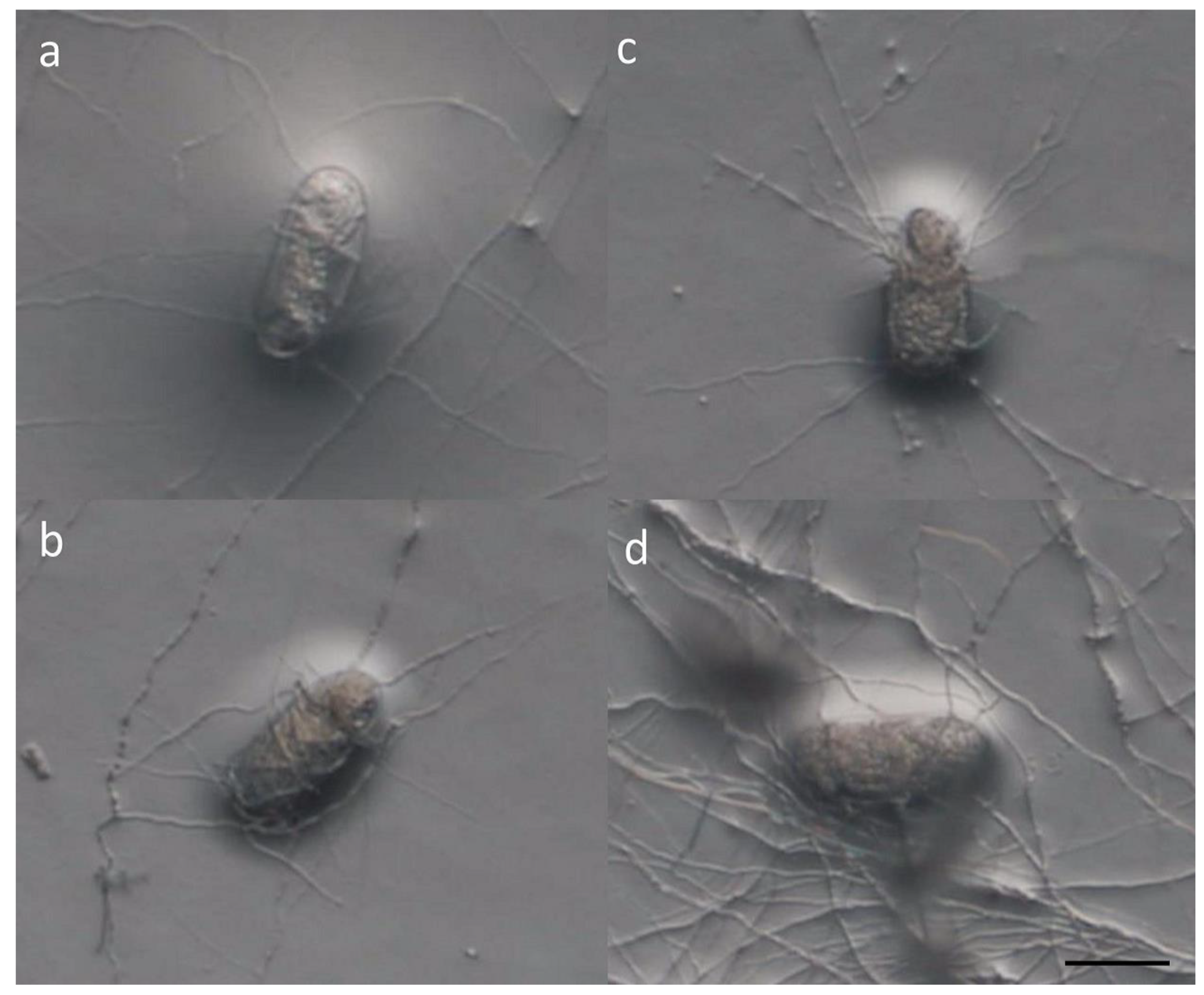
3.1. In Vitro Experiments

The fungal infection rate and egg hatching of M. enterolobii in different fungal cultures were recorded. Hyphal-wrapped eggs could easily be observed under a dissecting microscope and could be distinguished from the unimpacted ones (Figure 1). The eggs with disintegration contents and obviously penetrated and parasitized by fungal hyphae were considered infected by the inoculated fungi. The infection rate represents the percentage of eggs that were parasitized by the examined fungus. All studied fungi were equally capable of infecting about 50% of eggs (Table 1). Quickly after fungal parasitism, the eggs were dead and could no longer hatch. Compared to the no-fungal control, all examined fungi reduced the hatching rate significantly (Table 1). The lowest egg hatching rate was seen in the P. lilacinum PL251 treatment (40.33%), which was less than half of what was observed in the control (86.67%). A 41.83% hatching rate was observed in the H. oviparasitica DoUCR50 treatment. As for P. taiwanensis and Samsoniella sp. strain NTUCC 18–159, the egg hatching rate was 46.33% and 47.16%, respectively. The egg hatching rate reflects the growth potential of the nematode population after treatment and is highly associated with the future damage on the host. The results indicated that all examined fungi are capable of parasitizing M. enterolobii eggs through penetration, and the survival of the nematode eggs were seriously impacted by these biological processes, which then led to significantly lower egg hatching rates.

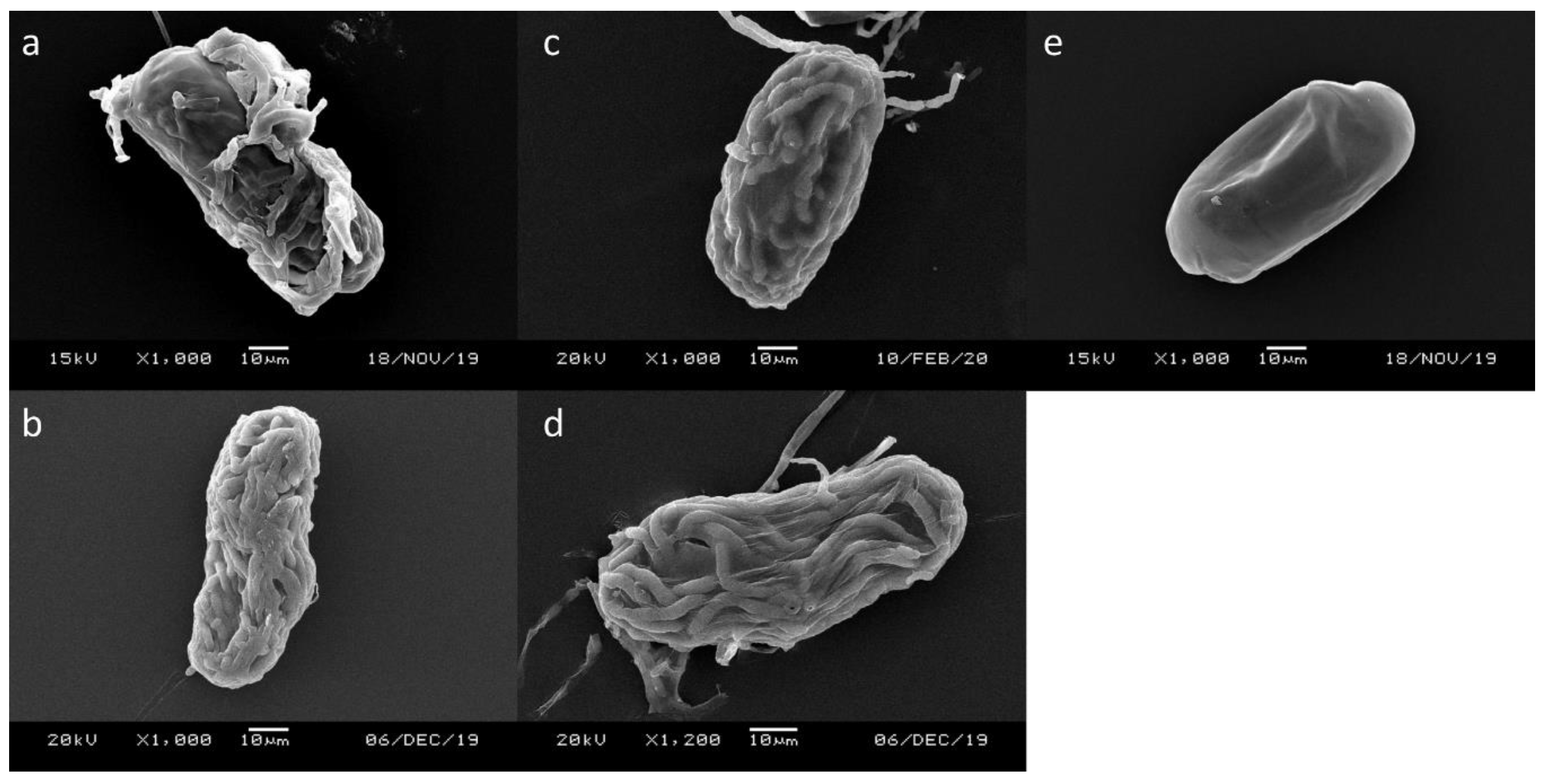
3.2. Scanning Electronic Microscope (SEM)

Conventional SEM was used to observe the eggs infected by different fungi. The surface of the control eggs was smooth, and the shape of the first stage juvenile (J1) was visible (Figure 2e). For the eggs infected by the examined nematophagous fungi, the hyphae originally wrapped around the eggshell may be washed away during the preparation process. Still, the eggshells filled with fungal hyphae were clearly observed (Figure 2a–d). The fungal hyphae grew into the eggs and colonized them without totally disrupting the eggshell. The original egg contents were seemingly lost, but the eggs remained in shape; some hyphae grew out of the eggs. Based on the observation under SEM, the four fungi likely shared the same egg-parasitism mechanism.

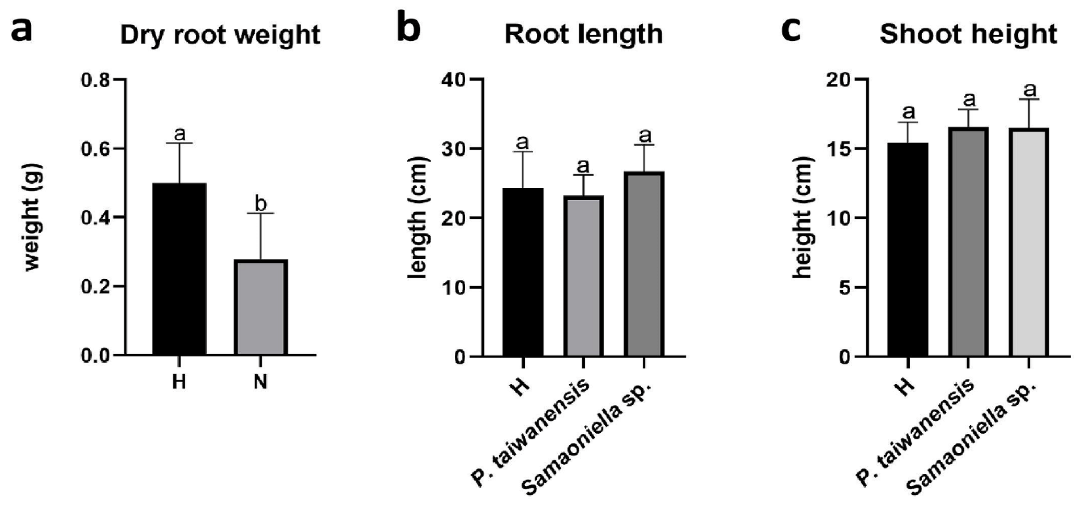
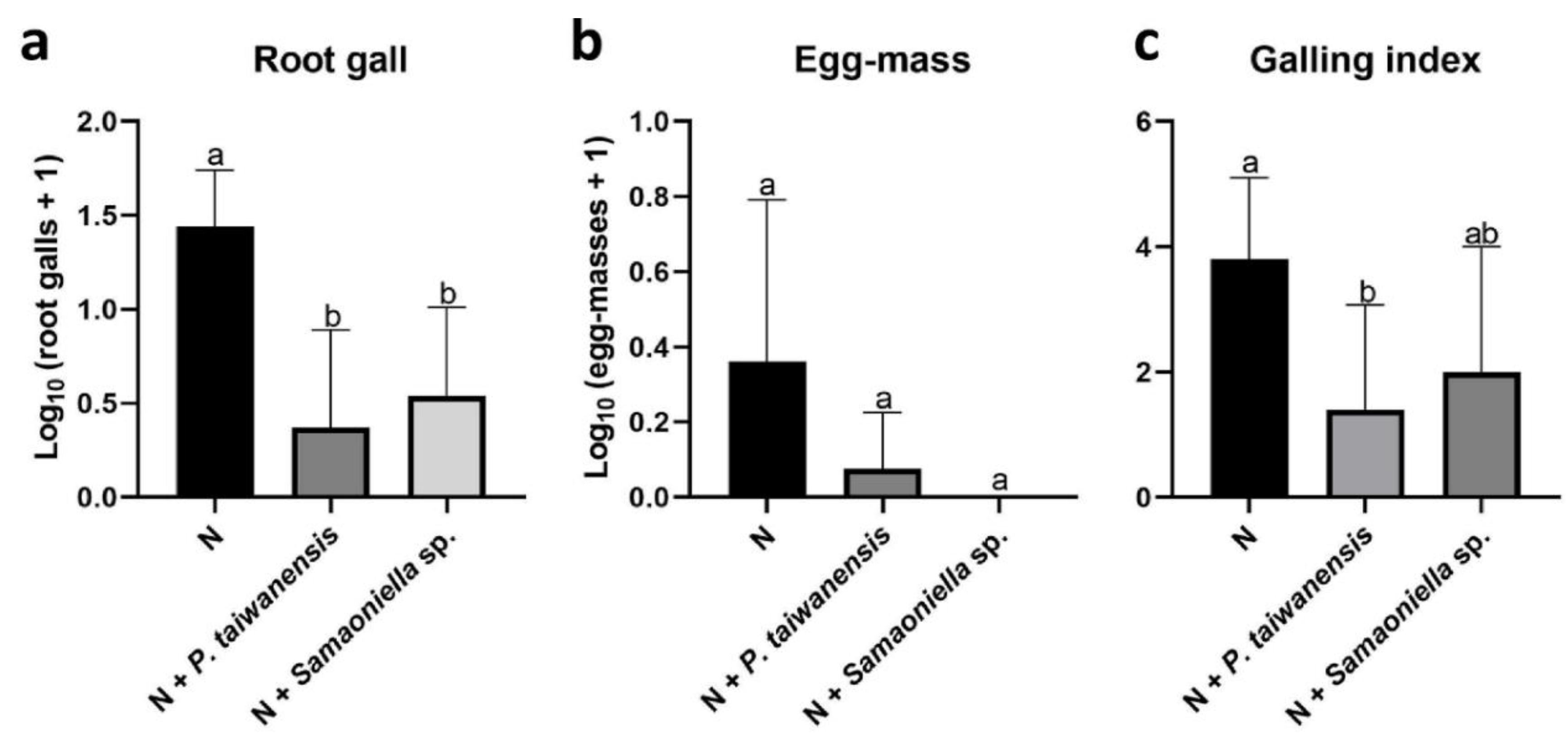
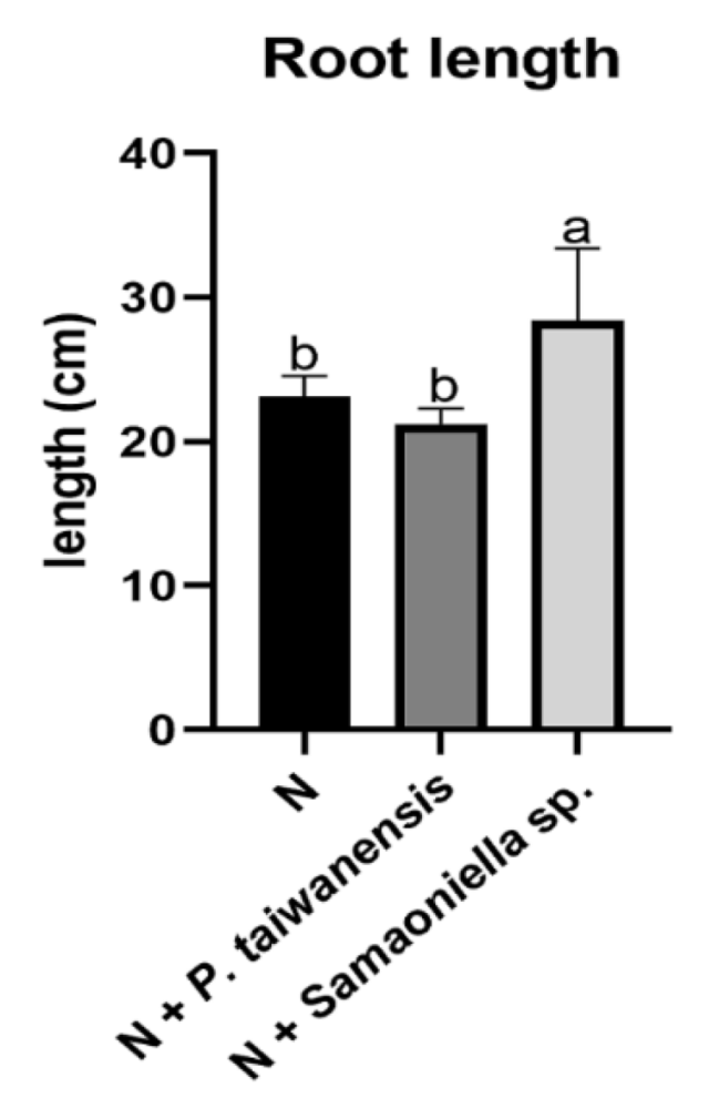
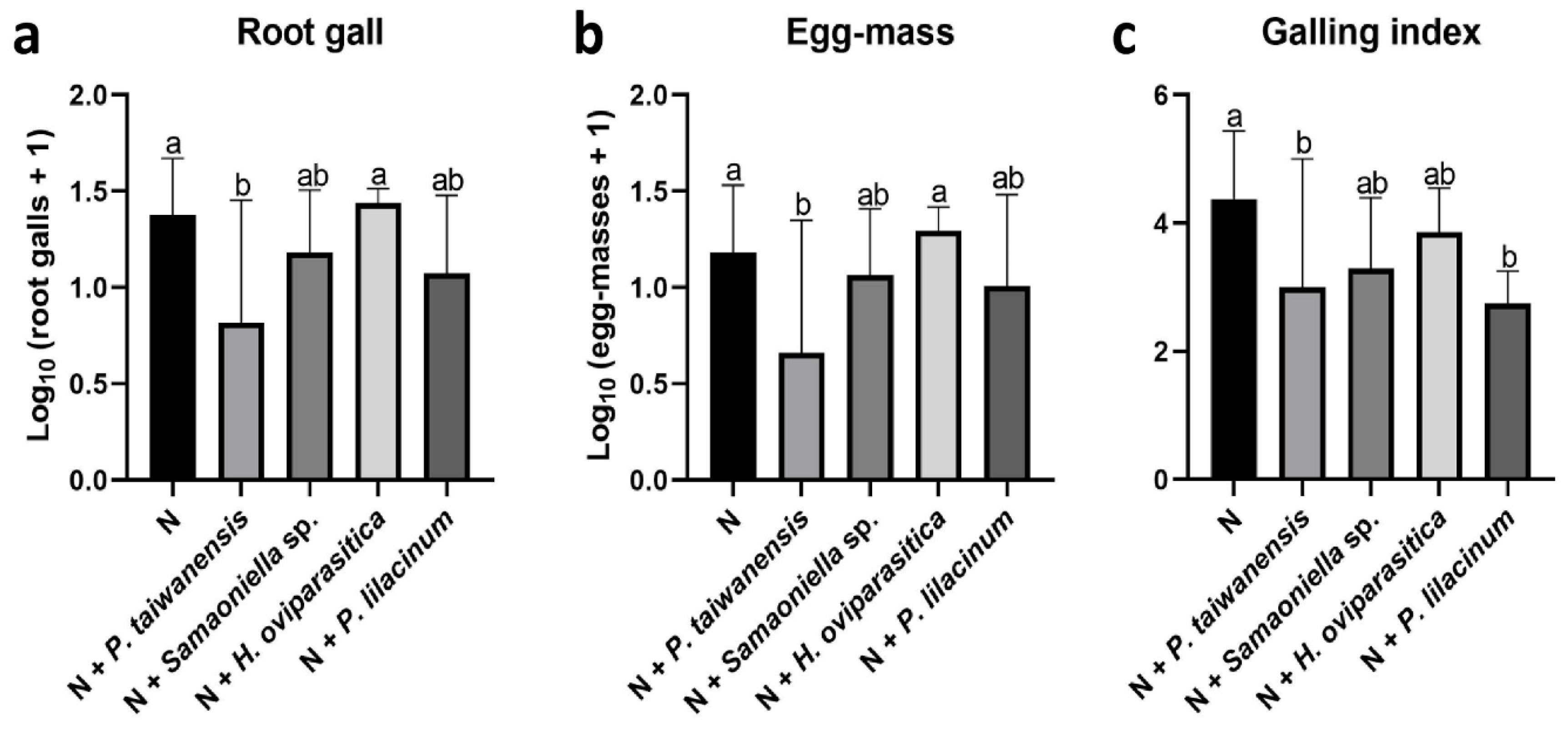
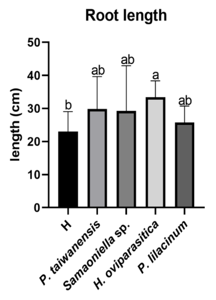
3.3. Greenhouse Trials

Dry root weights of the plant were significantly reduced when inoculated with nematodes (Figure 3a). No differences among all the treatments were observed in aboveground dry weight and shoot height. The examined fungi P. taiwanensis and Samsoniella sp. strain NTUCC 18–159 did not negatively affect the root length or shoot height when inoculated without the nematodes (Figure 3b,c).
However, both P. taiwanensis and Samsoniella sp. strain NTUCC 18–159 inhibited the nematode reproduction. In the first trial, significant reductions (P < 0.05) were observed in root gall numbers in P. taiwanensis and Samsoniella sp. strain NTUCC 18–159 treatments (Figure 4a). Due to the 10-day earlier harvesting caused by a fungal infection, only a few egg-masses were observed. No significant reduction of egg mass numbers in fungal treatments was recorded (Figure 4b). Root galling index was reduced in the P. taiwanensis treatment but not with Samsoniella sp. strain NTUCC 18–159 (Figure 4c). Surprisingly, a root growth-promoting effect was observed on the plants co-inoculated with Samsoniella sp. strain NTUCC 18–159 and M. enterolobii (Figure 5).
In the second trial, the M. enterolobii-suppression efficacy by P. taiwanensis and Samsoniella sp. strain NTUCC 18–159 were further compared with H. oviparasitica DoUCR50 and P. lilacinum PL251. Overall, the nematode-inhibition abilities of P. taiwanensis and Samsoniella sp. strain 180609-3 were similar to P. lilacinum PL251. The number of root galls significantly decreased in the P. taiwanensis treatment (Figure 6a). The reductions of root gall numbers were also observed in the two treatments inoculated with Samsoniella sp. strain NTUCC 18–159 and P. lilacinum, but were not significant by statistical standards (Figure 6a). The number of egg masses also showed a similar pattern (Figure 6b). A similarly galling index-reduction result was observed as in the first trial, the P. lilacinum treatment reduction was comparable to the P. taiwanensis treatment (Figure 6c). For the root and aboveground dry weights, there were no significant differences when compared to the no fungi-control. Similar results were seen in shoot height. For root length, no significant differences between healthy plants and plants infected by nematodes were observed, but H. oviparasitica alone increased the root length significantly (Figure 7).

4. Discussion

The M. enterolobii-suppression mechanism and ability of two newly identified fungi, P. taiwanensis and Samsoniella sp. strain NTUCC 18–159, were investigated through in vitro and in vivo experiments in the study. The overall results indicated the suppression efficacy was similar to P. lilacinum PL251. This particular strain had been widely commercialized and applied in fruit and vegetable cropping systems against root-knot nematodes, root-lesion nematodes, and citrus nematodes. Although there were some methodological differences, the egg hatching and infection rate in our study were similar to the previous in vitro research using P. lilacinum for controlling M. enterolobii [18]. As our data demonstrate, 40–50% M. enterolobii egg hatching rates with P. lilacinum PL251 in the in vitro experiments showed no significant differences with P. taiwanensis and Samsoniella sp. strain NTUCC 18–159. As for H. oviparasitica DoUCR50, previous studies reported its ability to parasitize primarily female sugar beet cyst nematodes [30], and a phylogenetically closely related fungal clade with similar nematophagous activity was identified [34]. This is the first study to reveal its ability to parasitize M. enterolobii eggs. However, in past observations, H. oviparasitica DoUCR50 hyphae would not penetrate eggs containing a juvenile (data not shown). This is consistent with Metarhizium guizhouense, another nematophagous fungi known to parasitize eggs of Meloidogyne incognita [19]. These phenomena suggest that the examined fungi are not capable of directly suppressing the egg hatching process. On the contrary, the fact that most of the non-hatched eggs in fungal treatments were penetrated by fungal hyphae indicates the principal parasitism mechanism was hyphae penetration. Further experiments on fungal secondary metabolites would provide valuable information on whether fungal secreted inhibitors, such as toxic substrates, are responsible of the gap between egg infection rate and reduction of egg hatching rate.
The interaction between M. enterolobii and the egg parasitic fungi was investigated with both a dissecting microscope and a scanning electron microscope (SEM), and the parasitism mechanism of the four nematophagous fungi was confirmed. With the dissecting microscope, the surfaces of the infected eggs were clearly seen wrapped by fungal hyphae. Through SEM, the hyphae growth and colonization inside the eggs were easily observed. The eggshells remained mostly in shape, but the contents within were nearly completely lost. This implied that these fungi secreted enzymes only to facilitate hyphae penetration while maintaining the egg contents inside the eggshell for the fungi to utilize the nutrients for later growth. After absorbing the nutrients, the hyphae grew out of the eggs and searched for a new host again. Similar mechanisms had been previously studied with the type strain of H. oviparasitica. Appressoria formed when the hyphae contacted the egg surface for attachment and penetration, and chitinase was known to be secreted by H. oviparasitica [35]. Moreover, Stirling and Mankau found that the hyphae of H. oviparasitica inside the eggs were swollen to 3–4 µm in diam compared to its normal width between 2–2.5 µm [36]. It was consistent with our observation that the H. oviparasitica DoUCR50 hyphae inside the eggs were about 4–4.5 µm in diam, which was thicker than hyphae located far from the eggs (around 2.5 µm in diam). It is worth mentioning that H. oviparasitica DoUCR50 is a strain with a different parasitic ability from Stirling and Mankau’s strain, but a similar mechanism was found when infecting Meloidogyne spp. However, such phenomena were not observed in the other three fungi in our investigation. Interestingly, entomopathogenic fungi shared similar mechanisms with egg-parasitic fungi when infecting insects. The appressoria structure and the enzymes secreted by fungi are considered as the crucial virulence factors. P. lilacinum had also been known to be both nematophagous and entomopathogenic [37,38,39]. The results from our previous [21] and this study proved that P. taiwanensis and Samsoniella sp. strain NTUCC 18–159 can both parasitize insects and nematodes, and the infection mechanisms appeared similar. Our findings not only showed the possibility of the two fungi being developed into biological control agents but also suggested that some entomopathogenic fungi might have a potential as biological control agents of nematodes.
The efficacies of the four fungi were further evaluated on Chinese cabbage in the greenhouse. Although the change of cabbage seeds might have been the reason why fewer egg masses were formed, the root gall numbers were reduced significantly, nevertheless. It clearly demonstrated the effectiveness of P. taiwanensis and Samsoniella sp. strain NTUCC 18–159 on inhibiting the nematodes’ feeding site establishment. The abilities of the two examined fungi to inhibit the M. enterolobii feeding site establishment and reproduction were as efficient as the already commercialized P. lilacinum PL251, despite the fact that only about 50% of eggs were parasitized by these fungi in vitro, which indicates the application potential as biological control agents. The galling index showed that the disease severity obviously decreased in P. taiwanensis and P. lilacinum treatments. However, the effects of H. oviparasitica DoUCR50 were not reflected in any of the nematode reproduction parameters. The weight and height of the Chinese cabbage indicated the actual market value of the crop, which was not impacted by any of the examined fungi. In summary, these fungi have the potential to become part of the integrated control strategies to root-knot nematode damage.
Many factors would influence the growth and control efficacy of biocontrol agents in planting materials or soil and therefore lead to efficacy gaps between in vitro study and greenhouse trials. Studies have shown that both inoculum level and application time affect the efficacy significantly [40]. Kiewnick and Sikora [41] mentioned that P. lilacinum cannot persist long in soil and it is likely necessary to apply 106 cfu/g soil multiple times to achieve the inhibition effect on M. incognita. The concentration may also be applicable on M. enterolobii. The interaction of the fungi with other microbes in soil, such as competition, may be a crucial issue in its persistence, population size and, eventually, the success of nematophagous fungi [42]. Our greenhouse trials used a sterilized growth medium, and only one fungus was inoculated in each treatment. These were rather uniform environments that excluded many biological factors such as microbial interaction effects. Increasing the temperature and soil pH may also affect the parasitic capability of nematophagous fungi [43]. The in vitro testing was conducted on a culture medium of pH close to eight at room temperature, around 28 °C. Meanwhile, the planting materials created for the greenhouse experiments were under the conditions of pH 5.5–6.5 and an average temperature of 23 °C. In addition, specifically for H. oviparasitica DoUCR50, the growth rate of the fungus may be an important factor. Based on our observation, H. oviparasitica needed more than one month to colonize half of the 9-cm Petri dish with PDA, which is much slower than P. taiwanensis, Samsoniella sp. strain NTUCC 18–159, and P. lilacinum PL251. Although the growth rate may be different in soil, possibly H. oviparasitica DoUCR50 needed less time to reach the egg-masses and infect the eggs before the J1 developed. Therefore, a higher amount of inoculum might make up for its slow-growing feature and thus enhance its efficacy in soil. In addition, egg hatching would also be influenced by factors such as humidity, root exudates, and host age, resulting in the differences between in vitro test and greenhouse trial [44,45]; yet no study has been done specifically on this emerging new species, M. enterolobii. Regarding the problems related to the use of nematophagous fungi mentioned above, a cocktail of several nematophagous species may be the solution. Previous studies have supported the feasibility of such a concept. For example, Yankova et al. combined Paecilomyces lilacinus strain 251 and Trichoderma viride strain T6 and discovered the control efficacy was greater than using either one alone [46]. Different combinations of strains of Pochonia chlamydosporia var. catenulata and Purpureocillium lilacinum were also tested in the study by Silva et al. [18].
In conclusion, the egg-parasitism mechanism of P. taiwanensis and Samsoniella sp. strain NTUCC 18–159 was revealed in this study. The fungi showed potential as biocontrol agents against M. enterolobii as part of an integrated pest management (IPM) approach. Both P. taiwanensis and Samsoniella sp. strain NTUCC 18–159 are indigenous isolates in Taiwan. Given that such strains are often more receptive than non-indigenous ones [47], the application potential of the two in Southeastern countries where climatic conditions are similar is worth noting. Furthermore, previous studies of fungi within the H. oviparasitica clade and Pochonia clamydospora have supported the hypothesis that suppression efficacy of nematophagous fungi varies among strains [34,48]. Strains of Purpureocillium lilacinum obtained from eggs have greater potential to suppress nematodes than the ones from soil [49]. The continuation of searching for more effective P. taiwanensis and Samsoniella sp. strains of high phylogenetic similarity with the help of molecular tools seems to be a reasonable strategy in the future. In the meantime, field micro-pot trials with inoculations of a cocktail of nematophagous fungi against M. enterolobii will be conducted to optimize the application options and evaluate the ultimate control efficacy under microbial competition impacts.

Author Contributions

Conceptualization, H.A.A. and J.-i.Y.; data curation, J.O.B.; formal analysis, J.-i.Y.; funding acquisition, J.-i.Y.; investigation, Y.-J.L.; methodology, Y.-J.L. and J.-i.Y.; resources, H.A.A., J.O.B., and J.-i.Y.; software, Y.-J.L.; supervision, J.O.B.; validation, H.A.A., J.O.B., and J.-i.Y.; visualization, Y.-J.L.; writing—original draft, Y.-J.L.; writing—review & editing, H.A.A., J.O.B., and J.-i.Y. All authors have read and agreed to the published version of the manuscript.

Funding

This study was funded by the Bureau of Animal and Plant Health Inspection and Quarantine, Council of Agriculture, Executive Yuan, Taiwan.

Acknowledgments

The authors thank Bayer Taiwan Company Ltd. for providing the BioAct products, Shiuh-Feng Shiao (Laboratory of Insect Systematics, National Taiwan University) and Shiang-Jiuun Chen (Department of Life Science, National Taiwan University) for SEM facility assistance, and Wei-yu Chuang for fungal sample preparation assistance.

Conflicts of Interest

The authors declare no conflict of interest. The funders had no role in the design of the study; in the collection, analyses, or interpretation of data; in the writing of the manuscript; or in the decision to publish the results.

References

  1. Rich, J.R.; Brito, J.A.; Kaur, R.; Ferrell, J.A. Weed species as hosts of Meloidogyne: A review. Nematropica 2009, 39, 157–185. [Google Scholar]
  2. Wesemael, W.; Viaene, N.; Moens, M. Root-knot nematodes (Meloidogyne spp.) in Europe. Nematology 2011, 13, 3–16. [Google Scholar] [CrossRef]
  3. Singh, S.; Singh, B.; Singh, A.P. Nematodes: A Threat to Sustainability of Agriculture. Procedia Environ. Sci. 2015, 29, 215–216. [Google Scholar] [CrossRef] [Green Version]
  4. Jenkins, W.R.; Taylor, D.P. Plant Nematology; Reinhold Publishing Corporation: New York, NY, USA, 1967. [Google Scholar]
  5. EPPO. An Emerging Root-Knot Nematode, Meloidogyne enterolobii: Addition to the EPPO Alert List. EPPO Reporting Service. 2008, p. 5. Available online: Gd.eppo.int/reporting/article-690 (accessed on 31 March 2020).
  6. Japan Plant Protection Station. Japan Plant Protection Act. List of the Plants Subject to Field Inspection in Exporting Countries. 2019. Available online: www.maff.go.jp/pps (accessed on 29 January 2020).
  7. Jindapunnapat, K.; Chinnasri, B.; Kwankuae, S. Biological control of root-knot nematodes Meloidogyne enterolobii in guava by the fungus Trichoderma harzianum. J. Dev. Sustain. Agric. 2013, 8, 110–118. [Google Scholar]
  8. Ye, W.M.; Koenning, S.R.; Zhuo, K.; Liao, J.L. First report of Meloidogyne enterolobii on cotton and soybean in North Carolina, United States. Plant Dis. 2013, 97, 1262. [Google Scholar] [CrossRef] [PubMed]
  9. Rutter, W.B.; Skantar, A.M.; Handoo, Z.A.; Mueller, J.D.; Aultman, S.P.; Agudelo, P. Meloidogyne enterolobii found infecting root-knot nematode resistant sweetpotato in South Carolina, United States. Plant Dis. 2018, 103, 775. [Google Scholar] [CrossRef]
  10. Carneiro, R.; Cirotto, P.; Quintanilha, A.; Silva, D.; Carneiro, R. Resistance to Meloidogyne mayaguensis in Psidium spp. accessions and their grafting compatibility with P. guajava cv. Paluma. Fitopatol. Bras. 2007, 32, 281–284. [Google Scholar] [CrossRef]
  11. Freitas, V.M.; Silva, J.G.P.; Gomes, C.B.; Castro, J.M.C.; Correa, V.R.; Carneiro, R.M.D.G. Host status of selected cultivated fruit crops to Meloidogyne enterolobii. Eur. J. Plant Pathol. 2017, 148, 307–319. [Google Scholar] [CrossRef]
  12. Kiewnick, S.; Karssen, G.; Brito, J.A.; Oggenfuss, M.; Frey, J.E. First report of root-knot nematode Meloidogyne enterolobii on tomato and cucumber in Switzerland. Plant Dis. 2008, 92, 1370. [Google Scholar] [CrossRef]
  13. Minuto, A.; Gullino, M.; Lamberti, F.; D’Addabbo, T.; Tescari, E.; Ajwa, H.; Garibaldi, A. Application of an emulsifiable mixture of 1,3-dichloropropene and chloropicrin against root knot nematodes and soilborne fungi for greenhouse tomatoes in Italy. Crop Prot. 2006, 25, 1244–1252. [Google Scholar] [CrossRef]
  14. Ibekwe, A.M.; Papiernik, S.K.; Gan, J.; Yates, S.R.; Yang, C.H.; Crowley, D.E. Impact of fumigants on soil microbial communities. Appl. Environ. Microbiol. 2001, 67, 3245–3257. [Google Scholar] [CrossRef] [PubMed] [Green Version]
  15. Brito, J.A.; Stanley, J.D.; Kaur, R.; Cetintas, R.; Di Vito, M.; Thies, J.A.; Dickson, D.W. Effects of the Mi-1, N and Tabasco genes on infection and reproduction of Meloidogyne mayaguensis on tomato and pepper genotypes. J. Nematol. 2007, 39, 327–332. [Google Scholar] [PubMed]
  16. Stirling, G.R. Biological Control of Plant Parasitic Nematodes: SOIL Ecosystem Management in Sustainable Agriculture, 2nd ed.; CAB International: Wallingford, UK, 2014. [Google Scholar]
  17. Lopez-Llorca, L.V.; Maciá-Vicente, J.G.; Jansson, H.B. Mode of action and interactions of nematophagous fungi. In Integrated Management of Plant Pests and Diseases; Ciancio, A., Mukerji, K.G., Eds.; Springer: Dordrecht, The Netherlands, 2008; Volume 2, pp. 51–76. [Google Scholar]
  18. Silva, S.D.; Carneiro, R.M.D.G.; Faria, M.; Souza, D.A.; Monnerat, R.G.; Lopes, R.B. Evaluation of Pochonia chlamydosporia and Purpureocillium lilacinum for suppression of Meloidogyne enterolobii on tomato and banana. J. Nematol. 2017, 49, 77–85. [Google Scholar] [CrossRef] [PubMed] [Green Version]
  19. Thongkaewyuan, A.; Chairin, T. Biocontrol of Meloidogyne incognita by Metarhizium guizhouense and its protease. Biol. Control 2018, 126, 142–146. [Google Scholar] [CrossRef]
  20. Kepenekci, I.; Saglam, H.D.; Oksal, E.; Yanar, D.; Yanar, Y. Nematicidal activity of Beauveria bassiana (Bals.-Criv.) Vuill. against root-knot nematodes on tomato grown under natural conditions. Egypt. J. Biol. Pest Control 2017, 27, 117–120. [Google Scholar]
  21. Yang, J.-I.; Stadler, M.; Chuang, W.-Y.; Wu, S.; Ariyawansa, H.A. In vitro inferred interactions of selected entomopathogenic fungi from Taiwan and eggs of Meloidogyne graminicola. Mycol. Prog. 2020, 19, 97–109. [Google Scholar] [CrossRef]
  22. Bidochka, M.J.; Khachatourians, G.G. Purification and properties of an extracellular protease produced by the entomopathogenic fungus Beauveria bassiana. Appl. Environ. Microbiol. 1987, 53, 1679–1684. [Google Scholar] [CrossRef] [Green Version]
  23. Leger, R.J.S.; Charnley, A.K.; Cooper, R.M. Characterization of cuticle-degrading proteases produced by the entomopathogen Metarhizium anisopliae. Arch. Biochem. Biophys. 1987, 253, 221–232. [Google Scholar] [CrossRef]
  24. Lei, Y.; Lü, L.; He, Y. Study on induction of enzyme activity of entomopathogenic fungus Isaria fumosorosea SCAU-IFCF01. Huan Jing Kun Chong Xue Bao 2017, 39, 1000–1006. (In Chinese) [Google Scholar]
  25. Díaz-Godínez, G.; Candia-Sanchez, L.F.; Díaz, R.; Sánchez, C.; Villegas, E.; Monroy, G.; Hernández Velázquez, V.M. Activity of lipases, chitinases and proteases of the entomopathogenic fungus, Metarhizium anisopliae developed in different culture media. In Biotechnology Summit 2016, 3rd ed.; Fernández-Luqueño, F., López-Valdez, F., Santos Villalobos, S.D.L., Eds.; Cinvestav: San Pedro Zacatenco, Mexico, 2016; pp. 155–159. [Google Scholar]
  26. Morton, O.; Hirsch, P.; Kerry, B. Infection of plant-parasitic nematodes by nematophagous fungi—A review of the application of molecular biology to understand infection processes and to improve biological control. Nematology 2004, 6, 161–170. [Google Scholar] [CrossRef]
  27. Bird, A.F.; McClure, M.A. The tylenchid (Nematoda) eggshell: Structure, composition and permeability. Parasitology 1976, 72, 19–28. [Google Scholar] [CrossRef]
  28. Baral, H.-O.; Weber, E.; Gams, W.; Hagedorn, G.; Liu, B.; Liu, X.; Marson, G.; Marvanová, L.; Stadler, M.; Weiß, M. Generic names in the Orbiliaceae (Orbiliomycetes) and recommendations on which names should be protected or suppressed. Mycol. Prog. 2018, 17, 5–31. [Google Scholar] [CrossRef] [Green Version]
  29. Kiewnick, S.; Sikora, R.A. Biological control of the root-knot nematode Meloidogyne incognita by Paecilomyces lilacinus strain 251. Biol. Control 2006, 38, 179–187. [Google Scholar] [CrossRef]
  30. Olatinwo, R.; Yin, B.; Becker, J.O.; Borneman, J. Suppression of the plant-parasitic nematode Heterodera schachtii by the fungus Dactylella oviparasitica. Phytopathology 2006, 96, 111–114. [Google Scholar] [CrossRef] [PubMed] [Green Version]
  31. Smith Becker, J.; Borneman, J.; Becker, J.O. Dactylella oviparasitica parasitism of the sugar beet cyst nematode observed in trixenic culture plates. Biol. Control 2013, 64, 51–56. [Google Scholar] [CrossRef]
  32. Hu, M.X.; Zhuo, K.; Liao, J.L. Multiplex PCR for the simultaneous identification and detection of Meloidogyne incognita, M. enterolobii, and M. javanica using DNA extracted directly from individual galls. Phytopathology 2011, 101, 1270–1277. [Google Scholar] [CrossRef] [Green Version]
  33. Thies, J.; Mueller, J.D.; Fery, R. Effectiveness of resistance to southern root-knot nematode in ‘Carolina Cayenne’ pepper in greenhouse, microplot, and field tests. J. Am. Soc. Hortic. Sci. 1997, 122, 200–204. [Google Scholar] [CrossRef]
  34. Yang, J.-I.; Benecke, S.; Jeske, D.R.; Rocha, F.S.; Smith Becker, J.; Timper, P.; Becker, J.O.; Borneman, J. Population dynamics of Dactylella oviparasitica and Heterodera schachtii: Toward a decision model for sugar beet planting. J. Nematol. 2012, 44, 237–244. [Google Scholar]
  35. Stirling, G.R.; Mankau, R. Mode of parasitism of Meloidogyne and other nematode eggs by Dactylella oviparasitica. J. Nematol. 1979, 11, 282–288. [Google Scholar]
  36. Stirling, G.R.; Mankau, R. Parasitism of Meloidogyne eggs by a new fungal parasite. J. Nematol. 1978, 10, 236–240. [Google Scholar]
  37. Kepenekci, I.; Oksal, E.; Saglam, H.D.; Atay, T.; Tulek, A.; Evlice, E. Identification of Turkish isolate of the entomopathogenic fungi, Purpureocillium lilacinum (syn: Paecilomyces lilacinus) and its effect on potato pests, Phthorimaea operculella (Zeller) (Lepidoptera: Gelechiidae) and Leptinotarsa decemlineata (Say) (Coleoptera: Chrysomelidae). Egypt. J. Biol. Pest Control 2015, 25, 121–127. [Google Scholar]
  38. Toledo-Hernández, R.A.; Holguín-Meléndez, F.; Huerta-Palacios, G.; Liedo, P.; Toledo, J.; Valle-Mora, J. Pathogenicity and virulence of Purpureocillium lilacinum (Hypocreales: Ophiocordycipitaceae) on Mexican fruit fly adults. Fla. Entomol. 2019, 102, 309–314. [Google Scholar] [CrossRef] [Green Version]
  39. Yüksel, E.; Açıkgöz, C.; Demirci, F.; Muștu, M. Effects of the entomopathogenic fungi, Beauveria bassiana, Isaria farinosa and Purpureocillium lilacinum, on eggs of Tuta absoluta (Meyrick) (Lepidoptera: Gelechiidae). /Entomopatojen funguslar Beauveria bassiana, Isaria farinosa ve Purpureocillium lilacinum’un Domates Yaprak galeri Güvesi Tuta absoluta’ nın yumurta dönemine etkileri. Türkiye Biyolojik Mücadele Dergisi 2017, 8, 39–47. [Google Scholar]
  40. Cabanillas, E.; Barker, K.R. Impact of Paecilomyces lilacinus inoculum level and application time on control of Meloidogyne incognita on tomato. J. Nematol. 1989, 21, 115–120. [Google Scholar]
  41. Kiewnick, S.; Sikora, R. Efficacy of Paecilomyces lilacinus (strain 251) for the control of root-knot nematodes. Commun. Agric. Appl. Biol. Sci. 2003, 68, 123–128. [Google Scholar]
  42. Heydari, A.; Pessarakli, M. A review on biological control of fungal plant pathogens using microbial antagonists. J. Biol. Sci. 2010, 10, 273–290. [Google Scholar] [CrossRef] [Green Version]
  43. Luambano, N.D.; Manzanilla-López, R.H.; Kimenju, J.W.; Powers, S.J.; Narla, R.D.; Wanjohi, W.J.; Kerry, B.R. Effect of temperature, pH, carbon and nitrogen ratios on the parasitic activity of Pochonia chlamydosporia on Meloidogyne incognita. Biol. Control 2015, 80, 23–29. [Google Scholar] [CrossRef]
  44. Takayuki, M.; Yuji, O. Tomato culture filtrate stimulates hatching and activity of Meloidogyne incognita juveniles. Nematology 2009, 11, 51–61. [Google Scholar]
  45. Wesemael, W.; Perry, R.; Moens, M.J.N.V. The influence of root diffusate and host age on hatching of the root-knot nematodes, Meloidogyne chitwoodi and M. fallax. Nematology 2006, 8, 895–902. [Google Scholar] [CrossRef]
  46. Yankova, V.; Markova, D.; Naidenov, M.; Arnaoudov, B. Management of root-knot nematodes (Meloidogyne spp.) in greenhouse cucumbers using microbial products. Türk Tarım Doğa Bilimleri Derg. 2014, 1 (Özel Sayı-2), 1569–1573. [Google Scholar]
  47. Monfort, E.; Lopez-Llorca, L.V.; Jansson, H.-B.; Salinas, J. In vitro soil receptivity assays to egg-parasitic nematophagous fungi. Mycol. Prog. 2006, 5, 18–23. [Google Scholar] [CrossRef]
  48. Yang, J.-I.; Loffredo, A.; Borneman, J.; Becker, J.O. Biocontrol efficacy among strains of Pochonia chlamydosporia obtained from a root-knot nematode suppressive soil. J. Nematol. 2012, 44, 67–71. [Google Scholar] [PubMed]
  49. Carneiro, R.M.D.G.; Hidalgo-Díaz, L.; Martins, I.; Silva, K.F.A.d.S.; Sousa, M.G.d.; Tigano, M.S. Effect of nematophagous fungi on reproduction of Meloidogyne enterolobii on guava (Psidium guajava) plants. Nematology 2011, 13, 721–728. [Google Scholar]
Figure 1. Observation of M. enterolobii eggs infected by nematophagous fungi, through a dissecting microscope. Bar = 45 μm. (a) Paraboeremia taiwanensis, (b) Samsoniella sp. strain NTUCC 18–159, (c) Hyalorbilia oviparasitica DoUCR50, (d) Purpureocillium lilacinum PL251.
Microorganisms 08 00828 g001
Figure 2. Observation of M. enterolobii eggs infected by nematophagous fungi through scanning electronic microscope (SEM). (a) Paraboeremia taiwanensis, (b) Samsoniella sp. strain NTUCC 18–159, (c) Hyalorbilia oviparasitica DoUCR50, (d) Purpureocillium lilacinum PL251, (e) No-fungi control.
Microorganisms 08 00828 g002
Figure 3. Plant growth parameters in greenhouse trial 1. (a) negative effect of M. enterolobii on dry root weight, (b) P. taiwanensis and Samsoniella sp. strain NTUCC 18–159 effects on root length, and (c) shoot height. H is the healthy control. N is the nematode-only treatment. Different letters indicate significant differences among treatments by Fisher’s LSD test (P < 0.05), respectively.
Microorganisms 08 00828 g003
Figure 4. Effects of P. taiwanensis, Samsoniella sp. strain NTUCC 18–159, H. oviparasitica, and P. lilacinum strain 251 (PL251) on M. enterolobii in greenhouse trial 1. (a) root gall, (b) egg mass, (c) root galling index. N is the nematode-only treatment. Different letters indicate significant differences among treatments by Fisher’s LSD test (P < 0.05), respectively.
Microorganisms 08 00828 g004
Figure 5. Root length promotion effect in greenhouse trial 1. N is the nematode-only treatment. Different letters indicate significant differences among treatments by Fisher’s LSD test (P < 0.05), respectively.
Microorganisms 08 00828 g005
Figure 6. Effects of P. taiwanensis, Samsoniella sp. strain NTUCC 18–159, H. oviparasitica DoUCR50, and P. lilacinum strain 251 (PL251) on M. enterolobii in the second greenhouse trial. (a) root gall, (b) egg mass, (c) galling index. N is the nematode-only treatment. Different letters indicate significant differences among treatments by Fisher’s LSD test (P < 0.05), respectively.
Microorganisms 08 00828 g006
Figure 7. Root length promotion effect in the second greenhouse trial. H is the healthy control. Different letters indicate significant differences among treatments by Fisher’s LSD test (P < 0.05), respectively.
Microorganisms 08 00828 g007
Table 1. Effects of the nematophagous fungi on M. enterolobii egg hatching and infection rate.
FungiEgg Hatching Rate (%) 1Egg Infection Rate (%) 1
Paraboeremia taiwanensis NTUCC 17–01346.33 ± 8.10 a47.83 ± 9.93 a
Samsoniella sp. NTUCC 18–15947.16 ± 8.13 a47.50 ± 9.18 a
Hyalorbilia oviparasitica strain DoUCR5041.83 ± 5.69 a51.17 ± 7.15 a
Purpureocillium lilacinum stain PL25140.33 ± 3.01 a55.83 ± 1.26 a
No fungi control86.67 ± 1.04 b-
1 Values refer to the means of 12 replicates. Means followed by the same letter in the same column showed no significant differences according to analysis of variance (ANOVA) with Fisher’s least significant difference (Fisher’s LSD) test (P ≥ 0.05).

