Article

Pan-Proteomic Analysis and Elucidation of Protein Abundance among the Closely Related Brucella Species, Brucella abortus and Brucella melitensis

1
Institute of Animal Hygiene and Environmental Health, Freie Universität Berlin, Centre for Infectious Medicine, Robert-von-Ostertag-Str. 7-13, 14163 Berlin, Germany
2
Department of Biotechnology, SRM University AP, Andhra Pradesh, Neerukonda, Mangalagiri, Guntur 522 502, India
3
Institute of Chemistry and Biochemistry, Freie Universität Berlin, Thielallee 63, 14195 Berlin, Germany
4
Sussex Neuroscience, School of Life Sciences, University of Sussex, Falmer, Brighton BN1 9QG, UK
5
Friedrich-Loeffler-Institute, Institute of Bacterial Infections and Zoonoses, Naumburger Str. 96a, 07743 Jena, Germany
6
Faculty of Veterinary Medicine, Benha University, Moshtohor, Toukh 13736, Egypt
*
Author to whom correspondence should be addressed.
Biomolecules 2020, 10(6), 836; https://doi.org/10.3390/biom10060836
Submission received: 10 March 2020 / Revised: 30 April 2020 / Accepted: 19 May 2020 / Published: 30 May 2020
(This article belongs to the Section Cellular Biochemistry)

Abstract

:
Brucellosis is a zoonotic infection caused by bacteria of the genus Brucella. The species, B. abortus and B. melitensis, major causative agents of human brucellosis, share remarkably similar genomes, but they differ in their natural hosts, phenotype, antigenic, immunogenic, proteomic and metabolomic properties. In the present study, label-free quantitative proteomic analysis was applied to investigate protein expression level differences. Type strains and field strains were each cultured six times, cells were harvested at a midlogarithmic growth phase and proteins were extracted. Following trypsin digestion, the peptides were desalted, separated by reverse-phase nanoLC, ionized using electrospray ionization and transferred into an linear trap quadrapole (LTQ) Orbitrap Velos mass spectrometer to record full scan MS spectra (m/z 300–1700) and tandem mass spectrometry (MS/MS) spectra of the 20 most intense ions. Database matching with the reference proteomes resulted in the identification of 826 proteins. The Cluster of Gene Ontologies of the identified proteins revealed differences in bimolecular transport and protein synthesis mechanisms between these two strains. Among several other proteins, antifreeze proteins, Omp10, superoxide dismutase and 30S ribosomal protein S14 were predicted as potential virulence factors among the proteins differentially expressed. All mass spectrometry data are available via ProteomeXchange with identifier PXD006348.

1. Introduction

Brucella represents a Gram-negative bacterial genus of the α-2 subgroup of Proteobacteria. Brucellae are highly adapted to their intracellular lifestyle and are the causative agents of human and animal brucellosis (“undulant fever”, “Malta fever”, “Mediterranean fever” or “Bang’s disease”) [1]. They are highly infective and 10–100 bacteria cause human infection [2,3]. The genus Brucella currently includes 12 accepted species that have been named according to their host specificity. To date, the mechanism behind the host specificity is not clear [4]. The classification of Brucella species is under debate due to the reported high degree of homology found in DNA-DNA hybridization studies. This results in the proposal that the genus Brucella is a single genomospecies and the species are only biovars of B. melitensis [5,6]. It is also believed that B. abortus and B. melitensis share a common ancestor that evolved from B. suis [7]. B. abortus (preferred host: cattle and other bovidae), B. melitensis (small ruminants such as goats and sheep) and B. suis (pigs) cause human brucellosis. The Centers for Disease Control and Prevention has listed Brucella species on the Federal Select Agent Program/Select Agents and Toxins List (https://www.selectagents.gov/SelectAgentsandToxinsList.html, assessed in March 2020) and on the Emergency Preparedness and Response Bioterrorism Agents/Diseases List (https://emergency.cdc.gov/agent/agentlist.asp, assessed in March 2020) as prepared by the National Center for Emerging and Zoonotic Infectious Diseases.
B. melitensis and B. abortus have striking similarities, i.e., two chromosomes, comparable gene sequence, organization and structure [7,8,9]. These two species are the most often sequenced species of Brucella with 256 genome assemblies of B. abortus and 254 genome assemblies of B. melitensis listed at the NCBI database (March 2020) [8,9]. The genome size of the B. abortus type strain 544 was reported to be 3,289,405 bp and was predicted to possess 3319 genes among which 3259 were protein-coding genes and 60 RNA genes [10]. The genome of B. melitensis strain 16M encompasses 3,294,935 bp distributed over two circular chromosomes and contains 3197 open reading frames (ORFs), potentially available for expression [11]. Comparison of the B. melitensis 16M genome with strains of five other Brucella species revealed alterations in ORFs and species-specific conservation in terms of genetic content deletion or missing genome islands [12]. The host specificity might be caused by the species-specific gene inactivation/activation that influences transcriptional regulators and outer membrane proteins [7,13,14]. Previous studies indicated that these two strains vary in their antigenicity, immunologic and genetic properties [15,16,17]. There is no clear information in the literature designating whether live attenuated B. abortus and B. melitens is strains provide cross-protection among bovines and small ruminants respectively [18,19]. These two species display differences in phenotype, e.g., dye sensitivity, CO2 requirement or H2S production, which are used for diagnosis and biotyping [4,20,21,22]. Differences in immuno-dominant proteins of field isolates were demonstrated using sera collected from naturally infected animals [23,24]. Although it is broadly accepted in the field that the Type IV secretion system (T4SS) is in some way tied to virulence in Brucella species, Brucella generally lacks classical virulence factors and in order to explain its virulence and host specificity, a better understanding at the proteome and metabolome level is needed. Proteomic analyses of various strains of B. abortus and B. melitensis have been reported [25,26,27,28,29,30,31,32,33,34], suffering from limitations in terms of technology and database coverage. In the present study, the label-free quantitative proteomic analysis includes the reference strains as well as strains isolated from infected animals analyzed in an earlier study [23]. The focus of this study was to investigate proteome level differences between B. abortus and B. melitensis cultured under laboratory conditions and relate them to possible factors involved in mechanisms of virulence and differences in host specificity.

2. Materials and Methods

2.1. Brucella Culture

Brucella type strains and field isolates as listed in Table 1 were from the culture collection of the Friedrich-Loeffler-Institut (FLI), Federal Research Institute for Animal Health, Institute of Bacterial Infections and Zoonoses (IBIZ), Jena, Germany. Each strain was independently cultivated 6 times in 50 mL of Tryptic Soy Broth at 37 °C in the presence of 5% CO2 with shaking until the CFU was around 5 × 10⁸ cells/mL. The cells were harvested by centrifugation at 11290× g for 5 min and after washing twice with phosphate buffer saline, the cells were inactivated and fixed by reconstituting the cell pellets with 300 µL of high performance liquid chromatography (HPLC) grade distilled water and 900 µL of absolute ethanol.

2.2. Whole-Cell Protein Extraction

In order to extract proteins from the ethanol-fixed cells, cells were centrifuged at 11,290× g for 2 min, the supernatant was discarded and the resultant cell pellets were air-dried for 20 min to remove ethanol traces. The cell precipitate was then reconstituted in 250 µL of lysis buffer (20 mM HEPES, pH 7.4), sonicated on ice for 1 min (duty cycle: 1.0, amplitude: 100%, UP100H; Hielscher Ultrasound Technology, Teltow, Germany), centrifuged at 11,290× g for 5 min at 4 °C and the supernatant collected. The protein content was measured using a modified Bradford’s method (Biorad, Munich, Germany). The values obtained were checked for consistency by Sodium Dodecyl Sulfate PolyAcrylamide Gel Electrophoresis (SDS-PAGE) [35]. A volume of the whole-cell extract containing 10 µg of protein was subjected to acetone precipitation, reconstituted in 10 μL sample loading buffer, heated for 5 min at 60 °C and subjected to gel electrophoresis (4% acrylamide concentration in the stacking and 12% acrylamide concentration in the separating gel); the protein bands were visualized using Coomassie staining [36].

2.3. In Solution Trypsin Digestion

The protein extract containing 10 µg of protein was subjected to acetone precipitation and trypsin digestion as described elsewhere [37]. In brief, following acetone precipitation, the precipitate was reconstituted with 10 µL of denaturation buffer (6 M urea/2 M thiourea in 10 mM HEPES, pH 8.0). All steps of in-solution trypsin digestion were carried out at room temperature. The reduction was carried out for 30 min by adding 0.2 µL of 10 mM dithiothreitol in 50 mM of ammonium bicarbonate (ABC). Subsequently, alkylation was performed for 30 min by adding 0.2 µL of 55 mM iodoacetamide in 50 mM ABC. Then, 0.4 µL of LysC (0.5 µg/µL; Wako, Neuss, Germany) in ABC solution was added and incubated overnight at room temperature. Next, 75 µL of ABC were added to decrease the urea concentration to <2 M to enable trypsin digestion. Trypsin digestion was carried out overnight at 37 °C after adding 0.4 µL of 0.5 µg/µL trypsin in 50 mM ABC and the reaction was arrested by adding 100 µL of 5% acetonitrile in 3% trifluoroacetic acid.

2.4. Liquid Chromatography–Electrospray Ionization–Tandem Mass Spectrometry (LC–ESI–MS/MS)

The trypsin-digested peptides were first desalted by solid-phase extraction, using the stage-tip procedure [38]. Nano liquid chromatography–tandem mass spectrometry (LC–MS/MS) analysis was carried out using a Dionex Ultimate 3000 nanoLC system (Dionex, Germering, Germany) coupled with an LTQ Orbitrap Velos mass spectrometer (Thermo Fisher Scientific, Bremen, Germany), operated in data-dependent acquisition mode with the Xcalibur software (version 21.0.1140, Thermo Fisher Scientific). The nanoLC system was used to load the peptides in 0.1% formic acid onto a C18 PepMap trap column (75 µm ID × 2 cm, Dionex). Then, separation was achieved with a 5–60% acetonitrile gradient (90 min) with 0.1% formic acid at a flow rate of 350 nL/min through a 25 cm fritless C18 microcolumn packed inhouse with ReproSil-Pur C18-AQ 3 μm resin (Dr. Maisch GmbH, Entringen, Germany). Online electrospray ionization with an electrospray voltage of 2 kV was used for direct ionization of the eluted peptides. The ions were then transferred into an LTQ Orbitrap Velos operated in the positive mode to record full scan MS spectra (from m/z 300–1700) at a resolution of R = 60,000 followed by isolation and fragmentation of the 20 most intense ions by collision-induced dissociation.

2.5. Protein Identification

All raw MS files were combined and processed with the MaxQuant software (version. 1.6.0.16/Max-Planck-Institute of Biochemistry, Martinsried, Germany) [39,40]. The following parameters were set for protein identification: minimum required peptide length, seven amino acids, enzymes, LysC and trypsin, both enzymes with two missed cleavages, fixed modification, cysteine carbamidomethylation and variable modifications, oxidation of methionine and protein N-terminal acetylation. The initial precursor and fragment ion maximum mass deviations were set to 7 ppm and 0.5 Da, respectively, for the search against forward and backward protein sequences of a combined Brucella database (B. abortus 2308 and B. melitensis M28) downloaded from the UniProt Knowledgebase. The target-decoy-based false discovery rate (FDR) for peptide and protein identification was set to 0.01 to ensure that the proteins identified with the lowest score had a probability of ≤1% of being a false identification. The most frequently observed laboratory contaminants were eliminated from the list of identified proteins and the proteins with at least one peptide unique to the protein sequence were considered as valid identifications. MS-based quantification of proteins was performed using the label-free quantification algorithm of the MaxQuant software package [41,42].

2.6. Data Analysis

The data analysis was carried out using the freely available software Perseus (version 1.4.1.3; Max-Planck-Institute of Biochemistry, Martinsried, Germany), after importing the label-free quantification (LFQ) intensities of the proteins from the MaxQuant analysis. The intensities were first transformed to a logarithmic scale with base two and the missing values were replaced (imputated) with the value of the lowest observed value in the dataset. Statistical analysis was carried out using a two-way Student t-test, error correction (p < 0.05) and FDR correction of the alpha error was carried out by the method of Benjamini–Hochberg [43]. The comparisons between the four different datasets (type and reference strains of each species) were carried out in different pairs. Heat map and hierarchical clustering of proteins (Euclidean distance and linkage) were calculated using z-score normalized and logarithmized intensities of identified proteins. For further visualization, volcano plots and principle component analysis (PCA) were performed. All the proteins that showed a fold-change of at least 1.5 and met p > 0.05 were considered differentially expressed.
The mass spectrometry data have been deposited to the ProteomeXchange Consortium via the PRIDE partner repository [44,45], with the dataset identifier PXD006348.

2.7. Functional Categorization and Pathways Analysis

The UniProt FASTA files of protein sequences were analyzed using http://eggnogdb.embl.de (assessed in February 2017) to achieve the functional annotation of the identified proteins in terms of clusters of orthologous group (COG) [46]. The canonical pathways were also analyzed using the Database for Annotation, Visualization and Integrated Discovery (DAVID) tool [47,48].

2.8. Screening for Virulence-Associated Proteins

Protein sequences downloaded from Uniprot in FASTA format were used to predict the virulence nature using the VirulentPred online analysis tool (http://bioinfo.icgeb.res.in/virulent/ assessed in Sep. 2019) [49].

2.9. Mass Spectrometry Data

The mass spectrometry proteomics data have been deposited to the ProteomeXchange Consortium [50] via the PRIDE partner repository with the dataset identifier PXD006348.

3. Results and Discussion

In the present study, the reference strains B. abortus (strain 544, ATCC 23448) and B. melitensis (strain 16M, ATCC 23456) [10,11,26] were chosen in an attempt to understand the proteomic differences between strains of closely related Brucella species. The strains isolated from infected animals that were previously used to demonstrate the existence of protein expression level differences [23] were also included for comprehension. Differences at the proteome level may also underlie differences in their phenotypes, pathogenicity and host specificity [22,51,52]. The vaccine strain Rev. 1 and the laboratory strain B115 of B. melitensis had comparable two-dimensional gel electrophoresis (2DE) protein patterns. However, the reference strain 16M displayed 50% fewer protein spots [53]. Strains of the same species possessing homologous genomes and displaying comparable phenotype or biochemical reactions [54] also displayed different proteomes in B. abortus strains, i.e., the virulent strain 2308 and the vaccine strain S19 [55]. Earlier genome-based suppressive subtractive hybridization studies had identified species-specific deletions which 2DE-based investigations could not confirm [27,28,56] due to the limitations in available protein identification coverage and of protein entries in the database. Factors such as heat, oxidative and acidic pH stress and culture media have influenced the 2DE based protein coverage of B. abortus and B. melitensis [32,57]. Application of LC–MS has enhanced the protein coverage as demonstrated in the case of reference strain B. abortus 2308 to create a dataset of 621 proteins among which 300 were not reported earlier and five were attributed to pseudogenes [29]. LC–MS-based quantitative proteomic comparison of the outer membrane fraction of virulent and avirulent strains of B. abortus results in the fact that Brucella virulence is based on extensive cell envelope-based modifications [26,30,55]. Therefore, the present study involving LC–MS-based quantitative proteomic analysis of whole-cell protein extracts was initiated to identify protein expression level differences between these two closely related bacterial species.

3.1. Brucella Whole-Cell Protein Extraction

Preliminary analyses using SDS-PAGE separation (Figure 1) revealed that both B. abortus and B. melitensis express very similar sets of proteins with regard to protein pattern and band intensities as well as the occurrence of few differences such as a distinct band around 35 kDa (B. abortus) and below 20 kDa (B. melitensis). The type strains and field strains displayed comparable bands but with varying intensities.

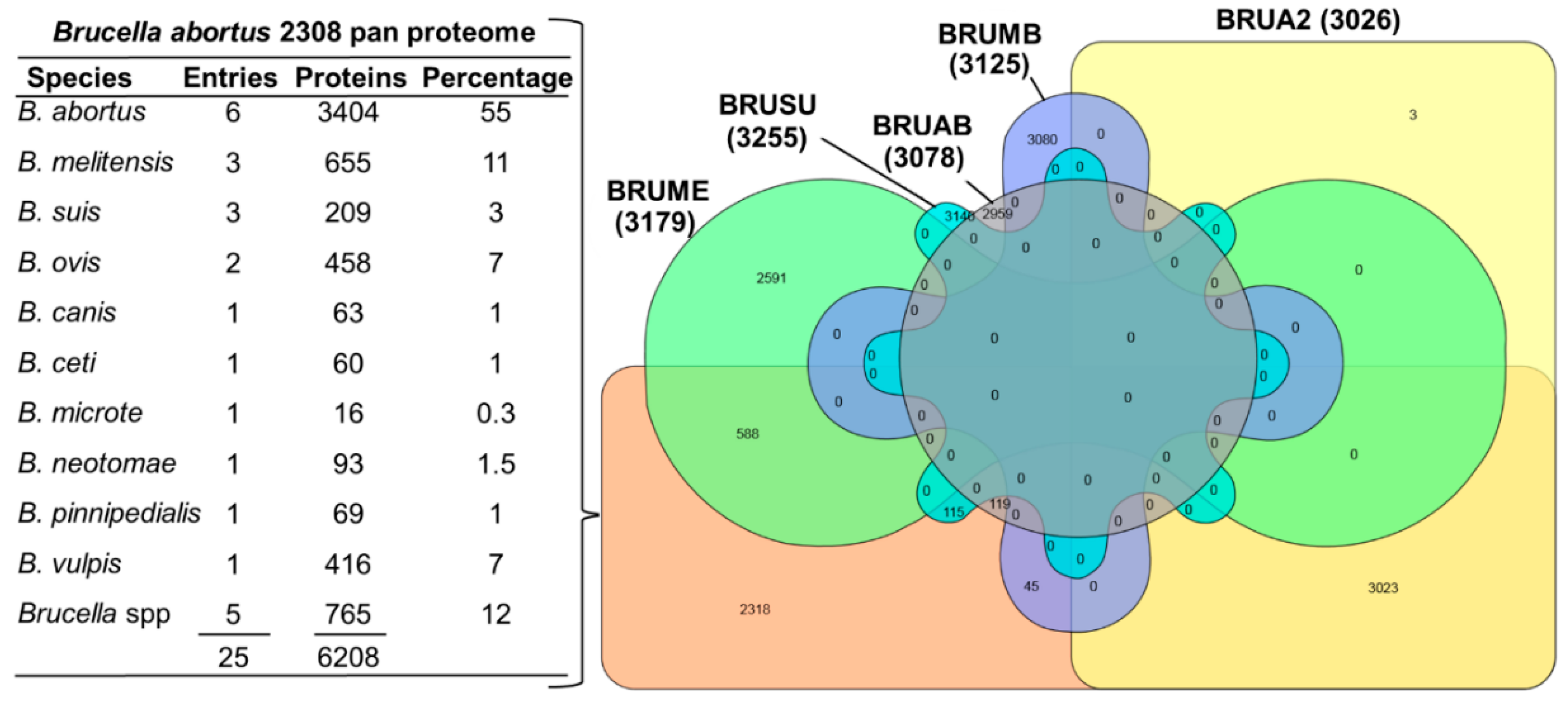
3.2. Databases and Protein Sequences

Protein identification by matching mass spectra against a database of known sequences is an important tool in proteomics. Despite the availability of a list of differentially expressed proteins and the influences of the physicochemical environment, missing or unknown sequences remain a limiting factor. UniProt listed as many as 458 proteome entries for Brucella, however, 329 entries of proteomic data were redundant and moved to the UniProt Archive (UniParc) database. The remaining nonredundant database represents 10 Brucella species and remains active in the UniProt KB database. UniProt introduced the new term “pan proteome” to describe the entire set of proteins thought to be expressed by a group of highly related organisms, e.g., multiple strains of a species. The pan database entries also included all sequences within a taxonomical group as well as unique sequences not found in the reference proteome [58]. Consequently, 25 proteomes representing 10 species of Brucella were used to create the B. abortus 2308 pan proteome and the B. abortus (strain 2308) proteome remained the reference proteome. An analysis of protein IDs of the pan proteome and five strains of Brucella sp. based on the online software tool InteractiVenn [59] revealed that the complete list of the Brucella reference proteome (except three entries) forms 47% of the pan proteome (Figure 2). Moreover, the bulk of protein entries in the pan proteome were from B. abortus, B. melitensis, B. suis, B. ovis and B. vulpis, respectively. The Brucella pan proteome contains 7266 protein entries. Their existence was mostly predicted (77%) or inferred from homology (21.6%) whereas evidence at the protein level (1%) and transcript level (0.1%) was scarce. The manual curation of protein entries was reported for 528 protein entries which correspond to approximately 7.3% of Brucella-specific protein entries. For MS-based proteome analysis of two species, pan proteome or inclusion of all 10 Brucella proteomes for protein identification might be inconvenient due to the difference in the entry IDs for each species. Therefore, for the sake of effective protein identification, the following two proteomes were combined: (1) B. abortus (strain 2308) with a protein count of 3023, distributed on chromosome I (1991) and II (1034, proteome UP000002719, organism ID: 359391. update: 11 February 2017) and (2) B. melitensis biotype 1 (strain 16M/ATCC 23456/NCTC 10094) with a protein count of 3178 distributed on chromosome I (2049) and II (1131, proteome UP000008511, organism ID: 224914. update: 04 February 2017). The latter was redundant and replaced with B. melitensis with a protein count of 3123 distributed on chromosome I (1075) and II (2048, proteome UP000290786, organism ID: 29459. update: 01 December 2019, accessed on March 2020). This choice was based on the designation of B. abortus (strain 2308) as reference proteome and the highest number of protein entries among the three available B. melitensis proteomes.

3.3. Protein Identification

A MaxQuant-Andromeda-based search against the combined database of B. abortus (strain 2308) and B. melitensis biotype 1 (strain 16M) resulted in the identification of 1202 proteins with at least one unique peptide specific for a protein. A total of 826 proteins were identified, after applying the filter that label-free quantification intensity (LFQ) of a protein was present in at-least four out of six replicates in each sample dataset and after removal of proteins matched toreverse sequences and those proteins identified “by site” (Supplementary Table S1). Among these identified proteins, 478 protein IDs belonged to the B. abortus reference proteome and the remaining 348 protein IDs could be allocated to the B. melitensis proteome. The distribution of the identified proteins with respect to the chromosomes was 360 on chromosome I (43.6%) and 118 on chromosome II (14%) of B. abortus and 161 proteins expressed on chromosome I (19.5%) and 56 on chromosome II (6.8%) of B. melitensis. The remaining 131 proteins (15.7%) belonged to B. melitensis unassembled WGS sequences, which are also part of the B. abortus 2308 pan proteome. Later, a database update resulted in the addition of one protein in chromosome I of B. abortus (strain 2308, update 05 December 2016). The proteome UP000008511 was moved to UniParc as it was identified as redundant (update 18 November 2016) and the majority of its protein entries were found to be matching with proteome UP000000419 (update 9 October 2016). As a result, 131 protein entries (belonging to proteome UP000008511: B. melitensis biotype 1) of the 826 identified proteins were found redundant (marked as removed in Supplementary Table S1) or obsolete and deposited at the UniParc database. Consequently, the remaining 695 identified proteins were considered for further analysis.

3.4. Comparative Proteomics of B. abortus and B. melitensis

Visualization through unsupervised hierarchical clustering of the proteomic data recapitulated the similarities between strains and species (Figure 3). All six replicates of each isolate clustered together and the species displayed a clear clustering. For comparative proteomics analysis, pairwise comparisons were carried out in six categories as follows,
  • Category I: B. melitensis 16M vs. B. abortus 544 (M vs. A);
  • Category II: B. melitensis C vs. B. abortus T (M2 vs. A2);
  • Category III: B. melitensis C vs. B. abortus 544 (M2 vs. A);
  • Category IV: B. melitensis 16M vs. B. abortus T (M vs. A2);
  • Category V: B. abortus T vs. B. abortus 544 (A2 vs. A);
  • Category VI: B. melitensis C vs. B. melitensis 16M (M2 vs. M).
As shown in Figure 3, the volcano plot displays the negative log10 t-test p-value over the log2 fold-change. Proteins with p-values above the dotted line (p < 0.05) were considered to be differentially expressed between the two groups. The left side of the plot represents the downregulated proteins and the right side represents the upregulated proteins. Table 2 lists the differentially expressed proteins in each of the above-described categories. The proteins identified as significantly regulated in at least three of the four comparison categories (I–IV) were considered to play a crucial role. As a result, 109 and 104 proteins were identified as up- or downregulated in B. melitensis when compared to B. abortus (Supplementary Table S2).

3.5. Geno Ontology and Clusters of Orthologous Groups

UniProt was used to cluster the proteins in accordance with their Gene Ontologies (GO) for understanding the functional role of the identified proteins. The GO mapping was possible only for those proteins that remained active at the UniProt database until the analysis. GO results presented in Figure 4 include 695 identified proteins among which 66 and 94 were up- or downregulated, respectively, in B. melitensis compared to B. abortus, while Clusters of Orthologous Groups (COGs) are indicated for all 826 identified proteins including 131 redundant proteins. Catalytic activity, binding properties, transporter and antioxidant activity, as well as ribosomal structural constitution, were different between these two species. Macromolecular, membrane and cellular components also varied. The major metabolic and cellular processes appeared to be similar between these species. The prediction of COGs revealed the distribution within four functional groups: cellular processes and signaling (D, M, O, T and U), information storage and processing (J, K and L), metabolism (C, E, F, G, H, I, P and G) and poorly characterized (S). Analysis of about 20% of the identified proteins identifies any COG. Based on the COG clustering, membrane proteins and proteins involved in biomolecular transport and protein synthesis mechanism were found to be enhanced in B. abortus in comparison to B. melitensis.

3.6. Bioinformatics Annotation of Differentially Expressed Proteins

As shown in Table 3, the DAVID analysis of differentially expressed proteins revealed involvement of several Kyoto Encyclopedia of Genes and Genomes (KEGG) pathways. Notably, histidine metabolism appeared to be different in all compared groups. As shown in Figure 5, among the top 10 pathways, the observed differences in the categories ABC transporters, aminoacyl-tRNA biosynthesis and oxidative phosphorylation merit further investigation to clarify, if these pathways influence biochemical diagnosis or host specificity. The role of ABC transporters in intracellular survival and virulence of Brucella was demonstrated in B. ovis [60]. It was also shown that about 9% of the coding ability of Brucella is devoted towards ABC transporters, but differences between these two Brucella species were reported [61]. Analysis using DAVID indicated that the proteins downregulated in B. abortus in comparison to B. melitensis occurred within three biological pathways, each with four of the proteins identified: pyruvate metabolism, histidine metabolism, arginine and proline metabolism and lysine degradation, and tryptophan metabolism. In contrast, the upregulated proteins were present in three other pathways: 11 proteins in metabolic pathways, four proteins in carbon metabolism and three proteins in pyrimidine metabolism.

3.7. Predicted Virulence-Associated Proteins

The prediction of protein pathogenicity was carried out using the freely available online Support Vector Machines (SVM)-based tool VirulentPred [49]. Of the 826 proteins identified, 102 proteins (12%) were predicted to be potentially virulence-associated (Supplementary Table 3) among which 22 proteins were identified as differentially expressed proteins (16 upregulated and six downregulated in B. abortus comparison to B. melitensis). With the exception of the proteins listed in Table 4, all other differentially expressed proteins predicted as virulence-associated were listed as uncharacterized proteins. The upregulation of potentially virulence-associated proteins and downregulation of ribosomal proteins indicate different degrees of control operations that prepare the bacterial agent for infection [62]. The identified proteins can be explored for further application in diagnostics.

3.8. Field Strains and Host Adaptability

Within the same species, the type and field strains also displayed significant variations in the protein abundances. As listed in Supplementary Table S1, the type strain and field strain of B. abortus showed differences in the expression of 180 proteins, among which 97 and 83 proteins were up- and downregulated, respectively, in the field strain when compared to that of the type strain. On the other hand, B. melitensis displayed differences in 224 proteins, among which 129 and 95 were found to be up- or downregulated in the field strain. Among these differentially expressed proteins, as listed in Table 5, 10 proteins including several binding proteins—appeared to be highly abundant among the type strains of both species, while 19 proteins—mostly belonging to the Type IV secretion system (T4SS)—were highly abundant among the field strains of B. abortus and B. melitensis. T4SS has been associated with increased host adaptability and described as an essential pathogenicity factor in several pathogens including Brucella spp., Helicobacter pylori, Legionella pneumophila and Bartonella spp [71,72]. T4SS influences the intracellular survival in the host [73,74]. Nine proteins identified as upregulated in the B. abortus field strain were found to be downregulated in the field strain of B. melitensis. On the other hand, seven proteins identified as upregulated in the B. melitensis field strain were found to be downregulated in the B. abortus field strain. These proteins are worth further analysis as they might play a role in the known host-species specificity and might be useful for designing species-specific diagnostic tools.

4. Conclusions

In conclusion, our quantitative proteomic analysis of reference and field-isolated strains of B. abortus and B. melitensis not surprisingly confirms the existence of proteome level differences between the strains. Besides differences in metabolic pathways, B. abortus and B. melitensis displayed differences in ABC transporters, which were shown to play a role in intracellular survival and virulence of Brucella. Field isolates displayed enhanced abundance in several binding proteins and Type IV secretion systems (T4SS), these have been associated with host adaptability and essential pathogenic factors. B. abortus field strain displayed a high abundance of 10 proteins and seven proteins were of high abundance in B. melitensis. These proteins might be playing a role in host specificity. With the exception of seven proteins, all other proteins (n = 15) identified as potentially virulence-associated were uncharacterized proteins. Problems arise from the existence of multiple redundant reference proteomes for different Brucella species. The benefits of the recently introduced pan proteome concept based on protein entries of 10 known Brucella species also remain limited, as long as the majority of protein entries at the UniProt database remain unreviewed/curated. Establishing a species-specific proteome would be useful for understanding the host specificity and infection of Brucella species. We suggest that in addition to improvements in the reference database, further in-depth proteomic analyses are performed on these strains cocultured with their respective host cell lines. This might lead to an understanding of the mechanism lying behind the described host specificity and pathogenicity.

Supplementary Materials

The following are available online at https://www.mdpi.com/2218-273X/10/6/836/s1, Table S1: List of identified proteins; Table S2: List of differentially expressed proteins; Table S3: Virulence-associated protein prediction result.

Author Contributions

Conceptualization, J.M., G.W. and H.N.; methodology, J.M., G.W., M.E. and C.W.; data analysis, J.M.; writing—original draft preparation, J.M.; writing—reviewing and editing, U.R., L.D.S. and H.N.; All authors have read and agreed to the published version of the manuscript.

Funding

This work is supported by an internal fund from the FLI. The author Murat Eravci was supported by the Deutsche Forschungsgemeinschaft (DFG, SFB 958).

Acknowledgments

The authors would like to thank Michael Kühl for excellent technical support. For mass spectrometry (Christoph Weise and Murat Eravci), we would like to acknowledge the assistance of the Core Facility BioSupraMol supported by the Deutsche Forschungsgemeinschaft (DFG). We would like to thank the Academy of Scientific Research and Technology (ASRT) Egypt for partial financial support. This work is part of the BrucMedNet project (ID: 698) funded by the ARIMNet2. The assistance in grammatical corrections by Marc Howard Rich, SRM University AP, Andhra Pradesh is greatly appreciated.

Conflicts of Interest

The authors declare no conflict of interest.

References

  1. Moreno, E.; Stackebrandt, E.; Dorsch, M.; Wolters, J.; Busch, M.; Mayer, H. Brucella abortus 16S rRNA and lipid A reveal a phylogenetic relationship with members of the alpha-2 subdivision of the class Proteobacteria. J. Bacteriol. 1990, 172, 3569–3576. [Google Scholar] [CrossRef] [PubMed] [Green Version]
  2. Cardoso, P.G.; Macedo, G.C.; Azevedo, V.; Oliveira, S.C. Brucella spp noncanonical LPS: Structure, biosynthesis, and interaction with host immune system. Microb. Cell Fact. 2006, 5, 13. [Google Scholar] [CrossRef] [PubMed] [Green Version]
  3. Christopher, S.; Umapathy, B.L.; Ravikumar, K.L. Brucellosis: Review on the recent trends in pathogenicity and laboratory diagnosis. J. Lab. Physicians 2010, 2, 55–60. [Google Scholar] [CrossRef] [PubMed]
  4. Whatmore, A.M. Current understanding of the genetic diversity of Brucella, an expanding genus of zoonotic pathogens. Infect. Genet. Evol. 2009, 9, 1168–1184. [Google Scholar] [CrossRef] [PubMed]
  5. Verger, J.M.; Grimont, F.; Grimont, P.A.D.; Grayon, M. Brucella, a Monospecific Genus as Shown by Deoxyribonucleic-Acid Hybridization. Int. J. Syst. Bacteriol. 1985, 35, 292–295. [Google Scholar] [CrossRef]
  6. Brenner, D.; Staley, J.; Krieg, N. Classification of Procaryotic Organisms and the Concept of Bacterial Speciation. In Bergey’s Manual® of Systematic Bacteriology; Brenner, D., Krieg, N., Staley, J., Garrity, G., Eds.; Springer US: New York, NY, USA, 2005; pp. 27–32. [Google Scholar]
  7. Chain, P.S.; Comerci, D.J.; Tolmasky, M.E.; Larimer, F.W.; Malfatti, S.A.; Vergez, L.M.; Aguero, F.; Land, M.L.; Ugalde, R.A.; Garcia, E. Whole-genome analyses of speciation events in pathogenic Brucellae. Infect. Immun. 2005, 73, 8353–8361. [Google Scholar] [CrossRef] [Green Version]
  8. Halling, S.M.; Peterson-Burch, B.D.; Bricker, B.J.; Zuerner, R.L.; Qing, Z.; Li, L.L.; Kapur, V.; Alt, D.P.; Olsen, S.C. Completion of the genome sequence of Brucella abortus and comparison to the highly similar genomes of Brucella melitensis and Brucella suis. J. Bacteriol. 2005, 187, 2715–2726. [Google Scholar] [CrossRef] [Green Version]
  9. Wang, Y.; Ke, Y.; Wang, Z.; Yuan, X.; Qiu, Y.; Zhen, Q.; Xu, J.; Li, T.; Wang, D.; Huang, L.; et al. Genome sequences of three live attenuated vaccine strains of Brucella species and implications for pathogenesis and differential diagnosis. J. Bacteriol. 2012, 194, 6012–6013. [Google Scholar] [CrossRef] [Green Version]
  10. Singh, D.K.; Kumar, A.; Tiwari, A.K.; Sankarasubramanian, J.; Vishnu, U.S.; Sridhar, J.; Gunasekaran, P.; Rajendhran, J. Draft Genome Sequence of Brucella abortus Virulent Strain 544. Genome Announc. 2015, 3. [Google Scholar] [CrossRef] [Green Version]
  11. DelVecchio, V.G.; Kapatral, V.; Redkar, R.J.; Patra, G.; Mujer, C.; Los, T.; Ivanova, N.; Anderson, I.; Bhattacharyya, A.; Lykidis, A.; et al. The genome sequence of the facultative intracellular pathogen Brucella melitensis. Proc. Natl. Acad. Sci. USA 2002, 99, 443–448. [Google Scholar] [CrossRef] [Green Version]
  12. Rajashekara, G.; Glasner, J.D.; Glover, D.A.; Splitter, G.A. Comparative whole-genome hybridization reveals genomic islands in Brucella species. J. Bacteriol. 2004, 186, 5040–5051. [Google Scholar] [CrossRef] [PubMed] [Green Version]
  13. Cloeckaert, A.; Verger, J.M.; Grayon, M.; Paquet, J.Y.; Garin-Bastuji, B.; Foster, G.; Godfroid, J. Classification of Brucella spp. isolated from marine mammals by DNA polymorphism at the omp2 locus. Microbes. Infect. Inst. Pasteur 2001, 3, 729–738. [Google Scholar] [CrossRef]
  14. Cloeckaert, A.; Vizcaino, N.; Paquet, J.Y.; Bowden, R.A.; Elzer, P.H. Major outer membrane proteins of Brucella spp.: Past, present and future. Vet. Microbiol. 2002, 90, 229–247. [Google Scholar] [CrossRef]
  15. Adone, R.; Muscillo, M.; La Rosa, G.; Francia, M.; Tarantino, M. Antigenic, immunologic and genetic characterization of rough strains B. abortus RB51, B. melitensis B115 and B. melitensis B18. PLoS ONE 2011, 6, e24073. [Google Scholar] [CrossRef] [PubMed] [Green Version]
  16. Yang, Y.; Wang, L.; Yin, J.; Wang, X.; Cheng, S.; Lang, X.; Wang, X.; Qu, H.; Sun, C.; Wang, J.; et al. Immunoproteomic analysis of Brucella melitensis and identification of a new immunogenic candidate protein for the development of brucellosis subunit vaccine. Mol. Immunol. 2011, 49, 175–184. [Google Scholar] [CrossRef] [PubMed]
  17. Al Dahouk, S.; Nöckler, K.; Scholz, H.C.; Tomaso, H.; Bogumil, R.; Neubauer, H. Immunoproteomic characterization of Brucella abortus 1119-3 preparations used for the serodiagnosis of Brucella infections. J. Immunol. Methods 2006, 309, 34–47. [Google Scholar] [CrossRef] [PubMed]
  18. Tabynov, K.; Ryskeldinova, S.; Sansyzbay, A. An influenza viral vector Brucella abortus vaccine induces good cross-protection against Brucella melitensis infection in pregnant heifers. Vaccine 2015, 33, 3619–3623. [Google Scholar] [CrossRef]
  19. Dorneles, E.M.; Sriranganathan, N.; Lage, A.P. Recent advances in Brucella abortus vaccines. Vet. Res. 2015, 46, 76. [Google Scholar] [CrossRef] [Green Version]
  20. Al Dahouk, S.; Tomaso, H.; Nockler, K.; Neubauer, H.; Frangoulidis, D. Laboratory-based diagnosis of brucellosis—A review of the literature. Part II: Serological tests for brucellosis. Clin. Lab. 2003, 49, 577–589. [Google Scholar]
  21. Al Dahouk, S.; Scholz, H.C.; Tomaso, H.; Bahn, P.; Gollner, C.; Karges, W.; Appel, B.; Hensel, A.; Neubauer, H.; Nockler, K. Differential phenotyping of Brucella species using a newly developed semi-automated metabolic system. BMC Microbiol. 2010, 10, 269. [Google Scholar] [CrossRef] [Green Version]
  22. Alton, G.G.; Forsyth, J.R.L. Brucella. In Medical Microbiology, 4th ed.; Baron, S., Ed.; University of Texas Medical Branch at Galveston: Galveston, TX, USA, 1996. [Google Scholar]
  23. Wareth, G.; Eravci, M.; Weise, C.; Roesler, U.; Melzer, F.; Sprague, L.D.; Neubauer, H.; Murugaiyan, J. Comprehensive identification of immunodominant proteins of Brucella abortus and Brucella melitensis using antibodies in the sera from naturally infected hosts. Int. J. Mol. Sci. 2016, 17, 659. [Google Scholar] [CrossRef] [Green Version]
  24. Wareth, G.; Melzer, F.; Weise, C.; Neubauer, H.; Roesler, U.; Murugaiyan, J. Proteomics-based identification of immunodominant proteins of Brucellae using sera from infected hosts points towards enhanced pathogen survival during the infection. Biochem. Biophys. Res. Commun. 2015, 456, 202–206. [Google Scholar] [CrossRef] [PubMed]
  25. Connolly, J.P.; Comerci, D.; Alefantis, T.G.; Walz, A.; Quan, M.; Chafin, R.; Grewal, P.; Mujer, C.V.; Ugalde, R.A.; DelVecchio, V.G. Proteomic analysis of Brucella abortus cell envelope and identification of immunogenic candidate proteins for vaccine development. Proteomics 2006, 6, 3767–3780. [Google Scholar] [CrossRef] [PubMed]
  26. DelVecchio, V.G.; Wagner, M.A.; Eschenbrenner, M.; Horn, T.A.; Kraycer, J.A.; Estock, F.; Elzer, P.; Mujer, C.V. Brucella proteomes—A review. Vet. Microbiol. 2002, 90, 593–603. [Google Scholar] [CrossRef]
  27. Eschenbrenner, M.; Horn, T.A.; Wagner, M.A.; Mujer, C.V.; Miller-Scandle, T.L.; DelVecchio, V.G. Comparative proteome analysis of laboratory grown Brucella abortus 2308 and Brucella melitensis 16M. J. Proteome Res. 2006, 5, 1731–1740. [Google Scholar] [CrossRef]
  28. Eschenbrenner, M.; Wagner, M.A.; Horn, T.A.; Kraycer, J.A.; Mujer, C.V.; Hagius, S.; Elzer, P.; DelVecchio, V.G. Comparative proteome analysis of Brucella melitensis vaccine strain Rev 1 and a virulent strain, 16M. J. Bacteriol. 2002, 184, 4962–4970. [Google Scholar] [CrossRef] [Green Version]
  29. Lamontagne, J.; Beland, M.; Forest, A.; Cote-Martin, A.; Nassif, N.; Tomaki, F.; Moriyon, I.; Moreno, E.; Paramithiotis, E. Proteomics-based confirmation of protein expression and correction of annotation errors in the Brucella abortus genome. BMC Genom. 2010, 11, 300. [Google Scholar] [CrossRef] [Green Version]
  30. Mujer, C.V.; Wagner, M.A.; Eschenbrenner, M.; Horn, T.; Kraycer, J.A.; Redkar, R.; Hagius, S.; Elzer, P.; Delvecchio, V.G. Global analysis of Brucella melitensis proteomes. Ann. N. Y. Acad. Sci. 2002, 969, 97–101. [Google Scholar] [CrossRef]
  31. Paredes-Cervantes, V.; Flores-Mejia, R.; Moreno-Lafont, M.C.; Lanz-Mendoza, H.; Tello-Lopez, A.T.; Castillo-Vera, J.; Pando-Robles, V.; Hurtado-Sil, G.; Gonzalez-Gonzalez, E.; Rodriguez-Cortes, O.; et al. Comparative proteome analysis of Brucella abortus 2308 and its virB type IV secretion system mutant reveals new T4SS-related candidate proteins. J. Proteom. 2011, 74, 2959–2971. [Google Scholar] [CrossRef]
  32. Wagner, M.A.; Eschenbrenner, M.; Horn, T.A.; Kraycer, J.A.; Mujer, C.V.; Hagius, S.; Elzer, P.; DelVecchio, V.G. Global analysis of the Brucella melitensis proteome: Identification of proteins expressed in laboratory-grown culture. Proteomics 2002, 2, 1047–1060. [Google Scholar] [CrossRef]
  33. Zai, X.; Yang, Q.; Yin, Y.; Li, R.; Qian, M.; Zhao, T.; Li, Y.; Zhang, J.; Fu, L.; Xu, J.; et al. Relative Quantitative Proteomic Analysis of Brucella abortus Reveals Metabolic Adaptation to Multiple Environmental Stresses. Front. Microbiol. 2017, 8, 2347. [Google Scholar] [CrossRef] [PubMed]
  34. Zai, X.; Yang, Q.; Liu, K.; Li, R.; Qian, M.; Zhao, T.; Li, Y.; Yin, Y.; Dong, D.; Fu, L.; et al. A comprehensive proteogenomic study of the human Brucella vaccine strain 104 M. BMC Genom. 2017, 18, 402. [Google Scholar] [CrossRef] [PubMed] [Green Version]
  35. Laemmli, U.K. Cleavage of structural proteins during the assembly of the head of bacteriophage T4. Nature 1970, 227, 680–685. [Google Scholar] [CrossRef]
  36. Candiano, G.; Bruschi, M.; Musante, L.; Santucci, L.; Ghiggeri, G.M.; Carnemolla, B.; Orecchia, P.; Zardi, L.; Righetti, P.G. Blue silver: A very sensitive colloidal Coomassie G-250 staining for proteome analysis. Electrophoresis 2004, 25, 1327–1333. [Google Scholar] [CrossRef]
  37. Murugaiyan, J.; Eravci, M.; Weise, C.; Roesler, U. Label-free quantitative proteomic analysis of harmless and pathogenic strains ofinfectious microalgae, Prototheca spp. Int. J. Mol. Sci. 2016, 18, 59. [Google Scholar] [CrossRef]
  38. Rappsilber, J.; Ishihama, Y.; Mann, M. Stop and go extraction tips for matrix-assisted laser desorption/ionization, nanoelectrospray, and LC/MS sample pretreatment in proteomics. Anal. Chem. 2003, 75, 663–670. [Google Scholar] [CrossRef] [PubMed]
  39. Cox, J.; Neuhauser, N.; Michalski, A.; Scheltema, R.A.; Olsen, J.V.; Mann, M. Andromeda: A peptide search engine integrated into the MaxQuant environment. J. Proteome Res. 2011, 10, 1794–1805. [Google Scholar] [CrossRef] [PubMed]
  40. Cox, J.; Matic, I.; Hilger, M.; Nagaraj, N.; Selbach, M.; Olsen, J.V.; Mann, M. A practical guide to the MaxQuant computational platform for SILAC-based quantitative proteomics. Nat. Protoc. 2009, 4, 698–705. [Google Scholar] [CrossRef] [PubMed]
  41. Luber, C.A.; Cox, J.; Lauterbach, H.; Fancke, B.; Selbach, M.; Tschopp, J.; Akira, S.; Wiegand, M.; Hochrein, H.; O’Keeffe, M.; et al. Quantitative proteomics reveals subset-specific viral recognition in dendritic cells. Immunity 2010, 32, 279–289. [Google Scholar] [CrossRef] [Green Version]
  42. Cox, J.; Hein, M.Y.; Luber, C.A.; Paron, I.; Nagaraj, N.; Mann, M. Accurate proteome-wide label-free quantification by delayed normalization and maximal peptide ratio extraction, termed MaxLFQ. Mol. Cell. Proteom. 2014, 13, 2513–2526. [Google Scholar] [CrossRef] [Green Version]
  43. Benjamini, Y.; Hochberg, Y. Controlling the false discovery rate—A practical and powerful approach to multiple testing. J. R. Stat. Soc. B Met. 1995, 57, 289–300. [Google Scholar] [CrossRef]
  44. Vizcaino, J.A.; Csordas, A.; Del-Toro, N.; Dianes, J.A.; Griss, J.; Lavidas, I.; Mayer, G.; Perez-Riverol, Y.; Reisinger, F.; Ternent, T.; et al. 2016 update of the PRIDE database and its related tools. Nucleic Acids Res. 2016, 44, 11033. [Google Scholar] [CrossRef] [PubMed] [Green Version]
  45. Vizcaino, J.A.; Deutsch, E.W.; Wang, R.; Csordas, A.; Reisinger, F.; Rios, D.; Dianes, J.A.; Sun, Z.; Farrah, T.; Bandeira, N.; et al. ProteomeXchange provides globally coordinated proteomics data submission and dissemination. Nat. Biotechnol. 2014, 32, 223–226. [Google Scholar] [CrossRef] [PubMed]
  46. Huerta-Cepas, J.; Szklarczyk, D.; Forslund, K.; Cook, H.; Heller, D.; Walter, M.C.; Rattei, T.; Mende, D.R.; Sunagawa, S.; Kuhn, M.; et al. eggNOG 4.5: A hierarchical orthology framework with improved functional annotations for eukaryotic, prokaryotic and viral sequences. Nucleic Acids Res. 2016, 44, D286–D293. [Google Scholar] [CrossRef] [Green Version]
  47. Huang, D.W.; Sherman, B.T.; Lempicki, R.A. Systematic and integrative analysis of large gene lists using DAVID bioinformatics resources. Nat. Protoc. 2009, 4, 44–57. [Google Scholar] [CrossRef]
  48. Huang, D.W.; Sherman, B.T.; Lempicki, R.A. Bioinformatics enrichment tools: Paths toward the comprehensive functional analysis of large gene lists. Nucleic Acids Res. 2009, 37, 1–13. [Google Scholar] [CrossRef] [Green Version]
  49. Garg, A.; Gupta, D. VirulentPred: A SVM based prediction method for virulent proteins in bacterial pathogens. BMC Bioinform. 2008, 9, 62. [Google Scholar] [CrossRef] [Green Version]
  50. Perez-Riverol, Y.; Csordas, A.; Bai, J.; Bernal-Llinares, M.; Hewapathirana, S.; Kundu, D.J.; Inuganti, A.; Griss, J.; Mayer, G.; Eisenacher, M.; et al. The PRIDE database and related tools and resources in 2019: Improving support for quantification data. Nucleic Acids Res. 2019, 47, D442–D450. [Google Scholar] [CrossRef]
  51. Meyer, M.E.; Morgan, W.J. Metabolic characterization of Brucella strains that show conflicting identity by biochemical and serological methods. Bull. World Health Organ. 1962, 26, 823–827. [Google Scholar]
  52. He, Y. Analyses of Brucella pathogenesis, host immunity, and vaccine targets using systems biology and bioinformatics. Front. Cell. Infect. Microbiol. 2012, 2, 2. [Google Scholar] [CrossRef] [Green Version]
  53. Teixeira-Gomes, A.P.; Cloeckaert, A.; Bezard, G.; Dubray, G.; Zygmunt, M.S. Mapping and identification of Brucella melitensis proteins by two-dimensional electrophoresis and microsequencing. Electrophoresis 1997, 18, 156–162. [Google Scholar] [CrossRef] [PubMed]
  54. Crasta, O.R.; Folkerts, O.; Fei, Z.; Mane, S.P.; Evans, C.; Martino-Catt, S.; Bricker, B.; Yu, G.; Du, L.; Sobral, B.W. Genome sequence of Brucella abortus vaccine strain S19 compared to virulent strains yields candidate virulence genes. PLoS ONE 2008, 3, e2193. [Google Scholar] [CrossRef] [PubMed]
  55. Lamontagne, J.; Forest, A.; Marazzo, E.; Denis, F.; Butler, H.; Michaud, J.F.; Boucher, L.; Pedro, I.; Villeneuve, A.; Sitnikov, D.; et al. Intracellular adaptation of Brucella abortus. J. Proteome Res. 2009, 8, 1594–1609. [Google Scholar] [CrossRef] [PubMed] [Green Version]
  56. Tsoktouridis, G.; Merz, C.A.; Manning, S.P.; Giovagnoli-Kurtz, R.; Williams, L.E.; Mujer, C.V.; Hagius, S.; Elzer, P.; Redkar, R.J.; Patra, G.; et al. Molecular characterization of Brucella abortus chromosome II recombination. J. Bacteriol. 2003, 185, 6130–6136. [Google Scholar] [CrossRef] [Green Version]
  57. Teixeira-Gomes, A.P.; Cloeckaert, A.; Zygmunt, M.S. Characterization of heat, oxidative, and acid stress responses in Brucella melitensis. Infect. Immun. 2000, 68, 2954–2961. [Google Scholar] [CrossRef] [Green Version]
  58. UniProt. UniProt: The universal protein knowledgebase. Nucleic Acids Res. 2017, 45, D158–D169. [Google Scholar] [CrossRef]
  59. Heberle, H.; Meirelles, G.V.; da Silva, F.R.; Telles, G.P.; Minghim, R. InteractiVenn: A web-based tool for the analysis of sets through Venn diagrams. BMC Bioinform. 2015, 16. [Google Scholar] [CrossRef]
  60. Silva, T.M.A.; Mol, J.P.S.; Winter, M.G.; Atluri, V.; Xavier, M.N.; Pires, S.F.; Paixao, T.A.; Andrade, H.M.; Santos, R.L.; Tsolis, R.M. The Predicted ABC Transporter AbcEDCBA Is Required for Type IV Secretion System Expression and Lysosomal Evasion by Brucella ovis. PLoS ONE 2014, 9. [Google Scholar] [CrossRef]
  61. Jenner, D.C.; Dassa, E.; Whatmore, A.M.; Atkins, H.S. ATP-Binding Cassette Systems of Brucella. Comp. Funct. Genom. 2009, 354649. [Google Scholar] [CrossRef] [Green Version]
  62. Smircich, P.; Eastman, G.; Bispo, S.; Duhagon, M.A.; Guerra-Slompo, E.P.; Garat, B.; Goldenberg, S.; Munroe, D.J.; Dallagiovanna, B.; Holetz, F.; et al. Ribosome profiling reveals translation control as a key mechanism generating differential gene expression in Trypanosoma cruzi. BMC Genom. 2015, 16. [Google Scholar] [CrossRef] [Green Version]
  63. Caswell, C.C.; Elhassanny, A.E.; Planchin, E.E.; Roux, C.M.; Weeks-Gorospe, J.N.; Ficht, T.A.; Dunman, P.M.; Roop, R.M., II. Diverse genetic regulon of the virulence-associated transcriptional regulator MucR in Brucella abortus 2308. Infect. Immun. 2013, 81, 1040–1051. [Google Scholar] [CrossRef] [PubMed] [Green Version]
  64. Tibor, A.; Wansard, V.; Bielartz, V.; Delrue, R.M.; Danese, I.; Michel, P.; Walravens, K.; Godfroid, J.; Letesson, J.J. Effect of omp10 or omp19 deletion on Brucella abortus outer membrane properties and virulence in mice. Infect. Immun. 2002, 70, 5540–5546. [Google Scholar] [CrossRef] [PubMed] [Green Version]
  65. Goolab, S.; Roth, R.L.; van Heerden, H.; Crampton, M.C. Analyzing the molecular mechanism of lipoprotein localization in Brucella. Front. Microbiol. 2015, 6. [Google Scholar] [CrossRef] [PubMed] [Green Version]
  66. Simborio, H.L.T.; Lee, J.J.; Reyes, A.W.B.; Hop, H.T.; Arayan, L.T.; Min, W.; Lee, H.J.; Yoo, H.S.; Kim, S. Evaluation of the combined use of the recombinant Brucella abortus Omp10, Omp19 and Omp28 proteins for the clinical diagnosis of bovine brucellosis. Microb. Pathog. 2015, 83–84, 41–46. [Google Scholar] [CrossRef]
  67. Gee, J.M.; Valderas, M.W.; Kovach, M.E.; Grippe, V.K.; Robertson, G.T.; Ng, W.L.; Richardson, J.M.; Winkler, M.E.; Roop, R.M. The Brucella abortus Cu,Zn superoxide dismutase is required for optimal resistance to oxidative killing by murine macrophages an wild-type virulence in experimentally infected mice. Infect. Immun. 2005, 73, 2873–2880. [Google Scholar] [CrossRef] [Green Version]
  68. Kim, S.W.; Joo, Y.J.; Kim, J. Asc1p, a ribosomal protein, plays a pivotal role in cellular adhesion and virulence in Candida albicans. J. Microbiol. 2010, 48, 842–848. [Google Scholar] [CrossRef]
  69. Du, Z.Q.; Li, X.; Wang, J.Y. Immunogenicity analysis of a novel subunit vaccine candidate molecule-recombinant L7/L12 ribosomal protein of Brucella suis. Appl. Biochem. Biotechnol. 2016, 179, 1445–1455. [Google Scholar] [CrossRef]
  70. Jain, S.; Kumar, S.; Dohre, S.; Afley, P.; Sengupta, N.; Alam, S.I. Identification of a protective protein from stationary-phase exoproteome of Brucella abortus. Pathog. Dis. 2014, 70, 75–83. [Google Scholar] [CrossRef] [Green Version]
  71. Atluri, V.L.; Xavier, M.N.; de Jong, M.F.; den Hartigh, A.B.; Tsolis, R.M. Interactions of the human pathogenic Brucella Species with their hosts. Annu. Rev. Microbiol. 2011, 65, 523–541. [Google Scholar] [CrossRef]
  72. Saenz, H.L.; Engel, P.; Stoeckli, M.C.; Lanz, C.; Raddatz, G.; Vayssier-Taussat, M.; Birtles, R.; Schuster, S.C.; Dehio, C. Genomic analysis of Bartonella identifies type IV secretion systems as host adaptability factors. Nat. Genet. 2007, 39, 1469–1476. [Google Scholar] [CrossRef]
  73. Boschiroli, M.L.; Ouahrani-Bettache, S.; Foulongne, V.; Michaux-Charachon, S.; Bourg, G.; Allardet-Servent, A.; Cazevieille, C.; Lavigne, J.P.; Liautard, J.P.; Ramuz, M.; et al. Type IV secretion and Brucella virulence. Vet. Microbiol. 2002, 90, 341–348. [Google Scholar] [CrossRef]
  74. Wattam, A.R.; Williams, K.P.; Snyder, E.E.; Almeida, N.F.; Shukla, M.; Dickerman, A.W.; Crasta, O.R.; Kenyon, R.; Lu, J.; Shallom, J.M.; et al. Analysis of ten Brucella genomes reveals evidence for horizontal gene transfer despite a preferred intracellular lifestyle. J. Bacteriol. 2009, 191, 3569–3579. [Google Scholar] [CrossRef] [PubMed] [Green Version]
Figure 1. SDS-PAGE separation of whole-cell extract: Lane (a) B. abortus 544, (b) B. abortus T (c) B. melitensis 16 M and (d) B. melitensis C. The strains display comparable bands. Arrows show distinct bands unique for the isolates with 35 kDa in B. abortus and below 20 kDa in B. melitensis.
Biomolecules 10 00836 g001
Figure 2. The table lists the Brucella species (n = 10) included in the B. abortus 2308 pan proteome. Entries: number of isolates; proteins: number of protein entries and percentage: indicates percentage found in the B. abortus 2308 pan proteome. InteractiVenn diagram of Brucella abortus 2308 pan proteome and the entries of five strains (BRUA2—B. abortus (strain 2308, yellow), BRUAB—B. abortus biovar 1 (strain 9-941, gray), BRUME—B. melitensis biotype 1 (strain 16M/ATCC 23456/NCTC 10094, green), BRUMB—B. melitensis biotype 2 (strain ATCC 23457, purple) and BRUSU—B. suis biovar 1 (strain 1330), blue).
Biomolecules 10 00836 g002
Figure 3. Protein expression profiling using label-free quantitative proteomics analysis. (A) Heat map analysis of 826 proteins among the four dataset groups and with six cultural replicates per group. The log10 value of the MS signal intensity is shown. Hierarchical clustering of proteins of all samples was performed using z-score protein intensities for the proteins with p < 0.05 and based on elucidation distance. Columns indicate the samples, and rows indicate the proteins. Protein expression values were log2-normalized (label-free quantification (LFQ)) intensities of all proteins quantified across the samples, where red and green indicate high and low intensity, respectively. (BG) Volcano plot for category I–VI, respectively. Ratios plotted for log 2-fold-change (x-axis) against negative log p-value (y-axis) of the Student’s t-test. Each dot represents a protein.
Biomolecules 10 00836 g003
Figure 4. Gene ontology clustering of identified up- and downregulated proteins. (A) Molecular Function (MF), (B) Cellular Component (CC), (C) Biological Process (BP) and (D) Cluster of Orthologous Groups (COGs).
Biomolecules 10 00836 g004
Figure 5. Top 10 KEGG pathways and the distribution of differentially expressed proteins. Number of proteins (x-axis) plotted against KEGG pathways (code) on the y-axis to show the number of proteins up- and downregulated when B. abortus compared to B. melitensis.
Biomolecules 10 00836 g005
Table 1. Brucella strains used in this study.
StrainDesignation/ID/NumberHostGeographical Region
FAOATCCNCTC
B. melitensisT16M2345610094GoatUSA
B. abortusT5442344810093CattleUK
B. melitensisC * SheepChina
B. abortusT * CattleTurkey
T Type strain, FAO—Food and Agriculture Organization * ID assigned to the field strains deposited at the culture collections of Friedrich-Loeffler-Institut, Jena, Germany (C: China, T: Turkey), ATCC—American Type Culture Collection, NCTC—National Collection of Type Cultures, UK.
Table 2. Number of regulated proteins among type and field strains.
RegulationCategory
I
M vs. A
II
M2 vs. A2
III
M2 vs. A
IV
M vs. A2
V
A2 vs. A
VI
M2 vs. M
Down-1731421662238396
Up-21619922124897129
Regulation, down- and up-: downregulated and upregulated, A—B. abortus, A2—B. abortus 544, M—B. melitensis and M2—B. melitensis 16M.
Table 3. KEGG pathways and the distribution of differentially expressed proteins.
KEGG PathwaysCategory
IIIIIIIVVVI
Metabolic pathways 34 ↑
Carbon metabolism 6 ↑
Glycolysis/gluconeogenesis 7 ↓
Pentose and glucuronate interconversions 3 ↓
Pyruvate metabolism 4 ↑
2-Oxocarboxylic acid metabolism4 ↓
Microbial metabolism in diverse environments 14 ↑10 ↑
Biosynthesis of amino acids11 ↓ 14 ↓
Histidine metabolism5 ↓5 ↓5 ↓6 ↓
Valine, leucine and isoleucine biosynthesis 4 ↓
Purine metabolism 7 ↑
Pyrimidine metabolism5 ↑6 ↑ 7 ↑
Biosynthesis of secondary metabolites19 ↓ 25 ↓
RNA polymerase 3 ↑
Ribosome17 ↑18 ↓14 ↓19 ↑ 22 ↓
Bacterial secretion system 5 ↑5 ↑ 4 ↑5 ↑
ABC transporters 14 ↑
Number: number of differentially expressed proteins, ↑—upregulated, ↓—downregulated.
Table 4. Differentially expressed proteins predicted as potentially virulence-associated.
Acc.Protein DescriptionRegSignificanceReference
D0B8I3*Antifreeze protein(+)Associated with MucR, a transcriptional regulator linked to Brucella virulence[63]
Q8YIA9Antifreeze protein (-)
Q2YM39Antifreeze protein type I(-)
D0B248 *LipA family protein(-)
Q2YIP8Lipoprotein Omp10(-)Reduced virulence in B. abortus with gene deletion and used in diagnostics[64,65,66]
Q2YKV9Superoxide dismutase [Cu-Zn] (-)intracellular survival and used as antigens for subunit vaccines[19,67]
Q2YRA830S ribosomal protein S14(-)role in cellular adhesion and virulence in Candida albicans[68]
ribosomal protein L7/L12 based subunit vaccines[69,70]
Acc. No is the UniProt ID, the protein ID marked with * were moved to UniPrac as they were found to be redundant proteins. Reg—status of protein regulation: (+) denotes upregulation and (-) indicates downregulation of proteins when B. abortus is compared with B. melitensis.
Table 5. Differentially expressed proteins among the field isolates in comparison to that of the respective type strains.
UniProt IDProtein
Downregulated in Field Isolates
Q8YBH7Bacterial extracellular solute-binding protein family 1
Q8YIX9Glyceraldehyde-3-phosphate dehydrogenase
Q2YMI0Phosphatidylserine decarboxylase proenzyme
Q2YLU2Peptidylprolyl isomerase
Q2YLF8Leu/Ile/Val-binding protein homolog 1
Q2YLG0Leu/Ile/Val-binding protein homolog 2
Q2YJA9Leu/Ile/Val-binding protein homolog 5
Q2YQQ6Glutelin:Lipoprotein YaeC family:NLPA lipoprotein
Q2YRP7Ribosome-recycling factor
Q2YR20Uncharacterized protein
Upregulated in Field Isolates
Q2YJ78Type IV secretion system protein virB8
Q2YJ79Type IV secretion system protein virB9
Q2YJ81Type IV secretion system protein virB10
Q2YJ83Type IV secretion system-outer membrane lipoprotein
Q2YJ77Type IV secretion system putative lipoprotein virB7
Q8YB25Alpha-methylacyl-CoA racemase
Q2YIG7NADH:flavin oxidoreductase/NADH oxidase
Q2YKS6Aminotransferase class IV
Q8YCZ2N-acetylglucosamine-6-phosphate deacetylase
Q8YI04ATP-dependent DNA ligase
Q2YK32Catalase
Q2YLX9Lipoprotein putative
Q2YN45Probable cytosol aminopeptidase
Q2YRJ0ATP/GTP-binding site motif A (P-loop)
Q2YRN7Uncharacterized protein
Q8YBU9Putative uroporphyrin-iii c-methyltransferase
Q2YKY2Uncharacterized protein
Q2YL40Uncharacterized protein
Q2YLM7Uncharacterized protein
Downregulated in B. abortus and Upregulated in B. melitensis
Q2YME1Threonylcarbamoyl-AMP synthase
Q2YNW9Dihydroxy-acid dehydratase
Q8YHA5Glutaryl-CoA dehydrogenase
Q2YP66Zinc-containing alcohol dehydrogenase
Q2YQE3Periplasmic binding protein
Q2YMW1Uncharacterized protein
Q2YPK6Uncharacterized protein
Upregulated in B. abortus and Downregulated in B. melitensis
Q2YQV750S ribosomal protein L20
Q2YR5650S ribosomal protein L28
Q2YRY0ABC-type glycine betaine transport system
Q8YBF5Maltose-binding periplasmic protein (Sugar ABC transporter)
Q8YCD1Cystine-binding periplasmic protein
Q8YGE8Cationic amino acid ABC transporter
Q2YKN7Uncharacterized protein
Q2YKN9Uncharacterized protein
Q2YQM2Uncharacterized protein

