Cytoprotection, Genoprotection, and Dermal Exposure Assessment of Chitosan-Based Agronanofungicides

 , , ,
, , ,
Abstract
:
1. Introduction
2. Materials and Methods
2.1. Materials
2.2. Preparation of Chitosan-Based Agronanofungicides
2.3. In Vitro Cytotoxicity and Genotoxicity Analysis
2.3.1. Cell Culture Conditions
2.3.2. Cell Viability 3-(4,5-Dimethyl-2-thiazolyl)-2,5-diphenyl-2H-tetrazolium Bromide (MTT) Assay
2.3.3. Alkaline Comet Assay
2.4. Cellular Death Assay
2.4.1. Cell Culture Conditions
2.4.2. FITC-Annexin V/PI Apoptosis Detection
2.4.3. Cell Morphology
2.5. Dermal Corrosion/Irritation Assay
2.5.1. Animals, Materials, and Experimental Design
2.5.2. Experimental Procedure
2.6. Statistical Analysis
3. Results
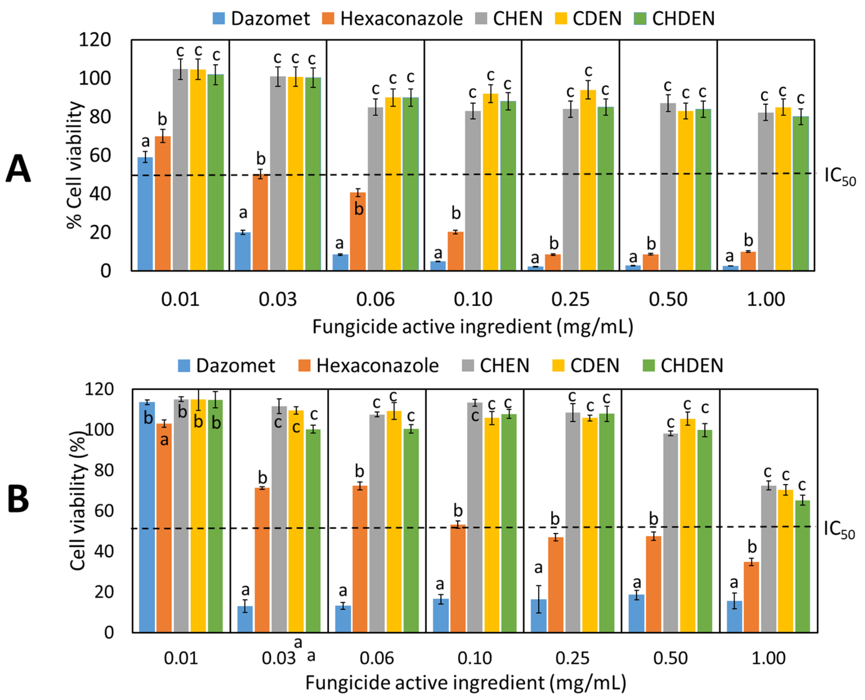
3.1. In Vitro Cytotoxicity Analysis
3.2. In Vitro Genotoxicity Analysis
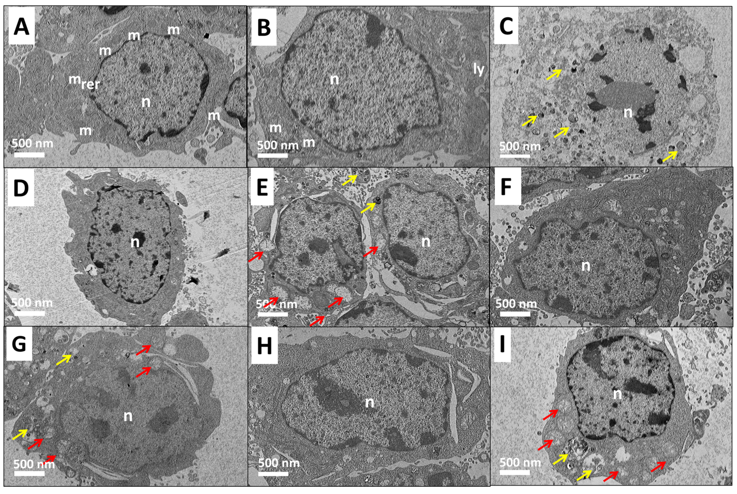
3.3. Cellular Death Assay
3.4. Dermal Irritation/Corrosion Analysis
4. Discussion
5. Conclusions
Author Contributions
Funding
Conflicts of Interest
References
- Aktar, W.; Sengupta, D.; Chowdhury, A. Impact of pesticides use in agriculture: Their benefits and hazards. Interdiscip. Toxicol. 2009, 2, 1–12. [Google Scholar] [CrossRef] [PubMed] [Green Version]
- Popp, J.; Pető, K.; Nagy, J. Pesticide productivity and food security. A review. Agron. Sustain. Dev. 2013, 33, 243–255. [Google Scholar] [CrossRef]
- Damalas, C.A.; Eleftherohorinos, I.G. Pesticide exposure, safety issues, and risk assessment indicators. Int. J. Environ. Res. Public Health 2011, 8, 1402–1419. [Google Scholar] [CrossRef] [PubMed]
- Jeyaratnam, J. Acute pesticide poisoning: A major global health problem. World Health Stat. Q. 1990, 43, 139–144. [Google Scholar]
- Eddleston, M.; Karalliedde, L.; Buckley, N.; Fernando, R.; Hutchinson, G.; Isbister, G.; Konradsen, F.; Murray, D.; Piola, J.C.; Senanayake, N. Pesticide poisoning in the developing world—A minimum pesticides list. Lancet 2002, 360, 1163–1167. [Google Scholar] [CrossRef]
- Bernkop-Schnürch, A.; Dünnhaupt, S. Chitosan-based drug delivery systems. Eur. J. Pharm. Biopharm. 2012, 81, 463–469. [Google Scholar] [CrossRef]
- Maluin, F.N.; Hussein, M.Z.; Idris, A.S. An overview of the oil palm industry: Challenges and some emerging opportunities for nanotechnology development. Agronomy 2020, 10, 356. [Google Scholar] [CrossRef] [Green Version]
- Oliveira, M.E.F.; Silva, É.C.G.; Câmara, C.A.; Souza, I.A.d.; Amorim, R.V.S. Evaluation of acute toxicity of β-lapachone associated with chitosan as a cytoprotective agent. J. Bras. Patol. Med. Lab. 2018, 54, 279–287. [Google Scholar] [CrossRef]
- Dudhani, A.R.; Kosaraju, S.L. Bioadhesive chitosan nanoparticles: Preparation and characterization. Carbohydr. Polym. 2010, 81, 243–251. [Google Scholar] [CrossRef]
- Hong, D.; Park, M.; Yang, S.H.; Lee, J.; Kim, Y.-G.; Choi, I.S. Artificial spores: Cytoprotective nanoencapsulation of living cells. Trends Biotechnol. 2013, 31, 442–447. [Google Scholar] [CrossRef]
- Park, J.H.; Hong, D.; Lee, J.; Choi, I.S. Cell-in-shell hybrids: Chemical nanoencapsulation of individual cells. Acc. Chem. Res. 2016, 49, 792–800. [Google Scholar] [CrossRef] [PubMed]
- Maluin, F.N.; Hussein, M.Z. Chitosan-based agronanochemicals as a sustainable alternative in crop protection. Molecules 2020, 25, 1611. [Google Scholar] [CrossRef] [PubMed] [Green Version]
- Kumari, A.; Yadav, S.K.; Yadav, S.C. Biodegradable polymeric nanoparticles based drug delivery systems. Colloids Surf. B Biointerfaces 2010, 75, 1–18. [Google Scholar] [CrossRef] [PubMed]
- Real, D.; Hoffmann, S.; Leonardi, D.; Salomon, C.; Goycoolea, F.M. Chitosan-based nanodelivery systems applied to the development of novel triclabendazole formulations. PLoS ONE 2018, 13, e0207625. [Google Scholar] [CrossRef]
- Manuja, A.; Kumar, B.; Kumar, R.; Chopra, M.; Dilbaghi, N.; Kumar, S.; Yadav, S.C. Encapsulated quinapyramine sulfate-loaded chitosan/mannitol nanoparticles: Biocompatibility and targeting efficiency in rabbit model of trypanosomosis. Antimicrob. Agents Chemother. 2018, 62, 00466-18. [Google Scholar] [CrossRef] [Green Version]
- Idris, A.; Maizatul, S. Stump Treatment with Dazomet for Controlling Ganoderma Disease in Oil Palm; MPOB: Jaya, Malaysia, 2012; Volume 107. [Google Scholar]
- Idris, A.; Arifurrahman, R.; Kushairi, A. Hexaconale as a Preventive Treatment for Managing Ganoderma in Oil Palm; MPOB: Jaya, Malaysia, 2010; Volume 75. [Google Scholar]
- Wang, D.; Fraedrich, S.W.; Juzwik, J.; Spokas, K.; Zhang, Y.; Koskinen, W.C. Fumigant distribution in forest nursery soils under water seal and plastic film after application of dazomet, metam-sodium and chloropicrin. Pest Manag. Sci. 2006, 62, 263–273. [Google Scholar] [CrossRef]
- Maznah, Z.; Halimah, M.; Ismail, S.; Idris, A.S. Dissipation of the fungicide hexaconazole in oil palm plantation. Environ. Sci. Pollut. Res. 2015, 22, 19648–19657. [Google Scholar] [CrossRef]
- Maluin, F.N.; Hussein, M.Z.; Yusof, N.A.; Fakurazi, S.; Idris, A.S.; Hilmi, Z.; Hailini, N.; Jeffery Daim, L.D. Preparation of chitosan-hexaconazole nanoparticles as fungicide nanodelivery system for combating Ganoderma disease in oil palm. Molecules 2019, 24, 2498. [Google Scholar] [CrossRef] [Green Version]
- Maluin, F.N.; Hussein, M.Z.; Yusof, N.A.; Fakurazi, S.; Idris, A.S.; Hilmi, N.H.Z.; Jeffery Daim, L.D. A potent antifungal agent for basal stem rot disease treatment in oil palms based on chitosan-dazomet nanoparticles. Int. J. Mol. Sci. 2019, 20, 2247. [Google Scholar] [CrossRef] [Green Version]
- Maluin, F.N.; Hussein, M.Z.; Yusof, N.A.; Fakurazi, S.; Abu Seman, I.; Zainol Hilmi, N.H.; Jeffery Daim, L.D. Enhanced fungicidal efficacy on Ganoderma boninense by simultaneous co-delivery of hexaconazole and dazomet from their chitosan nanoparticles. RSC Adv. 2019, 9, 27083–27095. [Google Scholar] [CrossRef] [Green Version]
- Maluin, F.N.; Hussein, M.Z.; Azah Yusof, N.; Fakurazi, S.; Idris, A.S.; Zainol Hilmi, N.H.; Jeffery Daim, L.D. Chitosan-based agronanofungicides as a sustainable alternative in the basal stem rot disease management. J. Agric. Food Chem. 2020, 68, 4305–4314. [Google Scholar] [CrossRef] [PubMed]
- Murni, N.; Dambatta, M.; Yeap, S.; Froemming, G.; Hermawan, H. Cytotoxicity evaluation of biodegradable Zn-3Mg alloy toward normal human osteoblast cells. Mater. Sci. Eng. C 2015, 49, 560–566. [Google Scholar] [CrossRef] [PubMed]
- Tice, R.R.; Agurell, E.; Anderson, D.; Burlinson, B.; Hartmann, A.; Kobayashi, H.; Miyamae, Y.; Rojas, E.; Ryu, J.C.; Sasaki, Y. Single cell gel/comet assay: Guidelines for in vitro and in vivo genetic toxicology testing. Environ. Mol. Mutagen. 2000, 35, 206–221. [Google Scholar] [CrossRef]
- OECD. Test No. 404: Acute Dermal Irritation; OECD: Paris, France, 2015. [Google Scholar]
- Buja, L.; Eigenbrodt, M.L.; Eigenbrodt, E.H. Apoptosis and necrosis. Basic types and mechanisms of cell death. Arch. Pathol. Lab. Med. 1993, 117, 1208–1214. [Google Scholar] [PubMed]
- Singh, N.P.; McCoy, M.T.; Tice, R.R.; Schneider, E.L. A simple technique for quantitation of low levels of DNA damage in individual cells. Exp. Cell Res. 1988, 175, 184–191. [Google Scholar] [CrossRef] [Green Version]
- Jiménez, C.; Capasso, J.M.; Edelstein, C.L.; Rivard, C.J.; Lucia, S.; Breusegem, S.; Berl, T.; Segovia, M. Different ways to die: Cell death modes of the unicellular chlorophyte Dunaliella viridis exposed to various environmental stresses are mediated by the caspase-like activity DEVDase. J. Exp. Bot. 2009, 60, 815–828. [Google Scholar] [CrossRef] [Green Version]
- Hong, J.; Hur, K.; Chung, J. Potentiation of early necrotic death of glucose-starved pheochromocytoma 12 cells by nerve growth factor. Mol. Cells 2000, 10, 443–451. [Google Scholar]
- Zhang, Y.; Chen, X.; Gueydan, C.; Han, J. Plasma membrane changes during programmed cell deaths. Cell Res. 2018, 28, 9. [Google Scholar] [CrossRef]
- WHO. The WHO Recommended Classification of Pesticides by Hazard and Guidelines to Classification 2009; WHO: Geneva, Switzerland, 2010. [Google Scholar]
- Authority, E.F.S. Conclusion on the peer review of the pesticide risk assessment of the active substance dazomet. EFSA J. 2010, 8, 1833. [Google Scholar] [CrossRef] [Green Version]
- Dourson, M.L.; Kohrman-Vincent, M.J.; Allen, B.C. Dose response assessment for effects of acute exposure to methyl isothiocyanate (MITC). Regul. Toxicol. Pharmacol. 2010, 58, 181–188. [Google Scholar] [CrossRef]
- Peluso, M.; Bolognesi, C.; Munnia, A.; Landini, E.; Parodi, S. In vivo studies on genotoxicity of a soil fumigant, dazomet. Environ. Mol. Mutagen. 1998, 32, 179–184. [Google Scholar] [CrossRef]
- Deguigne, M.B.; Lagarce, L.; Boels, D.; Harry, P. Metam sodium intoxication: The specific role of degradation products–methyl isothiocyanate and carbon disulphide–as a function of exposure. Clin. Toxicol. 2011, 49, 416–422. [Google Scholar] [CrossRef] [PubMed]
- Trösken, E.-R. Toxicological Evaluation of Azole Fungicides in Agriculture and Food Chemistry. Ph.D. Thesis, University of Würzburg, Würzburg, Germany, 2005. [Google Scholar]
- Marrs, T.C.; Ballantyne, B. Pesticide Toxicology and International Regulation; Wiley Online Library: Hoboken, NJ, USA, 2004; Volume 1. [Google Scholar]
- Chauhan, N.; Dilbaghi, N.; Gopal, M.; Kumar, R.; Kim, K.-H.; Kumar, S. Development of chitosan nanocapsules for the controlled release of hexaconazole. Int. J. Biol. Macromol. 2017, 97, 616–624. [Google Scholar] [CrossRef] [PubMed]
- Maruyama, C.R.; Guilger, M.; Pascoli, M.; Bileshy-José, N.; Abhilash, P.; Fraceto, L.F.; De Lima, R. Nanoparticles based on chitosan as carriers for the combined herbicides imazapic and imazapyr. Sci. Rep. 2016, 6, 19768. [Google Scholar] [CrossRef] [PubMed]


| Descriptions | Composition * (% w/w) | Nanoparticle Diameter ** (nm) | Appearance | Abbreviations |
|---|---|---|---|---|
| Pure hexaconazole | H (95%) | - | Yellowish-white powder | Hexaconazole |
| Pure dazomet | D (98%) | - | White powder | Dazomet |
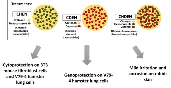
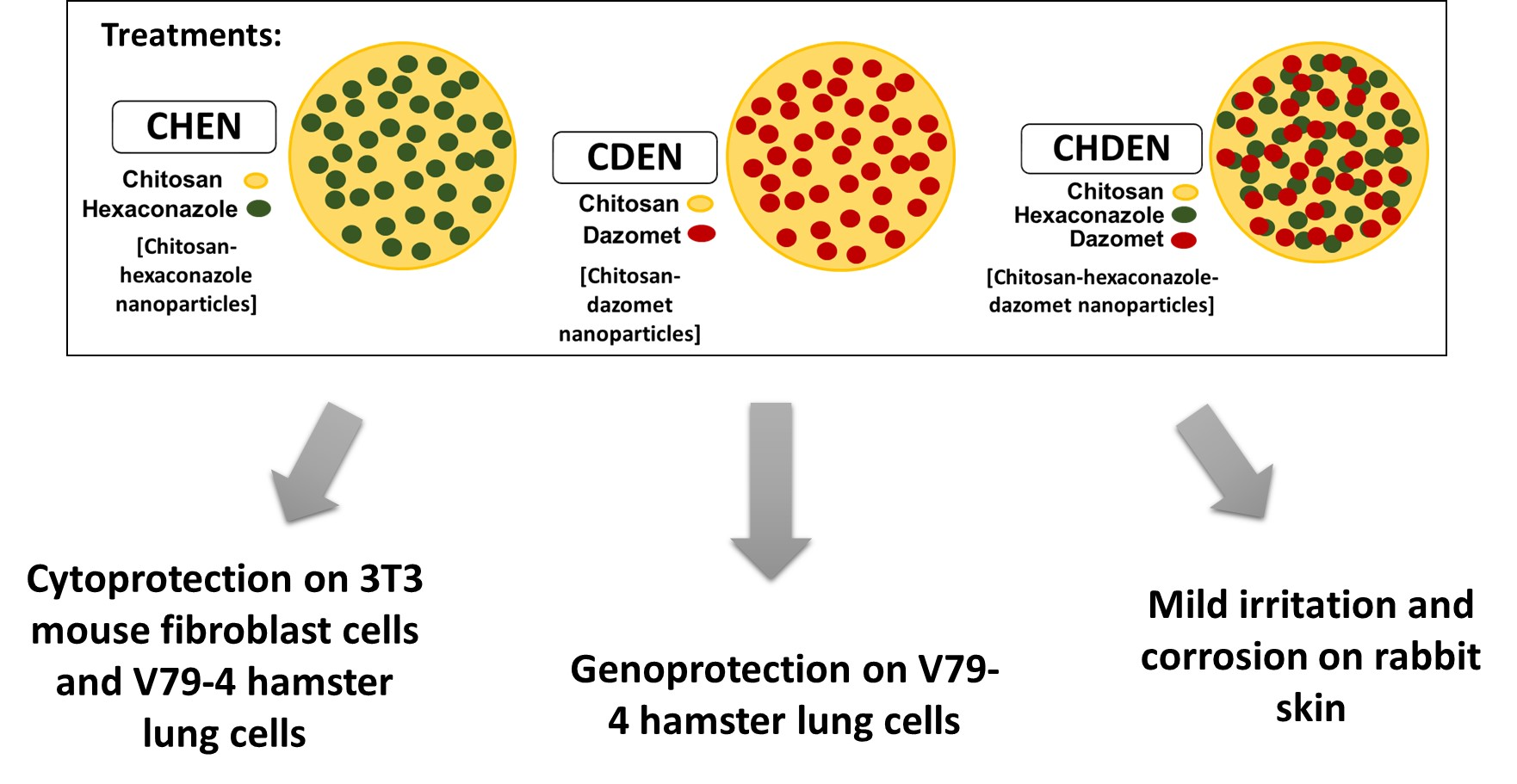
| Chitosan-hexaconazole nanoparticles | CS (85%) H (15%) | 18 | Yellowish-white powder | Chitosan-hexaconazole Nanoparticles (CHEN) |
| Chitosan-dazomet nanoparticles | CS (57%) D (33%) | 7 | Yellowish-white powder | Chitosan-dazomet Nanoparticles (CDEN) |
| Chitosan-hexaconazole-dazomet nanoparticles | CS (86%) H (7%) D (7%) | 5 | Yellowish-white powder | Chitosan-hexaconazole-dazomet Nanoparticles (CHDEN) |
| Reactions | Descriptions | Score |
|---|---|---|
| Erythema and eschar formation (E) | No erythema | 0 |
| Very slight erythema (barely perceptible) | 1 | |
| Well-defined erythema | 2 | |
| Moderate to severe erythema | 3 | |
| Severe erythema (beef-redness) to eschar formation | 4 | |
| Edema formation (O) | No edema | 0 |
| Very slight edema (barely perceptible) | 1 | |
| Slight edema | 2 | |
| Moderate edema (raised more than 1 mm) | 3 | |
| Severe edema (raised more than 1 mm and extending beyond exposure area) | 4 |
| DNA Damage Parameters | Negative Control (NC) | Positive Control (PC) | Vehicle Control (VC) | CHEN | CDEN |
|---|---|---|---|---|---|
| Average tail DNA (%) | 0.02 ± 0.00 a | 0.15 ± 0.03 b | 0.03 ± 0.01 a | 0.02 ± 0.01 a | 0.01 ± 0.01 a |
| Average tail length | 23.75 ± 3.28 a | 86.61 ± 3.97 b | 17.48 ± 1.22 c | 19.60 ± 1.65 c | 19.87 ± 1.88 c |
| Average tail moment value | 0.53 ± 0.09 a | 12.93 ± 0.51 b | 0.48 ± 0.13 a | 0.29 ± 0.06 c | 0.26 ± 0.12 c |
| Treatments | Healthy Cell (%) | Early Apoptosis (%) | Late Apoptosis (%) | Necrosis (%) |
|---|---|---|---|---|
| Control (nontreated) | 89.9 ± 6.0 a | 9.1 ± 3.5 a | 0.7 ± 0.5 a | 0.3 ± 0.5 a |
| Pure hexaconazole | 47.0 ± 3.5 b | 49.1 ± 5.6 b | 3.2 ± 1.5 a | 0.7 ± 0.3 a |
| Pure dazomet | 4.2 ± 2.0 c | 10.2 ± 4.5 a | 85.5 ± 5.5 b | 0.1 ± 0.4 a |
| CHEN | 56.4 ± 5.5 b | 28.7 ± 2.0 c | 4.7 ± 2.3 a | 0.3 ± 0.5 a |
| CDEN | 34.5 ± 4.0 d | 52.4 ± 3.0 b | 12.2 ± 1.5 c | 0.9 ± 0.2 a |
| (1) Initial Response | |||||||
| Time (h) | CHEN | CDEN | |||||
| Corrosion | Irritation | Corrosion | Irritation | ||||
| Erythema | Edema | Erythema | Edema | ||||
| 0 | No corrosive lesion observed | 1 | 1 | No corrosive lesion observed | 1 | 0 | |
| 1 | No corrosive lesion observed | 1 | 0 | No corrosive lesion observed | 1 | 0 | |
| 24 | Brownish line lesion seen (5 mm length) | 1 | 0 | No corrosive lesion observed | 2 | 0 | |
| 48 | - | - | - | Slight erosion on the skin | 1 | 0 | |
| (2) Confirmatory response | |||||||
| Time (h) | Rabbit No. | CHEN | CDEN | ||||
| Corrosion | Irritation | Corrosion | Irritation | ||||
| Erythema | Edema | Erythema | Edema | ||||
| 0 | 1 | No corrosive lesion observed | 1 | 1 | No corrosive lesion observed | 0 | 0 |
| 2 | No corrosive lesion observed | 0 | 0 | No corrosive lesion observed | 1 | 1 | |
| 1 | 1 | No corrosive lesion observed | 1 | 0 | No corrosive lesion observed | 0 | 0 |
| 2 | No corrosive lesion observed | 1 | 1 | No corrosive lesion observed | 1 | 0 | |
| Corrosion | |
| Categories | Criteria |
| Category 1 | Destruction of skin tissue in which necrosis of epidermis was observed in at least one tested animal after ≤4 h of exposure |
| Subcategory 1A | Skin corrosion in at least one tested animal at an exposure of ≤3 min upon removal of the test patch (observation period: ≤1 h) |
| Subcategory 1B | Skin corrosion in at least one tested animal at an exposure of >3 min and ≤1 h upon removal of the test patch (observation period: ≤14 days) |
| Subcategory 1C | Skin corrosion in at least one tested animal at an exposure of >1 h and ≤4 h upon removal of the test patch (observation period: ≤14 days) |
| Irritation | |
| Categories | Criteria |
| Irritation (Category 2) | Erythema and edema with a score of ≥2.3 and ≤4.0 in at least 2 out of 3 tested animals after 24, 48 and 72 h after removal of the test patch. |
| Mild irritation (Category 3) | Erythema and edema with a score of ≥1.5 and ≤2.3 in at least 2 out of 3 tested animals after 24, 48 and 72 h after removal of the test patch. |
© 2020 by the authors. Licensee MDPI, Basel, Switzerland. This article is an open access article distributed under the terms and conditions of the Creative Commons Attribution (CC BY) license (http://creativecommons.org/licenses/by/4.0/).
Share and Cite
Maluin, F.N.; Hussein, M.Z.; Yusof, N.A.; Idris, A.S.; Daim, L.D.J.; Sarian, M.N.; Rajab, N.F.; Ee Ling, S.; Rashid, N.; Fakurazi, S. Cytoprotection, Genoprotection, and Dermal Exposure Assessment of Chitosan-Based Agronanofungicides. Pharmaceutics 2020, 12, 497. https://doi.org/10.3390/pharmaceutics12060497
Maluin FN, Hussein MZ, Yusof NA, Idris AS, Daim LDJ, Sarian MN, Rajab NF, Ee Ling S, Rashid N, Fakurazi S. Cytoprotection, Genoprotection, and Dermal Exposure Assessment of Chitosan-Based Agronanofungicides. Pharmaceutics. 2020; 12(6):497. https://doi.org/10.3390/pharmaceutics12060497
Chicago/Turabian StyleMaluin, Farhatun Najat, Mohd Zobir Hussein, Nor Azah Yusof, Abu Seman Idris, Leona Daniela Jeffery Daim, Murni Nazira Sarian, Nor Fadilah Rajab, Siew Ee Ling, Noramiwati Rashid, and Sharida Fakurazi. 2020. "Cytoprotection, Genoprotection, and Dermal Exposure Assessment of Chitosan-Based Agronanofungicides" Pharmaceutics 12, no. 6: 497. https://doi.org/10.3390/pharmaceutics12060497