Article

Differentiation of Leishmania (L.) infantum, Leishmania (L.) amazonensis and Leishmania (L.) mexicana Using Sequential qPCR Assays and High-Resolution Melt Analysis

by
Marcello Ceccarelli
1,
Aurora Diotallevi
1,
Gloria Buffi
1,
Mauro De Santi
1,
Edith A. Fernández-Figueroa
2,3,
Claudia Rangel-Escareño
2,4,
Said A. Muñoz-Montero
2,
Ingeborg Becker
3,
Mauro Magnani
1 and
Luca Galluzzi
1,*
1
Department of Biomolecular Sciences, University of Urbino “Carlo Bo”, 61029 Urbino (PU), Italy
2
Department of Population Genomics, Computational and Integrative Genomics, National Institute of Genomic Medicine, Mexico City 14610, Mexico
3
Centro de Medicina Tropical, Unidad de Investigación en Medicina Experimental, Facultad de Medicina, Universidad Nacional Autónoma de México, Hospital General de México, México City 04360, Mexico
4
School of Engineering and Sciences, Tecnologico de Monterrey, Queretaro 76130, Mexico
*
Author to whom correspondence should be addressed.
Microorganisms 2020, 8(6), 818; https://doi.org/10.3390/microorganisms8060818
Submission received: 8 April 2020 / Revised: 14 May 2020 / Accepted: 28 May 2020 / Published: 29 May 2020
(This article belongs to the Special Issue New Advances in Leishnmaniasis)

Abstract

:
Leishmania protozoa are the etiological agents of visceral, cutaneous and mucocutaneous leishmaniasis. In specific geographical regions, such as Latin America, several Leishmania species are endemic and simultaneously present; therefore, a diagnostic method for species discrimination is warranted. In this attempt, many qPCR-based assays have been developed. Recently, we have shown that L. (L.) infantum and L. (L.) amazonensis can be distinguished through the comparison of the Cq values from two qPCR assays (qPCR-ML and qPCR-ama), designed to amplify kDNA minicircle subclasses more represented in L. (L.) infantum and L. (L.) amazonensis, respectively. This paper describes the application of this approach to L. (L.) mexicana and introduces a new qPCR-ITS1 assay followed by high-resolution melt (HRM) analysis to differentiate this species from L. (L.) amazonensis. We show that L. (L.) mexicana can be distinguished from L. (L.) infantum using the same approach we had previously validated for L. (L.) amazonensis. Moreover, it was also possible to reliably discriminate L. (L.) mexicana from L. (L.) amazonensis by using qPCR-ITS1 followed by an HRM analysis. Therefore, a diagnostic algorithm based on sequential qPCR assays coupled with HRM analysis was established to identify/differentiate L. (L.) infantum, L. (L.) amazonensis, L. (L.) mexicana and Viannia subgenus. These findings update and extend previous data published by our research group, providing an additional diagnostic tool in endemic areas with co-existing species.

1. Introduction

Leishmaniasis is caused by many Leishmania species belonging to subgenera Leishmania (Leishmania) and Leishmania (Viannia), creating a global public health problem with 0.2–0.4 million cases of visceral leishmaniasis (VL) and 0.7–1.2 million cases of cutaneous leishmaniasis (CL) per year [1]. In specific geographical regions, such as Central and South America, many Leishmania species are endemic and simultaneously present and, in some cases, can give rise to the same clinical form [2]. For instance, L. (L.) amazonensis and L. (L.) mexicana are responsible for the cutaneous manifestations and have a wide geographic distribution from Mexico to the north of Argentina. The epidemiological heterogeneity and difficulties in clinical approaches make species identification a critical step in clinical diagnosis and management, especially in case of co-infections. Therefore, an accurate diagnostic method allowing species identification is necessary [3]. Since the microscopic analysis does not provide information for species discrimination and the isoenzymatic characterization (i.e., multilocus enzyme electrophoresis) is a challenging and time-consuming technique, many biomolecular assays have been developed for Leishmania detection and species identification [4,5]. In particular, many qPCR assays have been designed to target the ribosomal DNA (rDNA) sequences and the kinetoplast DNA (kDNA) minicircle network that characterizes the Leishmania genus [6,7]. The rDNA sequence is repeated tens or hundreds of times per cell, allowing acceptable sensitivity also with DNA from clinical samples. Moreover, the variability of the internal transcribed spacer (ITS) sequences can be exploited for typing at the species level [8]. The kDNA minicircles are about 800 bp long and are present in several thousands of copies per cell, which makes them ideal targets for highly sensitive PCR-based assays. Minicircles are characterized by conserved regions in their replication origin [9], which allows the design of PCR primers with broad taxonomic coverage. On the other hand, the minicircle network is composed of different subclasses presenting high sequence variability, with exception of the conserved regions [10]. The number and identity of minicircle subclasses vary greatly among Leishmania species [11,12,13]. These features make the design of qPCR assays for identification at the species level difficult to perform, even using probes or melting analysis [14,15,16,17]. To differentiate L. (L.) infantum from L. (L.) amazonensis, we recently proposed an approach based on the evaluation of relative abundance of minicircle subclasses by using two qPCR assays [18,19,20]. However, our previous works did not include L. (L.) mexicana, which is closely related to L. (L.) amazonensis [21].
In this paper, we also applied this diagnostic approach to L. (L.) mexicana species, with the aim to extend the validity of our previous work. Results showed that, as previously demonstrated for L. (L.) amazonensis, L. (L.) mexicana can also be distinguished from L. (L.) infantum by exploiting the two qPCR assays designed on minicircle kDNA. Moreover, it was also possible to reliably distinguish L. (L.) mexicana from L. (L.) amazonensis species using a new high-resolution melt (HRM) assay designed on the ITS1 region (qPCR-ITS1).

2. Materials and Methods

2.1. Leishmania Strains, Clinical Samples and DNA Extraction

The Leishmania strains and isolates used in this study are listed in Table 1. The L. (L.) mexicana clinical isolates 2, 3 and 5 were from diffuse cutaneous leishmaniasis lesions, whereas the clinical isolates 14, 17 and MHOM/MX/2011/Lacandona were from localized cutaneous leishmaniasis lesions. The clinical samples were taken from individuals from Quintana Roo, an endemic area of leishmaniasis in Mexico, as well as from patients treated at the Tropical Medicine Center, Medical Faculty, National Autonomous University of Mexico (UNAM); all patients were clinically diagnosed as diffuse or localized cutaneous leishmaniasis by Giemsa-stained smears of the lesions and by ELISA test for Leishmania (Table 2).
The DNA was extracted from promastigote cultures and from clinical samples using phenol-chloroform standard procedures followed by ethanol precipitation and the High Pure PCR Template Preparation Kit (Roche, Mannheim, Germany). The DNA was quantified using a Qubit fluorometer (Life Technologies, Carlsbad, USA) and stored at −20 °C until being used.

2.2. ITS1-PCR RFLP

The L. (L.) mexicana strains and clinical samples were typed using ITS1-PCR RFLP according to Monroy-Ostria et al. [22]. Briefly, the PCR was performed using primers LITSR and L5.8S, following the amplification protocol—94 °C for 4 min followed by 36 cycles of 94 °C for 40 s, 54 °C for 30 s and 72 °C for 1 min and a final extension at 72 °C for 6 min. PCR products were nested using the same PCR conditions for 18 cycles. PCR products were digested with HaeIII for 3 h at 37 °C and for 20 min at 80 °C to inactivate the enzyme. The restriction fragments were subjected to electrophoresis on a 4% agarose gel.

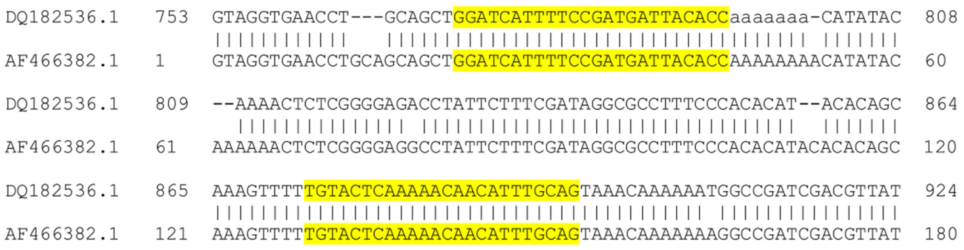
2.3. DNA Sequencing and Phylogenetic Analysis

The alanine aminotransferase (ALAT) gene was amplified in clinical samples px2, px3, px9, px10, pxJLC and in L. (L.) mexicana MHOM/MX/2011/Lacandona according to Marco et al. [23] using primers ALAT.F and ALAT.R. The amplification conditions were—94 °C for 3 min followed by 40 cycles of 94 °C for 30 s, 55 °C for 30 s and 72 °C for 30 s. PCR products were purified using the Agencourt AMPure XP kit (Beckman Coulter, Brea, CA, USA) and sequenced using the BigDye Terminator v3.1 Cycle Sequencing Kit (Thermo Fisher Scientific, Waltham, MA, USA) on ABI 3730xL DNA Analyzer (Thermo Fisher Scientific, Waltham, MA, USA). Chromatograms were visualized with ApE software and consensus sequences were generated and compared between them and with other validated species of L. (L.) mexicana deposited in GenBank using the Blastn tool available in the same platform. A phylogenetic reconstruction based on the Maximum Likelihood (ML) method was generated and a phylogenetic tree was constructed with 10,000 bootstrap replications, using the close-neighbor interchange method in Mega 6.0.

2.4. qPCR Assays

The qPCR-ML (amplifying kDNA minicircle subclass more represented in L. (L.) infantum) and qPCR-ama (amplifying kDNA minicircle subclass more represented in L. (L.) amazonensis) were performed as previously described [18]. The new assay qPCR-ITS1 was performed using the new primers ITS1mexama_F (5′-GGATCATTTTCCGATGATTACACC-3′) and ITS1mexama_R (5′-CTGCAAATGTTGTTTTTGAGTACA-3′), flanking a portion of ITS1 sequence containing differences between L. (L.) amazonensis and L. (L.) mexicana (Figure 1). The primers were designed using Primer BLAST and were verified against the ITS1 sequences of L. (L.) amazonensis (n = 32) and L. (L.) mexicana (n = 30) encompassing forward and/or reverse primers, available in the Genbank database.
For all assays, PCR reactions were carried out in a 25 μL volume with 1–3 μL of template DNA using SYBR green PCR master mix (Diatheva srl, Fano, Italy) or TB Green premix ex TaqII Mastermix (Takara Bio Europe, France) and 200 nM of each primer in a Rotor-Gene 6000 instrument (Corbett Life Science, Mortlake, Australia). The amplification conditions were—94 °C for 10 min, 40 cycles at 94 °C for 25 s, 60 °C for 20 s and 72 °C for 20 s. At the end of each run, a melting curve analysis was performed from 78 to 92 °C with a slope of 1 °C/s, and 5 s at each temperature. The reactions were performed in duplicate. Dilution curves (from 1.0 to 1 × 10⁻⁵ ng/reaction) were established using L. (L.) mexicana MHOM/MX/2011/Lacandona DNA for qPCR-ML, qPCR-ama and qPCR-ITS1. The threshold cycles (Cq) were determined using the quantitation analysis of the Rotor-Gene 6000 software, setting a threshold to 0.15. To evaluate the potential interference of host DNA as a background in the qPCR analysis, 30 ng of human DNA was spiked in the reaction tubes.

2.5. High-Resolution Melt (HRM) Analysis

The qPCR-ML, qPCR-ama and qPCR-ITS1 amplicons were analyzed by HRM protocol on a Rotor-Gene 6000 instrument as described previously [24] with few modifications. Briefly, HRM was carried out over the range from 80 to 90 °C (qPCR-ML, qPCR-ama) or 75 to 85 °C (qPCR-ITS1), rising at 0.1 °C/s and waiting for 2 s at each temperature. Each sample was run in duplicate, and the gain was optimized before melting on all tubes.

2.6. Ethics Approval

This study was conducted according to the principles expressed in the Declaration of Helsinki. This research was approved by the Institutional Ethics Committee of the Medical Faculty of the National Autonomous University of Mexico (FM/DI/013/SR/2019). Guidelines established by the Mexican Health Authorities were strictly followed. All patients received treatment and clinical care by health authorities and signed a written informed consent for the collection of samples and subsequent analysis.

2.7. Statistical Analysis

Statistical analysis was performed with GraphPad Prism 5.0 (GraphPad Software, San Diego, CA, USA). Normal distribution of data was assessed by D′Agostino and Pearson omnibus normality test (alpha = 0.05). Difference between Tm mean values was evaluated using the nonparametric Mann–Whitney test.

3. Results

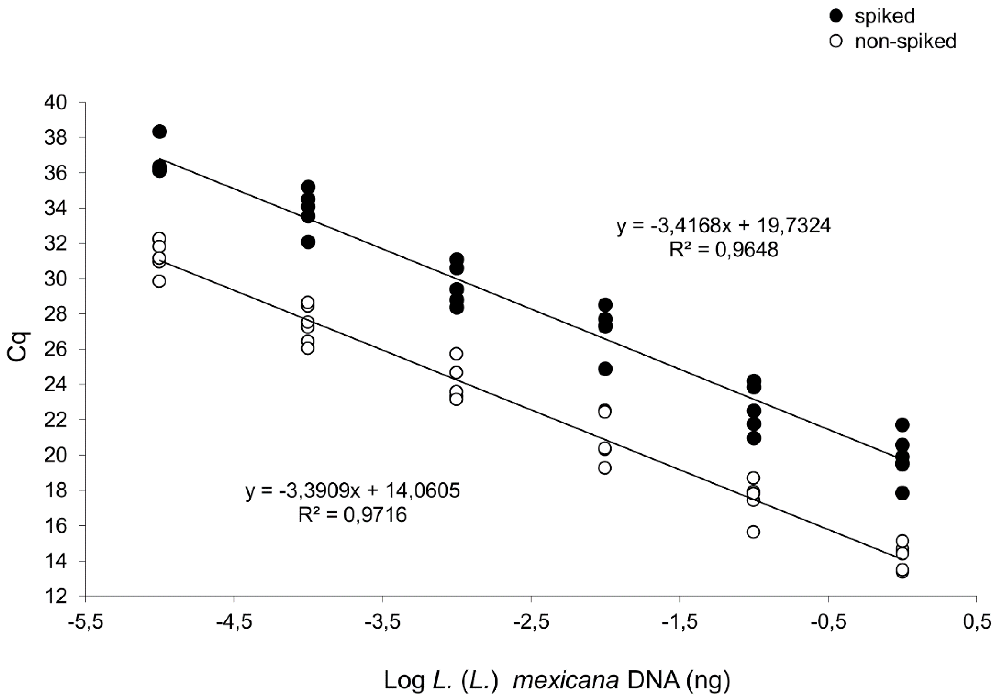
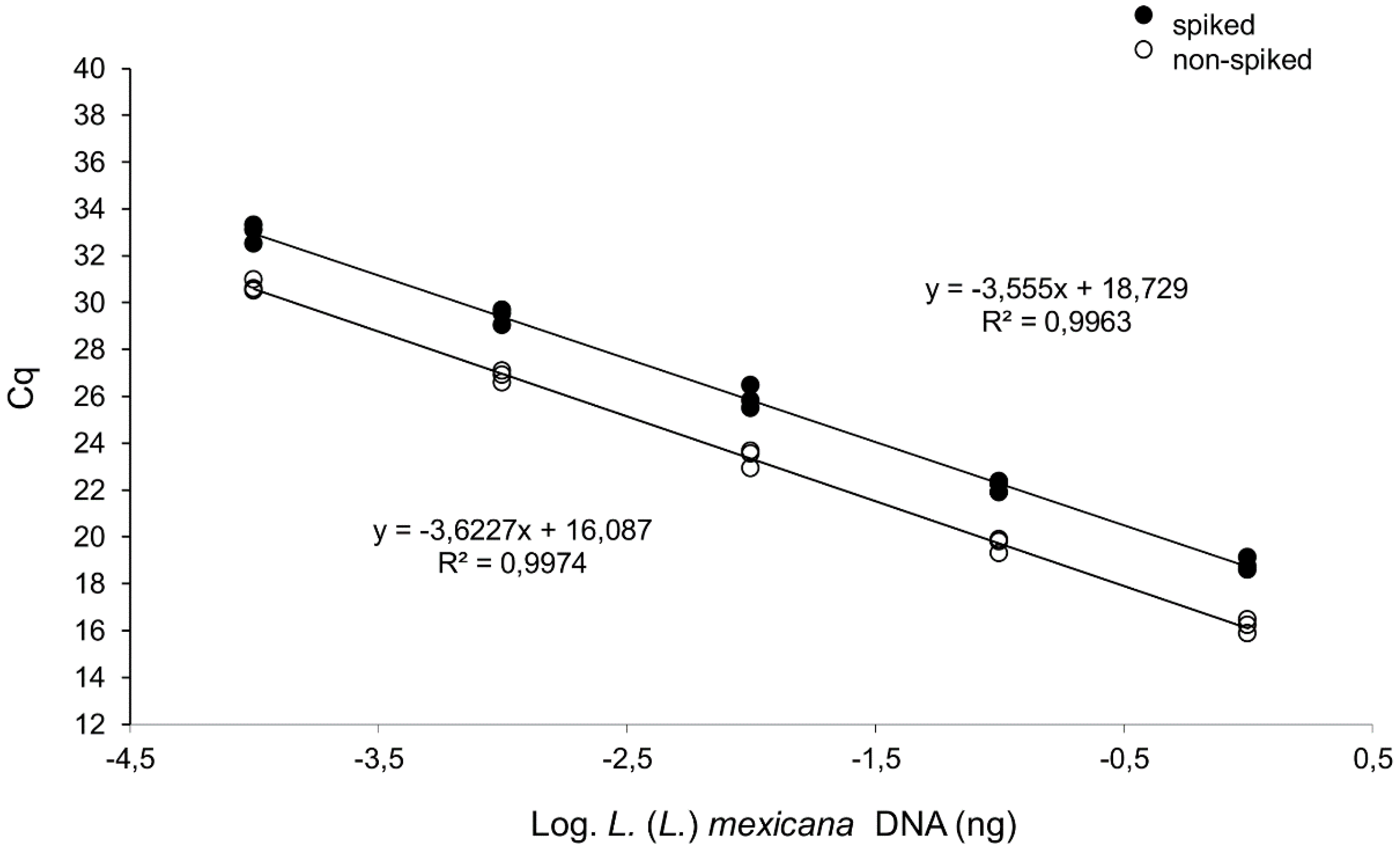
3.1. Both L. (L.) mexicana and L. (L.) amazonensis Can be Distinguished from L. (L.) infantum Exploiting A Differential qPCR Targeting Minicircle kDNA

Previously, we have shown that L. (L.) infantum and L. (L.) amazonensis can be distinguished by comparing the Cq values of two qPCR assays (qPCR-ML and qPCR ama). In this work, the qPCR-ML and qPCR-ama were sequentially performed using DNA from L. (L.) mexicana MHOM/MX/2011/Lacandona, isolate 2, isolate 3, isolate 5, isolate 14 and isolate 17 as templates. As already shown for L. (L.) amazonensis strains, Cq values obtained with qPCR-ama were much lower compared to those obtained with qPCR-ML (Table 3).
The Cq difference between qPCR-ama and qPCR-ML allowed us to include L. (L.) mexicana among the Leishmania (Leishmania) species that can be distinguished from L. (L.) infantum. Results from L. (L.) amazonensis MHOM/BR/00/LTB0016 and L. (L.) infantum MHOM/FR/78/LEM75 were included as representative results obtained previously. As a consequence of different minicircle subclass amplified, the qPCR-ML and qPCR-ama showed a different limit of detection, allowing to amplify up to 1.0 × 10⁻² and 1.0 × 10⁻⁵ ng of L. (L.) mexicana MHOM/MX/2011/Lacandona DNA, respectively (Table 3). In the qPCR-ML, the presence of 30 ng of purified human DNA delayed the limit of detection to 1.0 × 10⁻¹ ng (Figure S1). With regard to qPCR-ama, the efficiency and detection limit were evaluated using 10-fold L. (L.) mexicana MHOM/MX/2011/Lacandona DNA serial dilutions (from 1.0 to 1×10⁻⁵ ng) in three independent experiments. There was a linear correlation between the log of DNA concentration and Cq value (slope = −3.3909, R2 = 0.9716) with a reaction efficiency of 97%. In order to evaluate the interference of host DNA, the DNA dilutions were spiked with 30 ng of purified human DNA, showing a delay on the Cq values but with comparable efficiency and limit of detection (Figure 2). The efficiency and detection limit obtained with L. (L.) mexicana DNA were in agreement with previous results obtained using DNA template from L. (L.) amazonensis [18].
The qPCR-ML/qPCR-ama approach was also applied to 11 clinical samples. These samples were characterized as L. (L.) mexicana by ITS1-PCR RFLP (Figure S2), with the exception of pxCMU, for which a digestion profile could not be obtained. Moreover, the genotype of five clinical samples (px2, px3, px9, px10, pxJLC) were further confirmed as L. (L.) mexicana by sequencing and phylogenetic analysis of the alanine aminotransferase (ALAT) gene (Figure S3). All samples showed Cq qPCR-ama <Cq qPCR-ML (Table 4), confirming the presence of L. (L.) mexicana parasites.

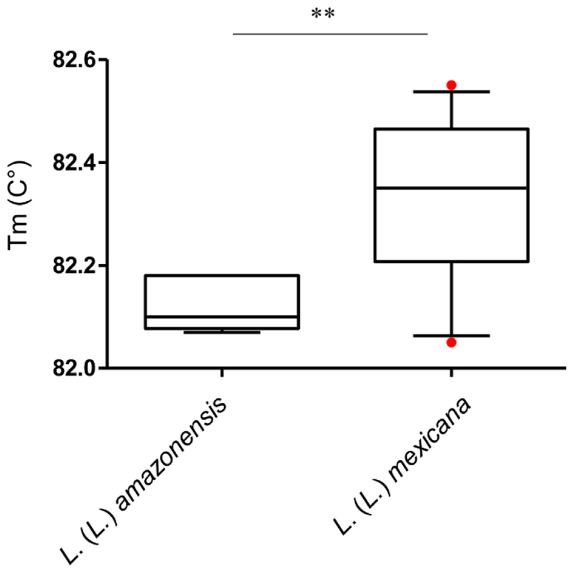
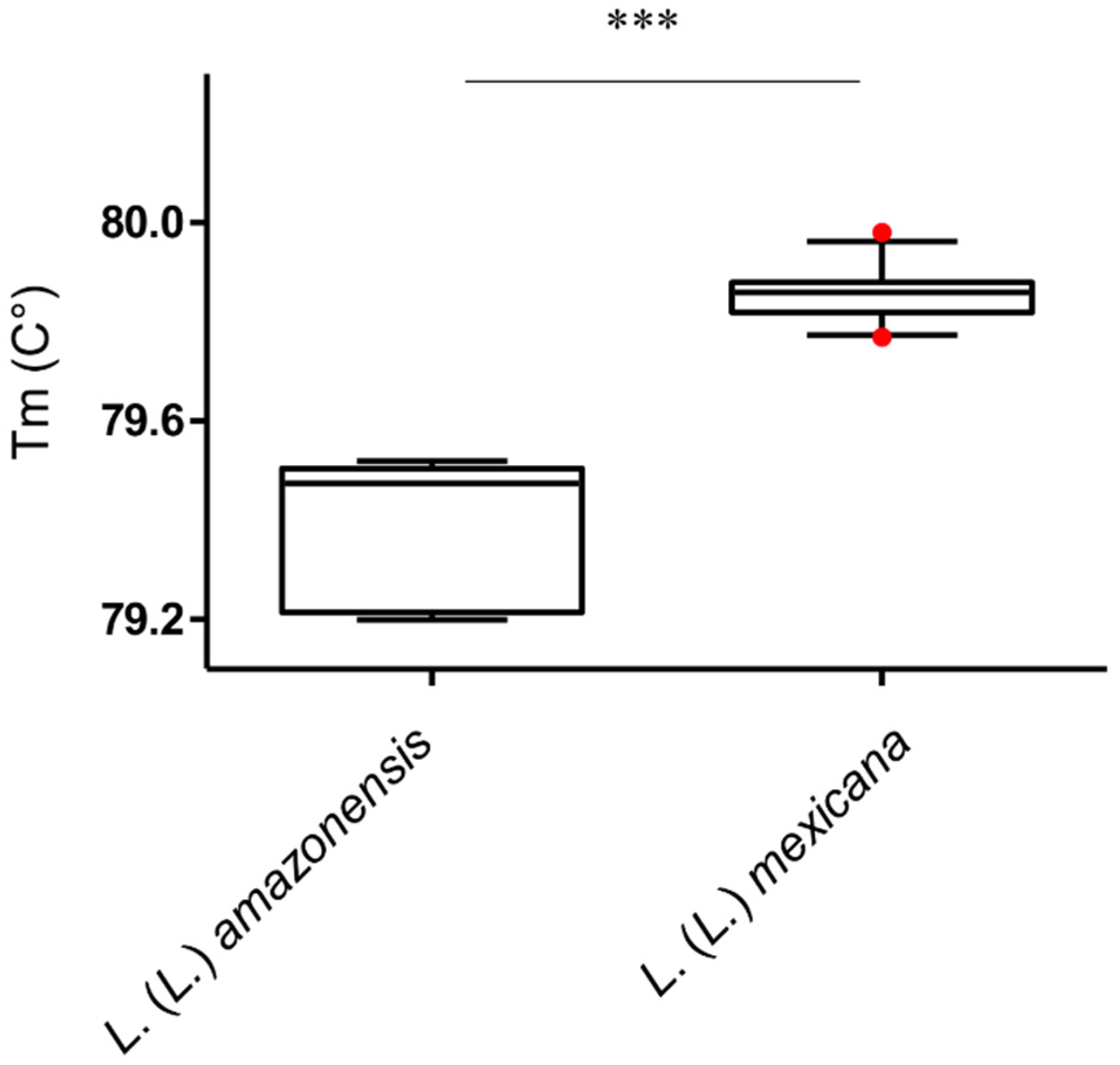
3.2. L. (L.) amazonensis Can be Differentiated from L. (L.) mexicana by qPCR-ITS1 HRM Analysis

In the attempt to differentiate L. (L.) mexicana and L. (L.) amazonensis, HRM analyses were performed after qPCR-ML and qPCR-ama using the L. (L.) mexicana and L. (L.) amazonensis samples indicated in Table 1 and Table 2. However, both assays did not allow us to distinguish the two species reliably. In particular, the qPCR-ML assay showed overly high Cq values (>30) in L. (L.) mexicana samples. Concerning the qPCR-ama assay, HRM analysis of all L. (L.) mexicana and L. (L.) amazonensis samples showed heterogeneous profiles (Figure S4). Moreover, despite that the mean Tm of PCR products from L. (L.) mexicana and L. (L.) amazonensis were significantly different (Mann–Whitney test, p < 0.01), the Tm value distributions partly overlapped, de facto making the distinction between the two species unreliable (Figure 3).
Therefore, a new qPCR assay and HRM analysis were designed on ITS1 sequences. The in silico analysis showed that PCR product lengths were 125–126 and 129–131 bp for L. (L.) amazonensis and L. (L.) mexicana, respectively. The qPCR-ITS1 efficiency and detection limit were evaluated using 10-fold L. (L.) mexicana MHOM/MX/2011/Lacandona DNA serial dilutions (from 1.0 to 1 × 10⁻⁴ ng). A linear correlation between the log of DNA concentration and Cq value was demonstrated (slope = −3.6227, R2 = 0.997), with a reaction efficiency of 89%. As shown for the qPCR-ama, spiking with 30 ng of purified human DNA induced a delay on the Cq values, but efficiency and limit of detection were not affected (Figure 4). The Tm analysis of qPCR-ITS1 amplicons obtained from all amplified L. (L.) amazonensis and L. (L.) mexicana samples allowed full discrimination between the two species (Mann–Whitney test, p < 0.001) (Figure 5) (Table S1). However, three clinical samples failed to amplify (Px7, PxGSF, PxCMU). Overall, the qPCR-ITS1 HRM assay for amazonensis/mexicana species discrimination showed 84.2% sensitivity and 100% specificity.

4. Discussion

The identification of Leishmania species is an important diagnostic aspect, especially in Latin America, not only for epidemiological studies but also for the accurate monitoring of clinical disease evolution. In fact, the only species causing VL in this geographical region is L. (L.) infantum (syn. chagasi), while cutaneous or mucocutaneous (MCL) manifestations can also be generated by Viannia subgenus and L. (L.) mexicana complex. In this epidemiological and clinical context, the species discrimination appears pivotal, e.g., to monitor a cutaneous lesion that could evolve in VL, MCL or disseminated CL, depending on the species. In this view, molecular diagnostic tools allowing species discrimination can be helpful. The kDNA minicircles are ideal targets for highly sensitive molecular detection of Leishmania spp. since they are present in thousands of copies per cell [25]. Since the pioneering work of Nicolas et al. [26], many qPCR assays have been designed on conserved regions of minicircles to detect/quantify Leishmania parasites. Moreover, several authors investigated the possibility to exploit minicircle sequences to discriminate Leishmania parasites at the species level, reaching only partial results due to the variability of minicircle subclasses [15,16]. Previously, we proposed an SYBR Green qPCR-based approach to distinguish L. (L.) infantum from L. (L.) amazonensis, exploiting the different abundance of minicircle subclasses rather than targeting a species-specific sequence. Using this approach, which relies on two qPCR assays (qPCR-ML and qPCR-ama) and evaluation of Cq values, we were able to distinguish the two species adequately [18].
In this work, we tested this approach with L. (L.) mexicana, which is genetically close to L. (L.) amazonensis. The comparison of Cq values of qPCR-ML and qPCR-ama confirmed results previously obtained with L. (L.) amazonensis, allowing us to include L. (L.) mexicana among the Leishmania (Leishmania) species that can be differentiated from L. (L.) infantum, therefore extending the conclusion of our previous work. Importantly, this approach was successfully applied to cutaneous lesions of 11 patients diagnosed with diffuse or localized cutaneous leishmaniasis. Notably, the clinical sample pxCMU, which was negative in ITS1-PCR RFLP, was identified as L. (L.) mexicana/amazonensis, evidencing the highest sensitivity of our qPCR assays targeting minicircles. These results further support the possibility of exploiting the relative abundance of minicircles for Leishmania species discrimination. Moreover, we confirmed the use of an adequate diagnostic approach based on consecutive qPCR assays to define species [18], as also proposed by other authors [27].
The distinction between L. (L.) amazonensis and L. (L.) mexicana is important for epidemiological studies and disease monitoring, but it can be challenging [28]. For instance, hsp70 analysis by Fraga et al. [29] did not resolve between these species. On the other hand, other authors were able to separate these species based on multilocus sequence typing (MLST) [30] or sequential real-time PCR assays [27].
The qPCR coupled with HRM analysis is considered as a good option in molecular diagnostics since it avoids the use of modified oligonucleotides, it is accurate, allows high-throughput applications and is faster and cheaper than other types of analysis such as MLST, RFLP or single-gene DNA sequencing. Moreover, since the qPCR is a closed-tube system, the potential for carryover contamination will be reduced. In the attempt to discriminate between L. (L.) amazonensis and L. (L.) mexicana, HRM profiles of amplicons from qPCR-ama were investigated; however, their heterogeneity did not us allow to distinguish these two species reliably. Since Schönian et al. demonstrated the possibility to discriminate the two species using ITS1-PCR RFLP [31], we designed an HRM-based assay exploiting differences in L. (L.) amazonensis and L. (L.) mexicana ITS1 sequences, in order to avoid restriction digestion and electrophoretic analysis. This process allows saving a considerable amount of time to perform the analysis and avoids possible difficulties in restriction fragment identification. As expected from the in silico sequence analysis, the observed HRM Tm values of all L. (L.) mexicana samples were significantly higher as those of all L. (L.) amazonensis samples, allowing a robust distinction between these two species. The fact that three clinical samples did not amplify (Px7, PxGSF, PxCMU) was probably due to the lower sensitivity of qPCR-ITS1 as compared to the assay targeting kDNA minicircles.

5. Conclusions

In the attempt to use a qPCR-based approach to differentiate Leishmania species co-existing in the New World, sequential qPCR assays and HRM analysis have been implemented. The results showed that—(i) L. (L.) infantum can be distinguished from L. (L.) mexicana comparing the Cq values of qPCR-ML and qPCR-ama, as previously shown for L. (L.) amazonensis; (ii) this distinction was possible not only using strains/isolates but also in clinical samples; (iii) the differentiation between L. (L.) amazonensis and L. (L.) mexicana was achieved by qPCR-ITS1 HRM analysis. Therefore, it was possible to design/update an algorithm that allows us to identify/differentiate L. (L.) infantum, L. (L.) amazonensis, L. (L.) mexicana and Viannia subgenus with sequential qPCR assays coupled with HRM analysis targeting minicircle kDNA and ITS1 sequence (Figure 6), which further extends our previous work.

Supplementary Materials

The following are available online at https://www.mdpi.com/2076-2607/8/6/818/s1, Figure S1: Electrophoretic analysis of qPCR-ML products, Figure S2: Digestion of ITS1 amplicons of clinical samples and reference Leishmania strains with the restriction endonuclease HaeIII, Figure S3: Maximum likelihood phylogenetic tree of ALAT amplicons, Figure S4: HRM analysis of qPCR-ama amplicons, Table S1: Results of qPCR-ITS1 HRM analysis from all L. (L.) amazonensis and L. (L.) mexicana samples.

Author Contributions

Conceptualization, M.C. and L.G.; methodology, M.C., A.D. and L.G.; validation, M.C., L.G., E.A.F.-F., C.R.-E., S.A.M.-M., I.B. and M.M.; formal analysis, L.G., A.D. and E.A.F.-F.; investigation, M.C., A.D., M.D.S., G.B. and E.A.F.-F.; resources, C.R.-E., I.B. and M.M.; writing—original draft preparation, M.C. and L.G.; writing—review and editing, A.D., G.B., E.A.F.-F., C.R.-E., S.A.M.-M., I.B., M.D.S. and M.M. All authors have read and agreed to the published version of the manuscript.

Funding

This research was partially funded by the Department of Biomolecular Sciences of University of Urbino and by INMEGEN-08/2013/I.

Acknowledgments

We thank Fabrizio Vitale and Christine Petersen for providing Leishmania spp. DNA. We also thank Marco Quintanilla for providing the clinical samples of patients from Quintana Roo.

Conflicts of Interest

The authors declare no conflict of interest. The funders had no role in the design of the study; in the collection, analyses, or interpretation of data; in the writing of the manuscript, or in the decision to publish the results.

References

  1. Alvar, J.; Vélez, I.D.; Bern, C.; Herrero, M.; Desjeux, P.; Cano, J.; Jannin, J.; den Boer, M. Leishmaniasis Worldwide and Global Estimates of Its Incidence. PLoS ONE 2012, 7, e35671. [Google Scholar] [CrossRef] [PubMed]
  2. Hashiguchi, Y.; Velez, L.N.; Villegas, N.V.; Mimori, T.; Gomez, E.A.L.; Kato, H. Leishmaniases in Ecuador: Comprehensive review and current status. Acta Trop. 2017, 166, 299–315. [Google Scholar] [CrossRef] [PubMed]
  3. Goto, H.; Lindoso, J.A. Current diagnosis and treatment of cutaneous and mucocutaneous leishmaniasis. Expert Rev. Anti Infect. Ther. 2010, 8, 419–433. [Google Scholar] [CrossRef] [PubMed]
  4. Akhoundi, M.; Downing, T.; Votýpka, J.; Kuhls, K.; Lukeš, J.; Cannet, A.; Ravel, C.; Marty, P.; Delaunay, P.; Kasbari, M.; et al. Leishmania infections: Molecular targets and diagnosis. Mol. Asp. Med. 2017, 57, 1–29. [Google Scholar] [CrossRef] [PubMed]
  5. Recalde, O.D.S.; Brunelli, J.P.; Rolon, M.S.; De Arias, A.R.; Aldama, O.; Gómez, C.V. First molecular report of Leishmania (Leishmania) amazonensis and Leishmania (Viannia) guyanensis in paraguayan inhabitants using high-resolution melt-PCR. Am. J. Trop. Med. Hyg. 2019, 101, 780–788. [Google Scholar] [CrossRef]
  6. Galluzzi, L.; Ceccarelli, M.; Diotallevi, A.; Menotta, M.; Magnani, M. Real-time PCR applications for diagnosis of leishmaniasis. Parasites Vectors 2018, 11, 273. [Google Scholar] [CrossRef]
  7. Medkour, H.; Varloud, M.; Davoust, B.; Mediannikov, O. New Molecular Approach for the Detection of Kinetoplastida Parasites of Medical and Veterinary Interest. Microorganisms 2020, 8, 356. [Google Scholar] [CrossRef] [Green Version]
  8. Schonian, G.; Kuhls, K.; Mauricio, I.L. Molecular approaches for a better understanding of the epidemiology and population genetics of Leishmania. Parasitology 2011, 138, 405–425. [Google Scholar] [CrossRef] [Green Version]
  9. Jensen, R.E.; Englund, P.T. Network News: The Replication of Kinetoplast DNA. Annu. Rev. Microbiol. 2012, 66, 473–491. [Google Scholar] [CrossRef]
  10. Brewster, S.; Barker, D. Analysis of minicircle classes in Leishmania (Viannia) species. Trans. R. Soc. Trop. Med. Hyg. 2002, 96, S55–S63. [Google Scholar] [CrossRef]
  11. Lee, S.-Y.; Lee, S.-T.; Chang, K.-P. Transkinetoplastidy-A Novel Phenomenon Involving Bulk Alterations of Mitochondrion-Kinetoplast DNA of a Trypanosomatid Protozoan 1, 2. J. Protozool. 1992, 39, 190–196. [Google Scholar] [CrossRef] [PubMed]
  12. Simpson, L. The genomic organization of guide RNA genes in kinetoplastid protozoa: Several conundrums and their solutions. Mol. Biochem. Parasitol. 1997, 86, 133–141. [Google Scholar] [CrossRef]
  13. Kocher, A.; Valière, S.; Bañuls, A.-L.; Murienne, J. High-throughput sequencing of kDNA amplicons for the analysis of Leishmania minicircles and identification of Neotropical species. Parasitology 2017, 145, 1–8. [Google Scholar]
  14. Mary, C.; Faraut, F.; Lascombe, L.; Dumon, H. Quantification of Leishmania infantum DNA by a real-time PCR assay with high sensitivity. J. Clin. Microbiol. 2004, 42, 5249–5255. [Google Scholar] [CrossRef] [Green Version]
  15. Pita-Pereira, D.; Lins, R.; Oliveira, M.P.; Lima, R.B.; Pereira, B.A.S.; Moreira, O.C.; Brazil, R.P.; Britto, C. SYBR Green-based real-time PCR targeting kinetoplast DNA can be used to discriminate between the main etiologic agents of Brazilian cutaneous and visceral leishmaniases. Parasites Vectors 2012, 5, 15. [Google Scholar] [CrossRef] [Green Version]
  16. Ceccarelli, M.; Galluzzi, L.; Migliazzo, A.; Magnani, M. Detection and Characterization of Leishmania (Leishmania) and Leishmania (Viannia) by SYBR Green-Based Real-Time PCR and High Resolution Melt Analysis Targeting Kinetoplast Minicircle DNA. PLoS ONE 2014, 9, e88845. [Google Scholar] [CrossRef] [Green Version]
  17. Losada-Barragán, M.; Cavalcanti, A.; Umaña-Pérez, A.; Porrozzi, R.; Cuervo-Escobar, S.; Vallejo, A.F.; Sánchez-Gómez, M.; Cuervo, P. Detection and quantification of Leishmania infantum in naturally and experimentally infected animal samples. Vet. Parasitol. 2016, 226, 57–64. [Google Scholar] [CrossRef]
  18. Ceccarelli, M.; Galluzzi, L.; Diotallevi, A.; Andreoni, F.; Fowler, H.; Petersen, C.; Vitale, F.; Magnani, M. The use of kDNA minicircle subclass relative abundance to differentiate between Leishmania (L.) infantum and Leishmania (L.) amazonensis. Parasites Vectors 2017, 10, 239. [Google Scholar] [CrossRef]
  19. Diotallevi, A.; Buffi, G.; Ceccarelli, M.; Neitzke-Abreu, H.C.; Gnutzmann, L.V.; da Costa Lima, M.S.; Di Domenico, A.; De Santi, M.; Magnani, M.; Galluzzi, L. Real-time PCR to differentiate among Leishmania (Viannia) subgenus, Leishmania (Leishmania) infantum and Leishmania (Leishmania) amazonensis: Application on Brazilian clinical samples. Acta Trop. 2020, 201, 105178. [Google Scholar] [CrossRef]
  20. Diotallevi, A.; Buffi, G.; Ceccarelli, M.; Di Domenico, A.; De Santi, M.; Magnani, M.; Galluzzi, L.; Neitzke-Abreu, H.C.; Gnutzmann, L.V.; da Costa Lima Junior, M.S. Data on the differentiation among Leishmania (Viannia) spp., Leishmania (Leishmania) infantum and Leishmania (Leishmania) amazonensis in Brazilian clinical samples using real-time PCR. Data Brief 2020, 28, 104914. [Google Scholar] [CrossRef]
  21. Tschoeke, D.A.; Nunes, G.L.; Jardim, R.; Lima, J.; Dumaresq, A.S.R.; Gomes, M.R.; De Mattos Pereira, L.; Loureiro, D.R.; Stoco, P.H.; De Matos Gliedes, H.L.; et al. The Comparative Genomics and Phylogenomics of Leishmania Amazonensis Parasite. Evol. Bioinform. 2014, 10, EBO.S13759. [Google Scholar] [CrossRef] [PubMed]
  22. Monroy-Ostria, A.; Nasereddin, A.; Monteon, V.M.; Guzmán-Bracho, C.; Jaffe, C.L. ITS1 PCR-RFLP Diagnosis and Characterization of Leishmania in Clinical Samples and Strains from Cases of Human Cutaneous Leishmaniasis in States of the Mexican Southeast. Interdiscip. Perspect. Infect. Dis. 2014, 2014, 607287. [Google Scholar] [CrossRef]
  23. Marco, J.D.; Barroso, P.A.; Locatelli, F.M.; Cajal, S.P.; Hoyos, C.L.; Nevot, M.C.; Lauthier, J.J.; Tomasini, N.; Juarez, M.; Estévez, J.O.; et al. Multilocus sequence typing approach for a broader range of species of Leishmania genus: Describing parasite diversity in Argentina. Infect. Genet. Evol. 2015, 30, 308–317. [Google Scholar] [CrossRef] [PubMed]
  24. Ceccarelli, M.; Diotallevi, A.; Andreoni, F.; Vitale, F.; Galluzzi, L.; Magnani, M. Exploiting genetic polymorphisms in metabolic enzymes for rapid screening of Leishmania infantum genotypes. Parasites Vectors 2018, 11, 572. [Google Scholar] [CrossRef] [PubMed]
  25. Quaresma, P.F.; Murta, S.M.F.; de Ferreira, E.C.; da Rocha-Lima, A.C.V.M.; Xavier, A.A.P.; Gontijo, C.M.F. Molecular diagnosis of canine visceral leishmaniasis: Identification of Leishmania species by PCR-RFLP and quantification of parasite DNA by real-time PCR. Acta Trop. 2009, 111, 289–294. [Google Scholar] [CrossRef]
  26. Nicolas, L.; Prina, E.; Lang, T.; Milon, G. Real-Time PCR for Detection and Quantitation of Leishmania in Mouse Tissues. J. Clin. Microbiol. 2002, 40, 1666–1669. [Google Scholar] [CrossRef] [Green Version]
  27. Hernández, C.; Alvarez, C.; González, C.; Ayala, M.S.; León, C.M.; Ramírez, J.D. Identification of Six New World Leishmania species through the implementation of a High-Resolution Melting (HRM) genotyping assay. Parasites Vectors 2014, 7, 501. [Google Scholar] [CrossRef]
  28. Zampieri, R.A.; Laranjeira-Silva, M.F.; Muxel, S.M.; Stocco de Lima, A.C.; Shaw, J.J.; Floeter-Winter, L.M. High Resolution Melting Analysis Targeting hsp70 as a Fast and Efficient Method for the Discrimination of Leishmania Species. PLoS Negl. Trop. Dis. 2016, 10, e0004485. [Google Scholar] [CrossRef] [Green Version]
  29. Fraga, J.; Montalvo, A.M.; De Doncker, S.; Dujardin, J.-C.; Van der Auwera, G. Phylogeny of Leishmania species based on the heat-shock protein 70 gene. Infect. Genet. Evol. 2010, 10, 238–245. [Google Scholar] [CrossRef]
  30. Van der Auwera, G.; Ravel, C.; Verweij, J.J.; Bart, A.; Schonian, G.; Felger, I. Evaluation of Four Single-Locus Markers for Leishmania Species Discrimination by Sequencing. J. Clin. Microbiol. 2014, 52, 1098–1104. [Google Scholar] [CrossRef] [Green Version]
  31. Schönian, G.; Nasereddin, A.; Dinse, N.; Schweynoch, C.; Schallig, H.D.F.; Presber, W.; Jaffe, C.L. PCR diagnosis and characterization of Leishmania in local and imported clinical samples. Diagn. Microbiol. Infect. Dis. 2003, 47, 349–358. [Google Scholar]
Figure 1. Alignment of ITS1 partial sequence of L. (L.) amazonensis MHOM/BR/73/M2269 (acc. n. DQ182536.1) and L. (L.) mexicana MHOM/MX/98/UNAM (acc. n. AF466382.1). Highlighted sequences represent primers ITS1mexama_F and ITS1mexama_R. Sequences are representative of L. (L.) amazonensis (n = 32) and L. (L.) mexicana (n = 30) ITS1 sequences available in the Genbank database.
Microorganisms 08 00818 g001
Figure 2. qPCR-ama curves constructed with serial dilutions of L. (L.) mexicana MHOM/MX/2011/Lacandona DNA. The curves were obtained with serial dilutions ranging from 1.0 to 1.0 × 10⁻⁵ ng/tube, spiked with 30 ng human DNA (upper curve, y = −3.4168x + 19.73; R2 = 0.9648) or nonspiked (lower curve, y = −3.3909x + 14.0605; R2 = 0.9716). Results were from three independent experiments in duplicate.
Microorganisms 08 00818 g002
Figure 3. Box and whisker plot with 5–95% confidence interval showing Tm values distribution obtained with high-resolution melt (HRM) analysis of qPCR-ama amplicons of L. (L.) mexicana (n = 32) and L. (L.) amazonensis (n = 6). Line within the box represents the median and the red dots above and below the whiskers represent the outliers that are either greater than 95th or less than 5th percentile. ** p < 0.01.
Microorganisms 08 00818 g003
Figure 4. qPCR-ITS1 curves constructed with serial dilutions of L. (L.) mexicana MHOM/MX/2011/Lacandona DNA. The curves were obtained with serial dilutions ranging from 1.0 to 1.0 × 10⁻⁴ ng/tube, spiked with 30 ng human DNA (upper curve, y = −3.555x + 18.729; R2 = 0.996) or nonspiked (lower curve, y = −3.623x + 16.087; R2 = 0.997). Triplicates of the PCR amplification are represented.
Microorganisms 08 00818 g004
Figure 5. Box and whisker plot with 5–95% confidence interval showing Tm value distribution obtained with HRM analysis of qPCR-ITS1 amplicons of L. (L.) mexicana (n = 26) and L. (L.) amazonensis (n = 6). Line within the box represents the median and the red dots above and below the whiskers represent the outliers that are either greater than 95th or less than 5th percentile. *** p < 0.001.
Microorganisms 08 00818 g005
Figure 6. Updated sequential qPCR and HRM approach targeting minicircle kDNA and ITS1 sequence for the identification of L. (L.) infantum, L. (L.) amazonensis, L. (L.) mexicana and Viannia subgenus. First, the qPCR-ML and qPCR-ama followed by HRM analysis are performed. The evaluation of Cq values and HRM profiles for both assays will allow discriminating among L. (Viannia) spp., L. (L.) infantum and the two species L. (L.) mexicana/amazonensis. Then, qPCR-ITS1 HRM analysis is performed to discriminate between L. (L.) amazonensis and L. (L.) mexicana.
Microorganisms 08 00818 g006
Table 1. Leishmania spp. reference strains/clinical isolates used in this study.
SpeciesStrain/Isolate
L. (L.) infantumMHOM/TN/80/IPT1
L. (L.) infantumMHOM/IT/86/ISS218
L. (L.) infantumMHOM/FR/78/LEM75
L. (L.) amazonensisMHOM/BR/00/LTB0016
L. (L.) amazonensisIFLA/BR/67/PH8
L. (L.) amazonensisClinical isolate
L. (V.) braziliensisMHOM/BR/75/M2904
L. (L.) mexicanaMHOM/MX/2011/Lacandona
L. (L.) mexicanaClinical isolate 2
L. (L.) mexicanaClinical isolate 3
L. (L.) mexicanaClinical isolate 5
L. (L.) mexicanaClinical isolate 14
L. (L.) mexicanaClinical isolate 17
Table 2. Clinical samples used in this study.
SampleSpecies Identification (ITS1-PCR RFLP)Species Identification (ALAT sequencing)CL Form
Px1L. (L.) mexicanan.a. 1LCL
Px2L. (L.) mexicanaL. (L.) mexicanaLCL
Px3L. (L.) mexicanaL. (L.) mexicanaLCL
Px4L. (L.) mexicanan.a. 1LCL
Px5L. (L.) mexicanan.a. 1DCL
Px7L. (L.) mexicanan.a. 1LCL
Px9L. (L.) mexicanaL. (L.) mexicanaDCL
Px10L. (L.) mexicanaL. (L.) mexicanaLCL
PxGSFL. (L.) mexicanan.a. 1LCL
PxCMUn.a. 1n.a. 1n.a. 1
PxJLCL. (L.) mexicanaL. (L.) mexicanaLCL
1 not available.
Table 3. qPCR-ML and qPCR-ama results in strains/clinical isolates.
Leishmania Species, Strain/IsolateDNA Template (ng)qPCR-ML (Cq ± SD)qPCR-ama (Cq ± SD)
L. (L.)mexicana MHOM/MX/2011/Lacandona1.031.61 ± 2.0314.25 ± 0.69
L. (L.)mexicana MHOM/MX/2011/Lacandona1.0 × 10−133.43 ± 2.0917.48 ± 1.13
L. (L.)mexicana MHOM/MX/2011/Lacandona1.0 × 10−237.53 ± 1.2720.68 ± 1.46
L. (L.)mexicana MHOM/MX/2011/Lacandona1.0 × 10−3n.d. 124.07 ± 1.09
L. (L.)mexicana MHOM/MX/2011/Lacandona1.0 × 10−4n.d. 127.37 ± 1.05
L. (L.)mexicana MHOM/MX/2011/Lacandona1.0 × 10−5n.d.131.36 ± 0.93
L. (L.)mexicana Isolate 21.033.19 ± 1.3416.78 ± 0.06
L. (L.)mexicana Isolate 31.033.62 ± 2.1418.62 ± 1.12
L. (L.)mexicana Isolate 51.038.19 ± 1.0120.14 ± 0.43
L. (L.)mexicana Isolate 141.034.59 ± 0.5116.54 ± 0.17
L. (L.)mexicana Isolate 171.035.94 ± 1.2019.15 ± 1.00
L. (L.)amazonensis MHOM/BR/00/LTB00161.0 × 10−133.95 ± 0.3421.1 ±1.02
L. (L.)infantum MHOM/FR/78/LEM751.014.42 ± 0.7528.02 ± 0.98
1 not detectable.
Table 4. qPCR-ML and qPCR-ama results in clinical samples.
Sample IDqPCR-ML (Cq ± SD)qPCR-ama (Cq ± SD)
Px1n.d. 127.71 ± 0.08
Px2n.d. 125.79 ± 0.55
Px3n.d. 124.57 ± 0.58
Px436.21 ± 1.9929.59 ± 0.80
Px5n.d. 128.99 ± 1.57
Px735.31 ± 1.4728.04 ± 0.25
Px9n.d. 128.35 ± 1.93
Px10n.d. 124.92 ± 0.87
PxGSF36.38 ± 1.7029.49 ± 0.53
PxCMU36.53 ± 1.3631.48 ± 0.36
PxJLCn.d. 134.87 ± 1.93
1 not detectable.

