Article

Evaluation of Experimental Multi-Particulate Polymer-Coated Drug Delivery Systems with Meloxicam

by
Mircea Hîrjău
1,
Dalia Simona Miron
2,
Valentina Anuța
3,*,
Dumitru Lupuliasa
1,
Mihaela Violeta Ghica
3,
Viorel Jinga
4 and
Cristina-Elena Dinu-Pîrvu
3
1
Department of Pharmaceutical Technology and Biopharmacy, Faculty of Pharmacy, “Carol Davila” University of Medicine and Pharmacy, 020956 Bucharest, Romania
2
Department of Pharmaceutical Physics and Informatics, Faculty of Pharmacy, “Carol Davila” University of Medicine and Pharmacy, 020956 Bucharest, Romania
3
Department of Physical and Colloidal Chemistry, Faculty of Pharmacy, “Carol Davila” University of Medicine and Pharmacy, 020956 Bucharest, Romania
4
Department of Clinical Sciences no.3, Faculty of Medicine, “Carol Davila” University of Medicine and Pharmacy, 020021 Bucharest, Romania
*
Author to whom correspondence should be addressed.
Coatings 2020, 10(5), 490; https://doi.org/10.3390/coatings10050490
Submission received: 19 March 2020 / Revised: 10 May 2020 / Accepted: 13 May 2020 / Published: 20 May 2020

Abstract

:
The objectives of this study are the development and evaluation of modified release multi-particulate drug delivery systems containing a BCS class II drug (meloxicam), formulated as polymer-coated pellets. Inert seeds containing microcrystalline cellulose, lactose monohydrate, and polyvinylpyrrolidone were prepared by extrusion-spheronization. The obtained cores were loaded with meloxicam using the drug layering technique, by spray coating in a fluidized bed with a liquid dispersion of the drug. The resulting drug pellets were film-coated with various polymers (Acryl-EZE® 93O, Eudragit® RS 30-D as well as experimental composite obtained by adding Methocel™ E5 Premium LV as pore forming agent to the extended release polymer Eudragit® RS 30-D). All experimental systems were evaluated by scanning electron microscopy and in vitro release testing, in an attempt to investigate the characteristics of the film coatings and their influence on drug release from the multi-particulate systems. The in vitro release study was performed in two stages, using two media with pH values corresponding to the gastric and intestinal environment (HCl 0.1N, pH = 1.2 for the first two hours of the test and phosphate buffer 50 mM, pH 6.8 for the next 4 h). The in vitro release data have highlighted the impact of the formulation factors on the drug release.

1. Introduction

Pharmaceutical pellets are small, spherical, free-flowing, and high-density granules, with size ranging from 0.1 to 1.5 mm. When an active pharmaceutical ingredient is formulated into these multi-particulate drug delivery systems, the total dose is divided into the subunits represented by the pellets [1,2,3]. For various pharmaceutical applications, drug-loaded pellets can be coated with polymeric films [4] and subsequently filled into hard capsules or compressed into multiple unit pellet systems (MUPS) [1,5,6].
These multi-particulate drug delivery systems display a number of technological and clinical advantages, compared to single-unit dosage forms (with conventional or modified release), such as simplicity of coating, lower intra- and inter-subject variability of drug plasma levels, stable plasma concentrations and reduced bowel irritations, due to the uniform distribution of the pellets in the gastrointestinal tract [7,8,9,10,11,12]. Furthermore, pellets are less susceptible to allow a sudden and complete release of the drug (dose dumping effect), as each pellet acts as an individual drug reservoir [2,13].
Extrusion-spheronization is one of the most employed pelletization techniques [14,15]. This might be attributed to the fact that by using this method, product quality is controlled mainly by the formulation factors, while technological parameters (machine settings) are less critical [16]. Microcrystalline cellulose (MCC) is usually the main excipient used for the pellet base [17], acting as a spheronization aid due to its ability to retain large amounts of water in its structure, providing an elasto-plastic wet mass that is suitable for successful extrusion and good product quality [2,18,19,20].
Pellets can be coated with polymeric coatings, in order to mask the unpleasant taste of certain active pharmaceutical ingredients (APIs), to improve their stability, to avoid interactions between other formulation components and the drug, but, more often, in order to achieve a target release profile for the drug [21,22].
In the formulation of advanced drug delivery systems, functional coatings which lead to an extended or delayed release of the drug are often employed [23].
Such films allow formulators to control the rate of drug release as a function of time (in the case of modified release coatings) or a response to the environment (e.g., depending on the pH, in the case of enteric coatings) [24]. Enteric-coated pellets are particularly suitable for the administration of drugs that are unstable in the gastric fluid or those that cause the irritation of the gastric mucosa or that are absorbed at the duodenal or intestinal level [3,4].
Application of functional coating can be performed by using various techniques such as powder layering or spray coating of suspension containing dissolved polymer [25].
Meloxicam (MX) is a preferential inhibitor of cyclooxygenase 2 (COX-2) non-steroidal, anti-inflammatory, anti-rheumatoid, and analgesic drug, belonging to the oxicam class [26]. Its oral formulations are widely used to treat rheumatoid arthritis, ankylosing spondylitis, as well as other various pain syndromes of skeletomuscular origin [27].
According to the Biopharmaceutical Classification System (BCS), it is included in Class II because of its low, pH dependent solubility but good permeability [28,29]. Its low water solubility (12 µg/mL) and wettability lead to poor drug dissolution and consequently variations in bioavailability [30].
As other NSAIDs, meloxicam has some significant side-effects, causing potentially serious dose-dependent gastrointestinal (GI) complications such as upper GI bleeding [31]. Therefore, there is a considerable interest in developing new forms to prevent its delivery in the gastric compartment and allow its release at the proximal intestinal lumen, preventing therefore the gastric lesions resulting from the direct contact with the gastric mucosa. Thus, it would seem plausible that the formulation of meloxicam as enteric release pellets would overcome some of these disadvantages by avoiding gastrointestinal adverse reactions [32].
Also, pursuing an extended release profile would result in the maintenance of therapeutic plasma concentrations over longer periods of time, which would ultimately provide a safer and more convenient dosing schedule for the patient. Thus, an orally administered extended-release meloxicam formulation provided therapeutic levels of the drug for durations ranging from 48 to 72 h [33], that would make meloxicam an ideal candidate for administration for chronic pain management. A similar approach was considered by Auriemma G. et al. who have developed floating gastro-retentive gel-beads with sustained release properties containing piroxicam in order to overcome fluctuations in plasma levels [34].
The aim of the present study is to design and develop a polymer-coated modified release multiunit dosage form containing meloxicam able to provide an extended release of the drug, while reducing to a minimum the potential upper GI side effects of the drug due to its direct contact with the gastric mucosa.
For the experimental dosage forms we aim to also investigate the correlations between the film surface characteristics and the in vitro drug dissolution profile. The film properties that were assessed were the morphology of the surface and cross-section of coating pellets.

2. Materials and Methods

2.1. Materials

Microcrystalline cellulose (commercial grade Avicel™ PH 101, FMC, Cork, Ireland), lactose monohydrate 200 mesh (Meggle GmbH, Wasserburg am Inn, Germany), polyvinylpyrrolidone (commercial grade Kollidon® 30, BASF AG GmbH, Ludwigshafen, Germany) were used for the laboratory-scale manufacture of the inert pellets by extrusion-spheronization. Meloxicam (Cipla Ltd., Mumbai, India) was kindly provided as a gift sample by Labormed Pharma S.R.L., Bucharest, Romania.
A dispersion containing meloxicam, polyvinylpyrrolidone (PVP), sodium dodecyl sulfate (≥99.0%, Sigma-Aldrich Inc., Saint Louis, MO, USA), talc (Imerys Talc Luzenac, Luzenac, France), and distilled water was used for the drug-layering of the pellets. The film-coating of the meloxicam pellets was performed using Acryl-EZE® 93O (Colorcon, West Point, PA, USA), Eudragit® RS 30-D (Evonik Industries AG, Essen, Germany), Methocel™ E5 Premium LV (Dow Chemical Company, Midland, MI, USA). PEG 400 (BASF AG GmbH, Ludwigshafen, Germany) was associated as a plasticizer in the Eudragit® coating dispersions. The film-coated pellets were filled into hard gelatin capsules, size 00 (Capsugel, Bornem, Belgium).
Meloxicam sodium salt hydrate (≥98%, Sigma-Aldrich, St. Louis, MO, USA) was used for the construction of the calibration curve. Hydrochloric acid (37%, Honeywell Riedel-de Haën AG, Seelze, Germany) and trisodium phosphate dodecahydrate (≥98.0%, Honeywell Riedel-de Haën AG, Seelze, Germany) were used for the preparation of the dissolution media. CAPWHT-XL helix wire sinkers (QLA, Telford, PA, USA) were used in order to prevent the capsules from floating.

2.2. Methods

2.2.1. Obtaining and Characterization of the of Inert Pellets by Extrusion-Spheronization

The composition of the inert pellet cores included microcrystalline cellulose (40.0%, w/w of the moist mass) as spheronization aid and filler and lactose monohydrate (25.0%, w/w of the moist mass) as filler. The powder ingredients were dry mixed in a cubic mixer (Erweka KU1 drive with a cube mixer made of acrylic glass, 3.5 l volume, Erweka GmbH, Frankfurt am Main, Germany) for 20 min at 20 rpm. A binding solution (2.5%, w/w aqueous solution of povidone) was gradually added by spraying (in five sequences) to the homogenous powder blend, over a 20 min timespan. Each addition of a fraction of the binder solution was followed by a new mixing sequence (15 min, 20 rpm). The total amount of binder solution added was equivalent to 35.0%, w/w of the resulting moist mass.
This mass was further processed by extrusion and spheronization, using benchtop equipments: an extruder (Model 25, Caleva Process Solutions Ltd., Dorset, UK) and a spheronizer (Model 120, Caleva Process Solutions Ltd., Dorset, UK). The process parameters (spheronization speed and time) are presented in Table 1.
Following manufacture, the resulting pellets were collected and dried in a dry air tray oven (Memmert GmbH + Co. KG, Schwabach, Germany), at 40 °C for 24 h, in order to remove the residual water.
Several characteristics of resulting pellets, deemed relevant for subsequent processing, were evaluated: particle size distribution, shape descriptors, and Hausner ratio.
Particle size distribution was determined by sieving a sample of pellets through a set of standard sieves using a CISA Sieve Shaker (CISA Cedaceria, Barcelona, Spain). The amplitude of the electromagnetic shaker was set at 1.5 mm and a sample of 100 g of inert pellets was sieved for 10 min. The fractions of pellets remaining on the sieves with 1.18 mm, 0.8 mm, 0.6 mm, 0.315 mm, and 0.250 mm eye openings were determined by weighting and expressed as percentages of the total weight of the sample. During the formulation and process development of the inert pellets, it was intended to obtain a large fraction of particles with sizes between 0.8 and 1.8 mm. Such a size distribution was considered to be optimal for ulterior coating and encapsulation of the pellets.
The pellets were also evaluated for several shape descriptors, such as circularity, aspect ratio, roundness, and sphericity. These parameters were considered relevant as a high degree of sphericity is desired. This characteristic offers certain advantages to the pellets, such as a minimal contact, improved flowing behavior and would promote the deposition of uniform polymeric films on the surface of the pellets.
According to scientific literature, circularity alone is incapable of differentiating between particles with very different aspect ratios, but may, however, be of value when it is used in conjunction with aspect ratio or with other factors sensitive to aspect ratio [35]. Aspect ratio describes the ratio between the width and length of the pellet and is related to the elongation of the particle, whereas sphericity is described by the value of roundness [36].
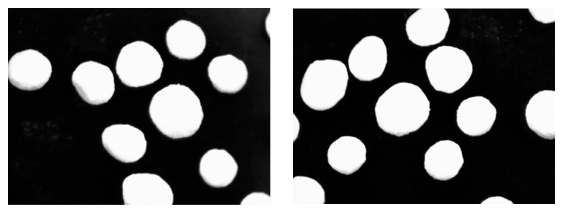
The shape of the pellets was assessed by the following procedure: Individual photographs were taken for 200 pellets laid out on a dark background using a Visioscan® VC 98 (Courage + Khazakha GmbH, Köln, Germany) camera, with a magnification degree of 30×. Each image was analyzed using the ImageJ software (U.S. National Institute of Health, Bethesda, Maryland, USA). The software allows the calculation of certain shape descriptors of the photographed objects based on the following equations:
C i r c u l a r i t y = 4 π × S h a p e   a r e a S h a p e   p e r i m e t e r 2
A s p e c t   R a t i o = M a j o r   A x i s M i n o r   a x i s
R o u n d n e s s = 4 × S h a p e   a r e a π × M a j o r   a x i s 2
Also, given that the software reports the area and perimeter of the photographed objects, it was possible to calculate the sphericity of the pellets (a measure of their roundness) according to the equation:
S p h e r i c i t y = P E Q P C P r e a l = 2 π A P r e a l
where PEQPC is the perimeter of a circle that has an area equivalent to that of the pellet image and Preal represents the perimeter of the pellet image [37].
According to the ImageJ software manual and to scientific literature [35,36,37,38], for either circularity or aspect ratio, values closer to 1 indicate that the photographed objects have a circular shape. Thus, it can be assumed that the closer the values of these two parameters are closer to 1, the more spherical the pellets are.
Hausner ratio is an expression of the friction forces between particles during particle flow, thus being considered an important parameter for the uniform filling of the pellets into hard gelatin capsules. In order to determine the values of this parameter, tapped and bulk densities of the pellets after 300 taps were determined using a Erweka SVM 101 tapped density tester (Erweka GmbH, Heusenstamm, Germany), fitted with a 25 mL cylinder. Based on these results, the Hausner ratio was determined according to the following equation:
H a u s n e r   r a t i o = ρ T ρ B
where ρ T is the tapped density and ρ B is the bulk density of the solid material. A Hausner ratio value lower than 1.2 is an indicator of good flowing characteristics [39].
The fraction of inert pellet cores with sizes between 800 and 1180 μm, separated by sieving, was used for further processing (drug loading).

2.2.2. Loading of the Inert Pellets with Meloxicam by Drug Layering

The inert pellets previously obtained were loaded with meloxicam by drug layering. This technique involved spraying of a meloxicam dispersion on the inert pellet cores in a fluidized bed coating equipment.
First, a meloxicam dispersion was prepared. The composition of this dispersion is shown in Table 2.
Samples of 30 g inert pellets were preheated (at 40 °C) in a fluidized bed coating equipment (Caleva Mini Coater/Drier 2, Caleva Process Solutions Ltd., Dorset, UK).
The meloxicam dispersion, maintained under constant stirring at 800 rpm (Variomag Mono Direct magnetic stirrer, Thermo Fischer Scientific Inc., Waltham, MA, USA) during the coating process, was sprayed on the preheated pellet cores in a fluidized bed coating equipment (Caleva Mini Coater/Drier 2, Caleva Process Solutions Ltd., Dorset, UK). The process parameters used during fluid bed coating of the pellets are shown in Table 3.
For the experimental batches (30 g of inert pellet cores), the amount of meloxicam dispersion used was of 55 g, and the duration of the drug loading stage was of 2 h. The duration of the drug-loading stage was established based of meloxicam assay results obtained for small samples of pellets that were periodically taken out of the coating chamber.
The assay of drug content was performed on the resulting drug-loaded pellets, by UV spectrophotometry, using a Jasco V-530 UV-Vis spectrophotometer (Jasco International Co., Ltd., Tokyo, Japan). Approximately 3 g of pellets were triturated and 80 mg of pellet powder were accurately weighted into a 25 mL volumetric flask, and methanol was added to make up the volume. The samples were dispersed by sonication at room temperature (sonication bath Elma S30H, Elma Schmidbauer GmbH, Singen, Germany) for 10 min. Subsequently, samples of 5 mL were filtered through 0.45 μm regenerated cellulose non-sterile filters (Phenomenex, Torrance, CA, USA). Each sample was diluted with methanol (1:9 v/v) and its absorbance was recorded at 361.4 nm.
A calibration curve generated for dilutions of meloxicam reference standard in methanol, in the concentration range 0.05–400 μg/mL was used for drug quantification.

2.2.3. Film Coating of Meloxicam Pellets

The meloxicam pellets were subsequently subjected to a film-coating process.
Three coating materials were selected, namely Acryl-EZE® 93O white (Colorcon, West Point, PA, USA), Eudragit® RS 30-D (Evonik Industries AG, Essen, Germany), and an experimental composite film made up from Eudragit® RS 30-D and Methocel™ E5 Premium LV (Dow Chemical Company, Midland, MI, USA).
Acryl-EZE® 93O white (further referred to as ”Acryl-EZE®”) is a fully formulated, ready-to-use product, used for enteric coating, in which the film-forming agent is represented by an anionic methacrylic acid—ethyl acrylate (1:1) copolymer, type C [40]. The free carboxylic groups of the copolymer form salts in an alkaline environment, thus the polymer is soluble in solutions with a pH value greater than 5.5.
Eudragit® RS 30-D (Eudragit®) is a liquid dispersion containing a copolymer of ethyl acrylate, methyl metacrylate, and small amounts of methacrylic acid esters with quaternary ammonium groups, which are present as salts, thus making the polymer permeable to aqueous solutions. Film-coating with a dispersion of this product leads to insoluble films, but which are slightly permeable to water, the swelling of the film being independent of the environmental pH values. Drug release from oral preparations film-coated with this product is usually time-dependent. The amount of drug released can be controlled by the film thickness [21,41], this parameter influencing the duration of the drug diffusion stage. Another means of ensuring a prolonged release is to incorporate pore-forming agents in the polymeric film formula.
Methocel™ E5 Premium LV (Methocel™) is a commercial grade of hypromellose, a water-soluble film-forming agent made up of methyl and hydroxypropyl mixed ether of cellulose, with pH-independent solubility profile. In this study, taking into consideration the characteristics of the Eudragit® films (insoluble and slightly permeable), Methocel™ was used as a pore-forming agent, with the aim of adjusting the permeability of the Eudragit® film [42].
The polymeric film coatings were applied using the same equipment that was used for the coating of the pellets with the meloxicam layer.
Three experimental formulations with different type of polymer coating were subsequently obtained.
The first formulation, (coded “MXA”), was obtained by spray coating of the meloxicam pellets with a 20% (w/w) dispersion of Acryl-EZE® in distilled water.
The pellets were preheated in the fluid bed equipment at 35 °C and the Acryl-EZE® dispersion was sprayed onto the meloxicam pellets, using the settings of the technological parameters described in Table 4.
The dispersion was sprayed onto the pellet bed until a weight gain of 8% of the pellets, taking into consideration that the product manufacturer (Colorcon, West Point, PA, USA) recommends a 7–12% weight gain of the substrate, in order to obtain enteric properties. After the spraying stage, the pellets were kept in the fluidized bed, with an air flow temperature of 40 °C, in order to allow the film to cure.
For the second formulation (coded “MXE”) meloxicam pellets were coated with a dispersion of Eudragit® in water, prepared by the formula presented in Table 5 and the technological parameters described in Table 6.
It should be noted that the amount of Eudragit® product used in the formulation translates into a 10% solid content of the final coating dispersion.
The dispersion was sprayed until a 5% weight gain of the pellets, followed by curing of the film by keeping the pellets in fluidized bed, at 42 °C, for 20 min.
Finally, a third formulation (coded MX-EM) was coated with a film in which Eudragit® (as a film-forming agent) and Methocel™ (as a pore-forming agent) were combined. The composition of this film was based on the formula of the Eudragit® dispersion previously used, where 5% of Methocel™ was associated. No significant increase of the viscosity of the coating dispersion could be observed (usually, 2% Methocel™ aqueous solutions have a nominal viscosity of 4–6 mPa · s). The formula of the coating solution is depicted in Table 7. The same process parameters as those presented in Table 6 were used in this experiment.
The formula was sprayed onto the pellets until a weight increase of 5% was determined. The same film curing conditions were applied (the pellets were kept in the fluid bed air current, at 42 °C, for 20 min).
The three variants of film coatings would lead to an enteric release of the BCS class II drug (for the pellets coated with Acryl-EZE®) or to a controlled, extended release (for the Eudragit® coated pellets).

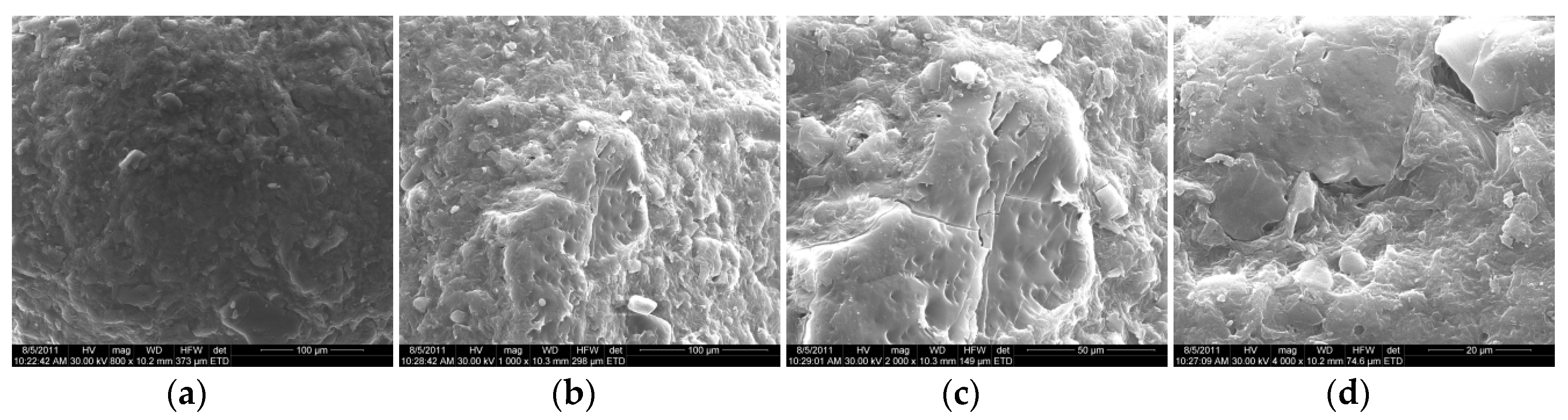
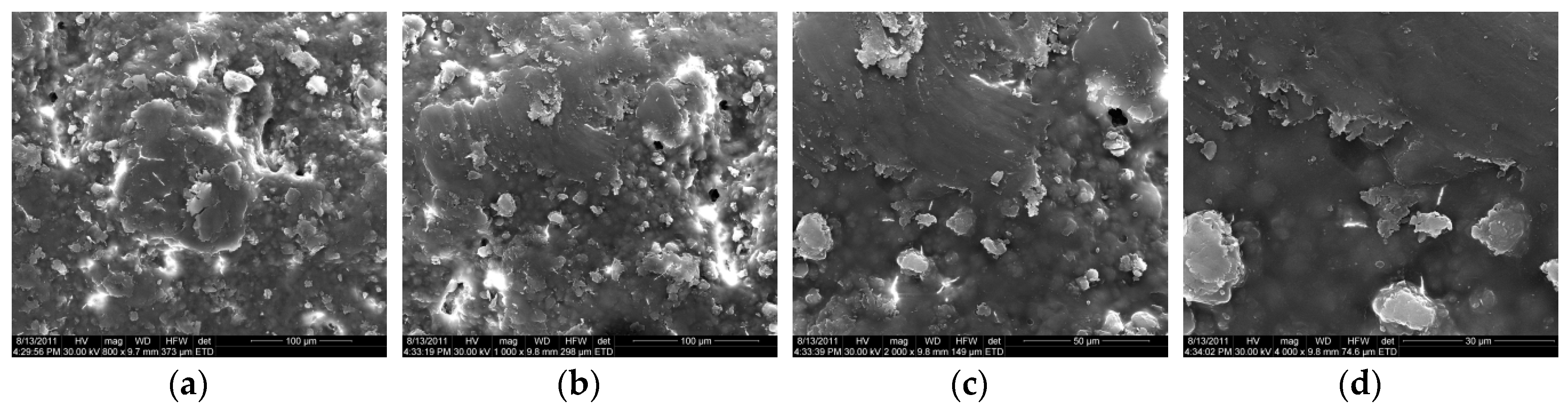
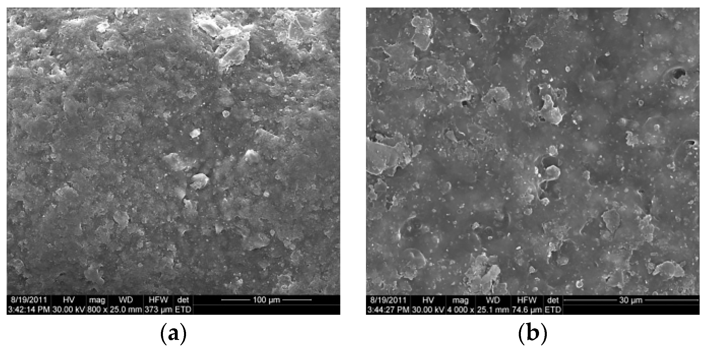
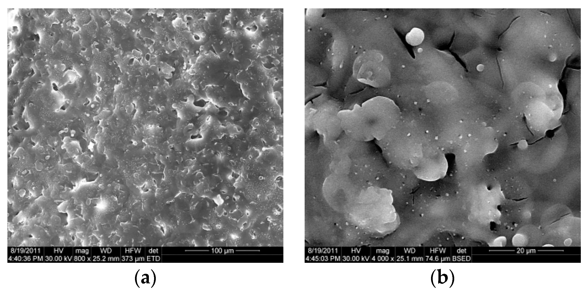
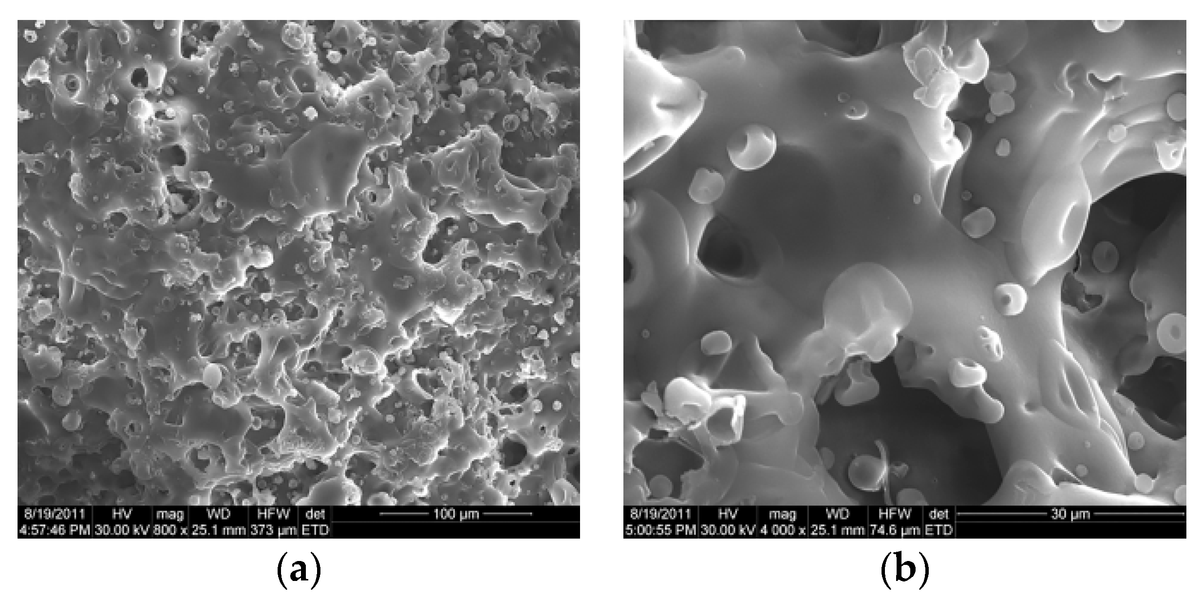
2.2.4. Evaluation of Shape and Surface Morphology of Coated and Uncoated Meloxicam Pellets by Scanning Electron Microscopy (SEM)

In order to evaluate the shape and surface morphology of the meloxicam pellets (both uncoated and film-coated) and to study and better understand the drug layering and film-coating processes, samples of inert pellets (“IP”), uncoated meloxicam pellets (“MXU”), and film-coated meloxicam pellets (film-coated with Acryl-EZE®-sample “MXA”, coated with Eudragit®-sample “MXE”, and coated with the Eudragit® and Methocel™ mixture sample “MX-EM”), respectively, were analyzed by using a Quanta Inspect F scanning electron microscope (FEI Co., Eindhoven, The Netherlands), operated in high vacuum mode (<6 ×   10 4 Pa).
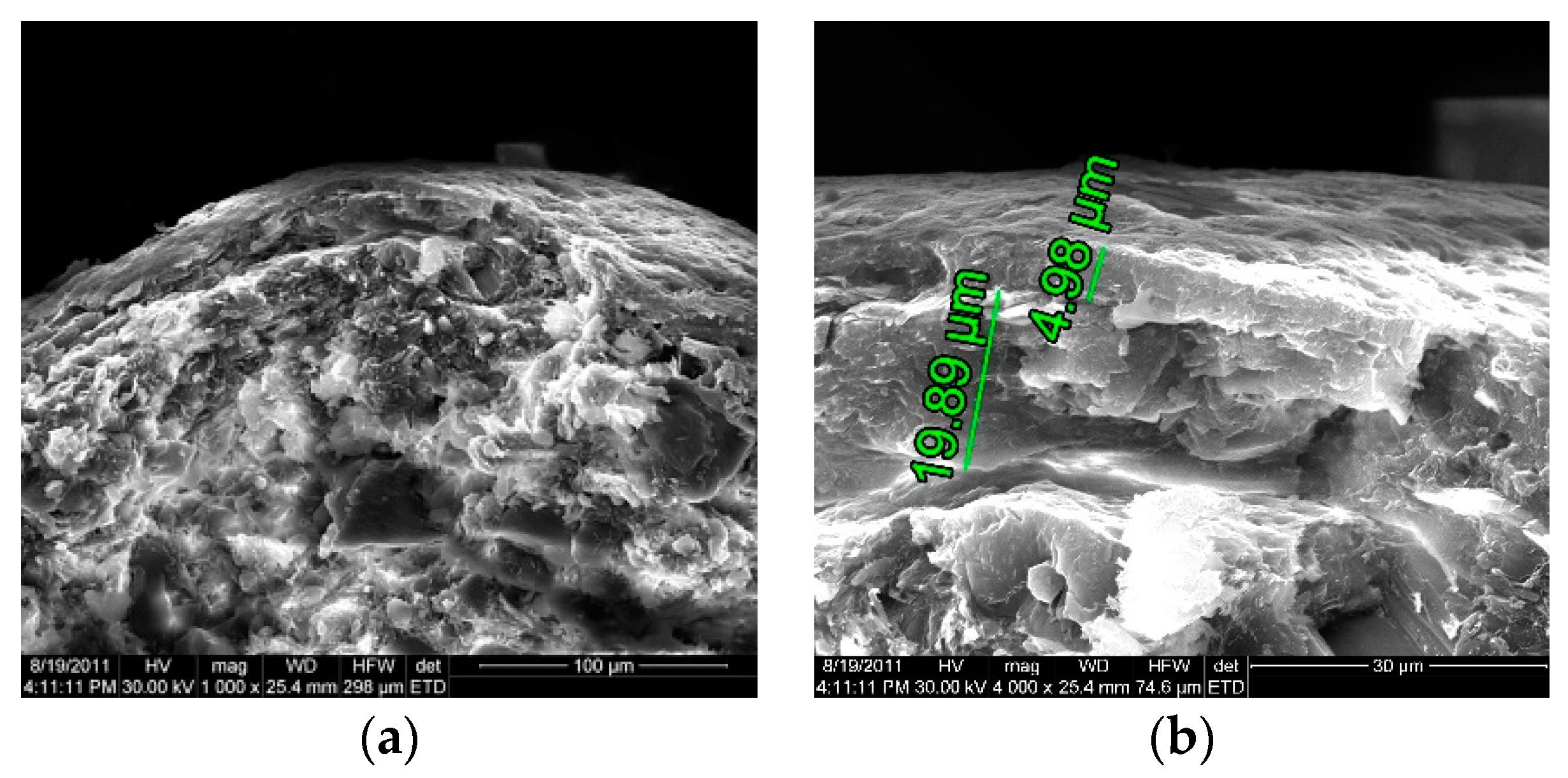
Cross-section images of the pellet samples were also taken. For this, pellet samples were lightly crushed with pestle in a mortar, resulting fractured pellet samples, which were subsequently processed for SEM analysis.
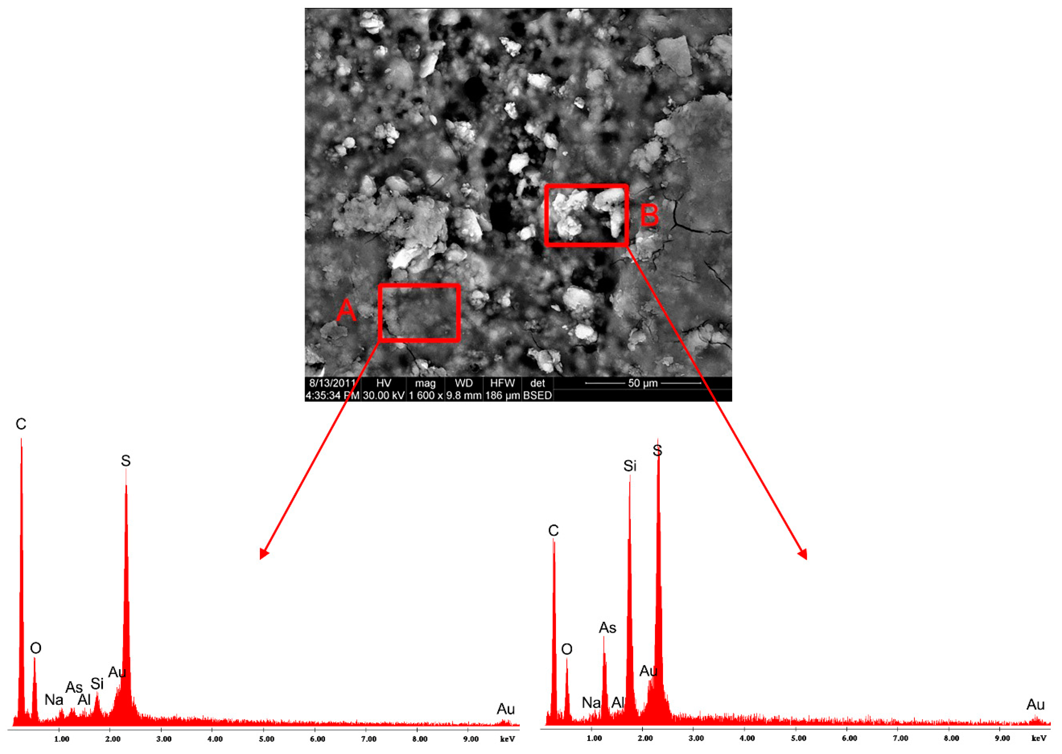
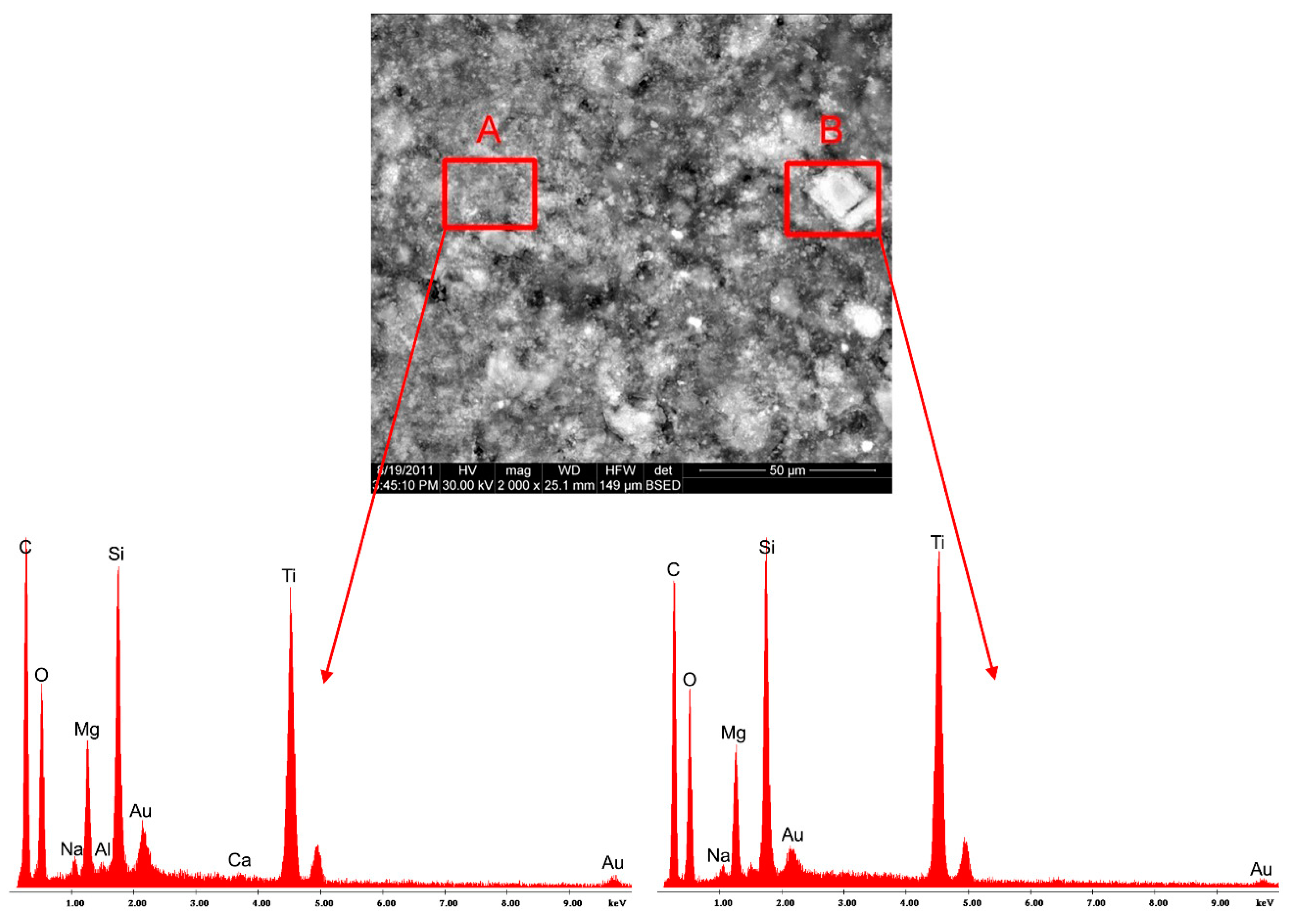
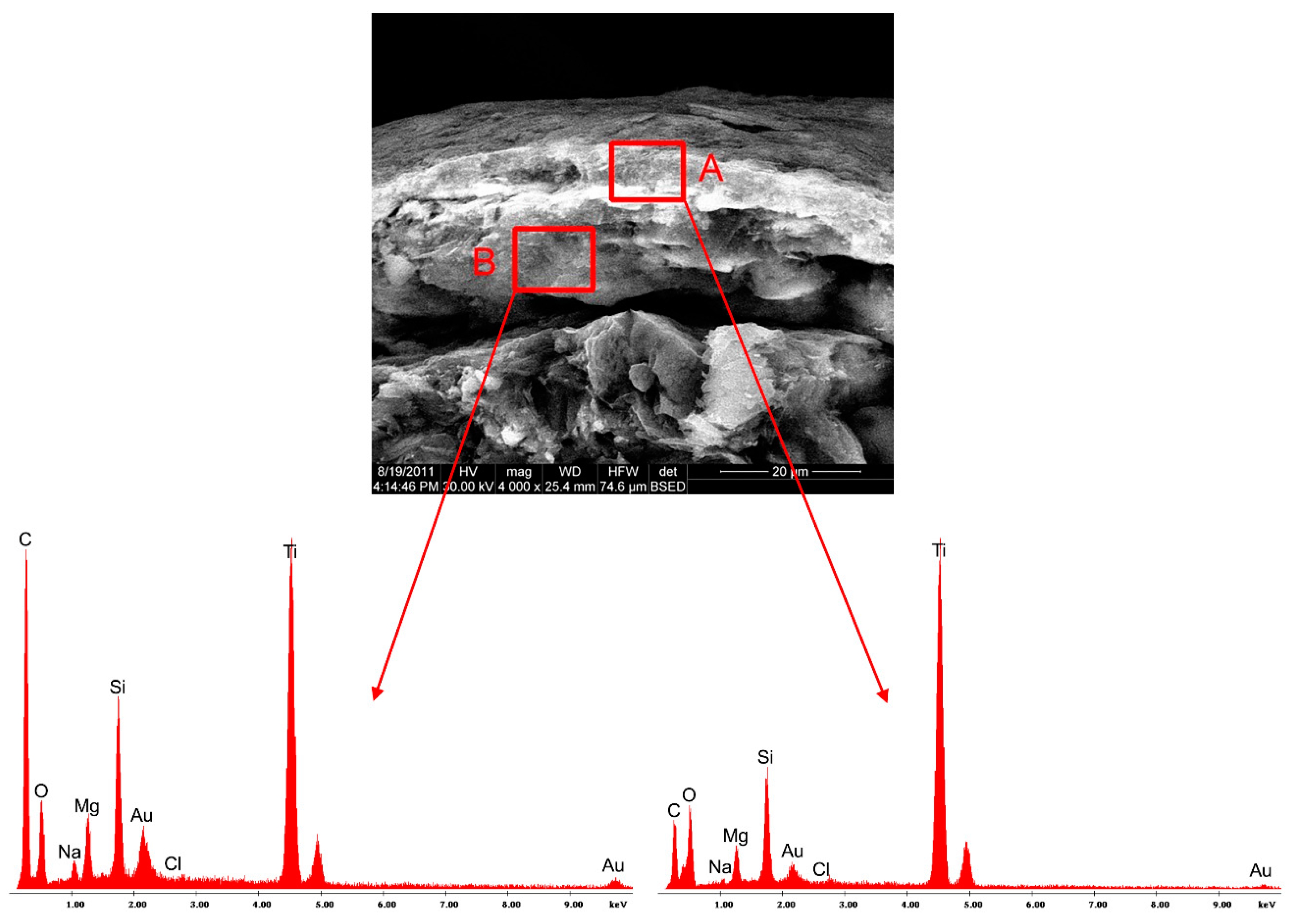
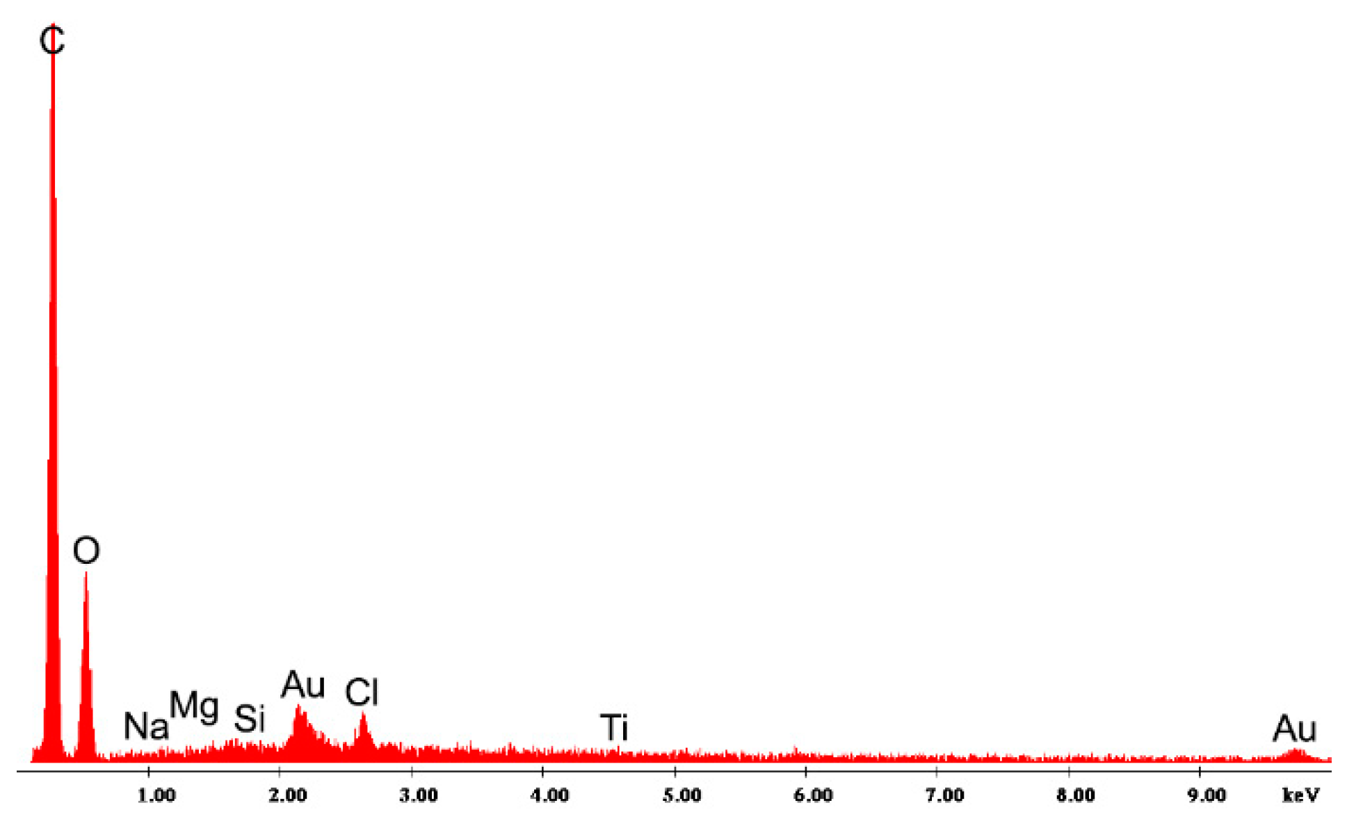
Before each SEM experiment, the surface of the samples was gold coated in argon atmosphere by means of an E5400 Sputter Coater (Bio-RAD Polaron, Kennett Square, PA, USA), thus avoiding the charging of the pellet samples during the analysis. The electron microscope has two detectors—a Everhart-Thornley detector (ETD) for secondary electrons and a back-scatter detector (BSED) and a magnification range of 50× to 1,000,000×. Also, the equipment has an energy dispersive X-ray analyzer (EDAX), which allows the local chemical composition and the distribution of significant elements on the pellet surface to be determined.
SEM images with 800×, 1000×, 2000×, and 4000× degree of magnification were used for the study of the surface morphology. Furthermore, X ray spectra were recorded for samples of meloxicam pellets, using the EDAX detector of the electron microscope, with the aim of elucidating the nature of the formations that could be observed on the surface of the pellets in the images with a high degree of magnification.

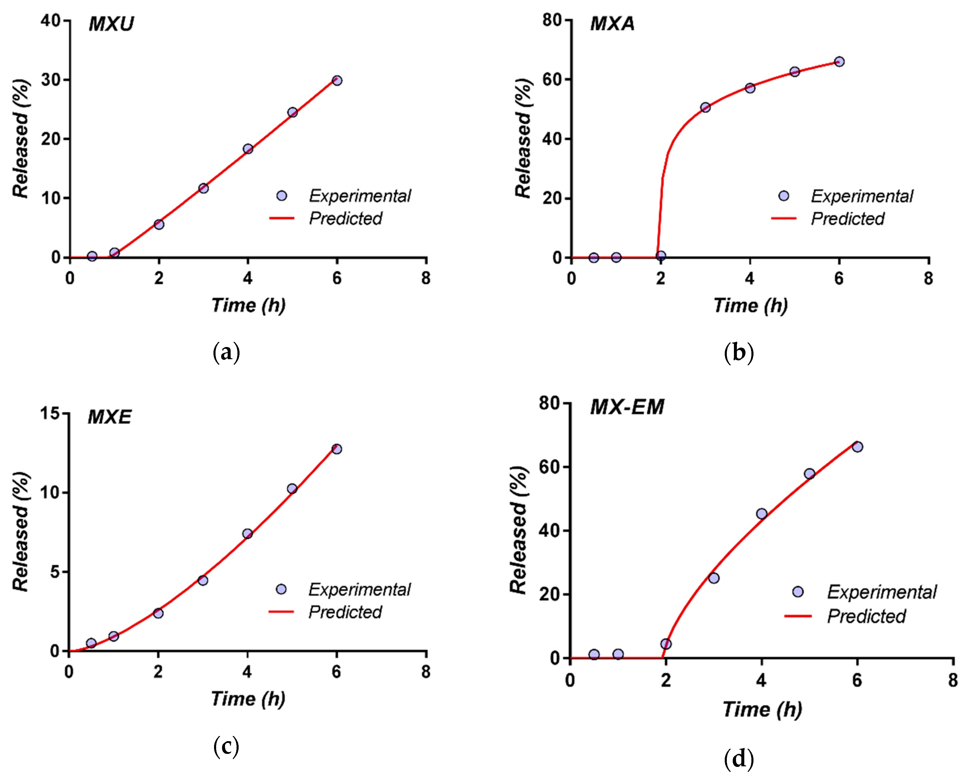
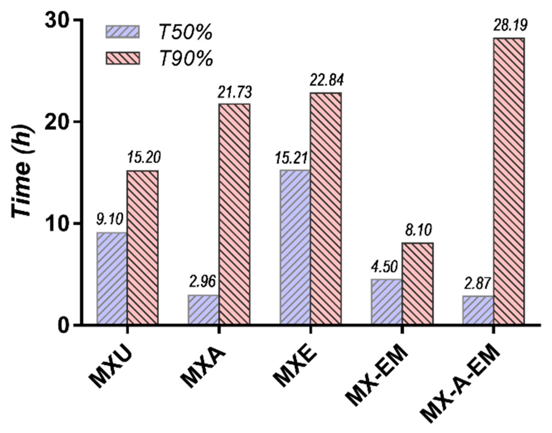
2.2.5. Study of the In Vitro Release Profile of Meloxicam from the Experimental Multi-Particulate Systems

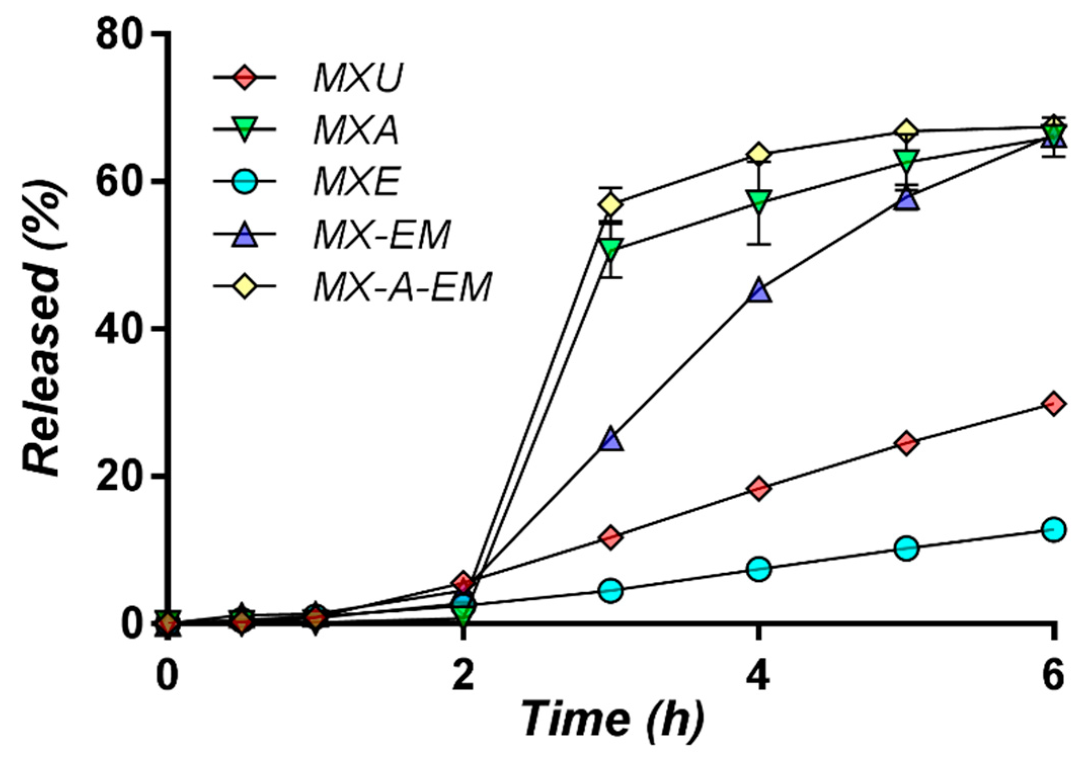
Evaluation of the in vitro release profiles of meloxicam from uncoated and film-coated pellets was performed in order to study the impact of the polymeric coating on the API release profile and subsequently the potential therapeutic performances of the experimental pellets.
In order to study meloxicam release kinetics, the experimental pellets were formulated as single-unit dosage forms (size 00 hard gelatin capsules, Capsugel, Bornem, Belgium).
Accurately weighed pellets (0.625 g), equivalent of 15 mg of meloxicam were manually loaded into the gelatin capsules, according to the method described by Manda A. et al. [43].
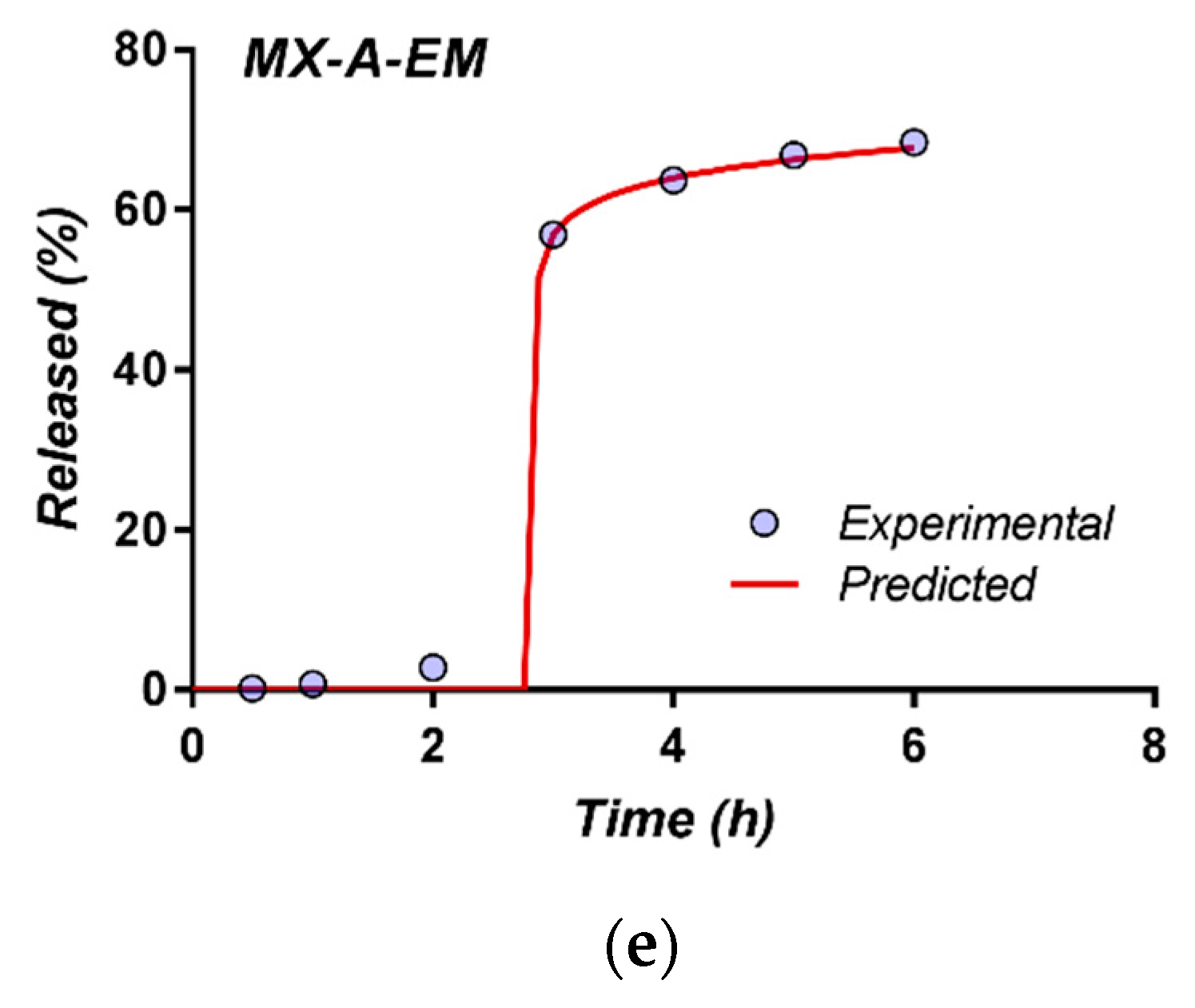
Capsules filled with the previously obtained meloxicam pellets (MXU, MXA, MXE, and MX-EM) were prepared and included in this study. Furthermore, in an additional experiment, the capsule content was represented by a combination of meloxicam pellets coated with Acryl-EZE® (MXA) and pellets coated with the composite film in which Eudragit® was associated with Methocel™ (MX-EM). By combining the two fractions of pellets into the same dosage unit, it was intended to achieve a reduced rate of release for the first two hours of the dissolution test (from the fraction of pellets coated with Eudragit® associated with Methocel™). The amount of drug released from these pellets in the pH 6.8 dissolution media was supplemented by the one yielded from the pellets coated with Acryl-EZE®, following the dissolution of this film at changing the pH of the medium. The two fractions of pellets were combined in a 1:1 ratio so that the dosage unit strength would be of 15 mg meloxicam per capsule. This sample was coded “MX-A-EM.”
The in vitro release of meloxicam from the experimental pellet formulations was performed in a Hanson SR8 Dissolution Test System (Teledyne Hanson Research, Chatsworth, CA, USA) using USP Apparatus 2 (paddle) at 75 rpm and 37 ± 0.5 °C. In vitro drug release testing was performed following the recommendations for delayed-release formulations (method A) from the United States Pharmacopoeia [44] and comprised of two stages: An acid stage (2 h) in simulated gastric conditions (750 mL of 0.1 N hydrochloric acid) followed by a 4-h buffer stage (1000 mL of 50 mM pH = 6.8 phosphate buffer), for testing the drug release rate in intestinal conditions. After the completion of the acidic stage, a volume of 250 mL of preheated 0.20 M tribasic sodium phosphate was added, and the pH adjusted, if necessary, to a value of 6.8 ± 0.05.
Aliquots of 5 ± 0.1 mL were withdrawn at 0.5, 1, 2, 3, 4, 5, and 6 h, and replaced with blank medium at the same temperature. Six replicates were performed for each of the tested formulations. Dissolution profile of pure drug in both media as was used for reference.
The samples were filtered through a 0.45 mm regenerated cellulose non-sterile filters (Phenomenex, Torrance, CA, USA), diluted as needed, and analyzed by UV spectroscopy versus blank medium, using a Jasco V-530 spectrophotometer (Jasco International Co., Tokio, Japan) at 334 nm for pH 1.2 and 361.4 nm for pH 6.8.

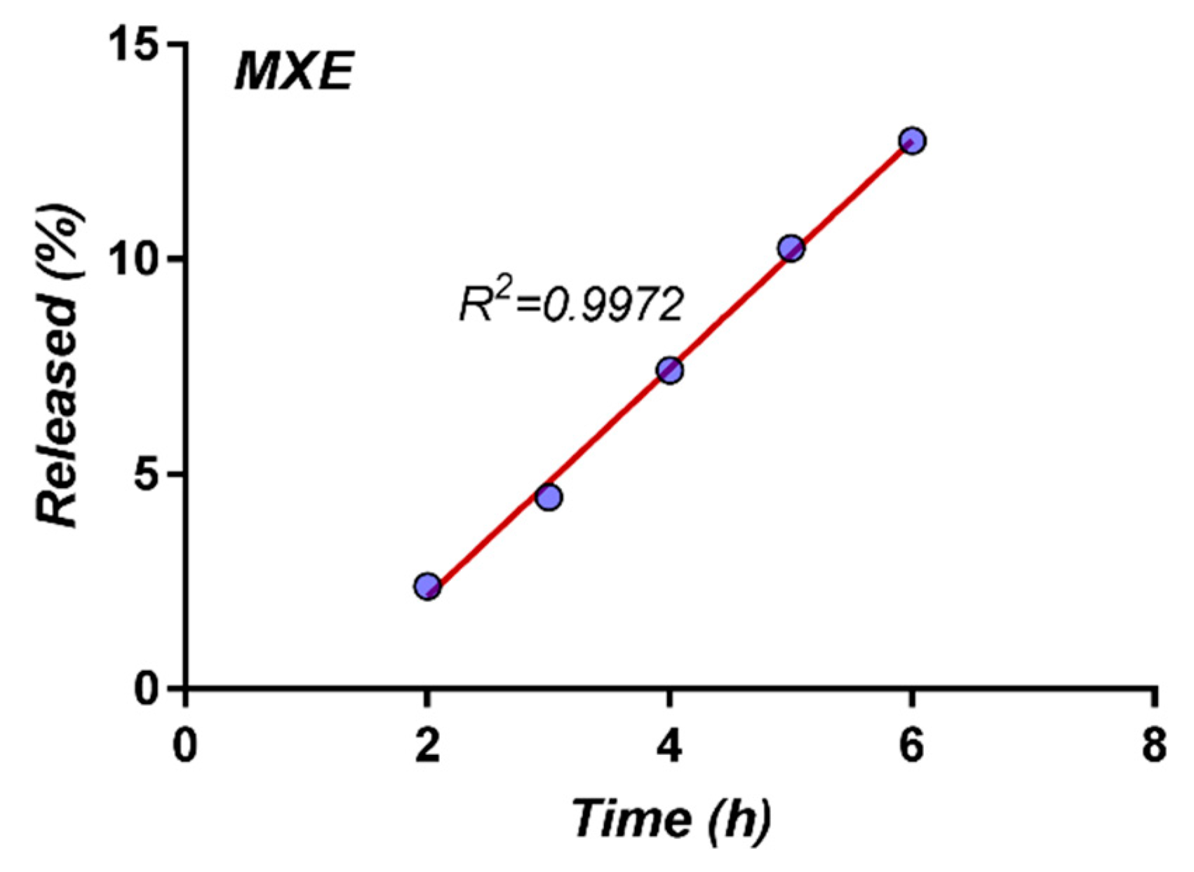
2.2.6. Mathematical Modeling of the Meloxicam In Vitro Release Kinetics

One method of elucidating the mechanism of API release from dosage forms is based on the best fit of experimental data on different mathematical models. [45].
The modeling of meloxicam release profiles was performed by using the DDSolver Excel add-in software package [46]. In order to elucidate the mechanism of meloxicam release from the experimental multi-particulate systems, first 60% drug release data were fitted in Korsmeyer-Peppas model [47]:
F = k K P × t n
where F is the percentage of drug released at time t, k K P is the release constant incorporating structural and geometric characteristics of the dosage form, and n is the diffusional exponent indicating the drug-release mechanism.

3. Results

3.1. The Evaluation of the Characteristics of Inert Pellets

The results for pellet size distribution are presented in Table 8.
The data indicate that, for a large fraction of the sample (almost 95%), the pellet size is within the 0.800–1.18 mm range.
The visual assessment of the photographs used for the evaluation of the shape descriptors have revealed that the pellets appear as slightly elongated spheroids (Figure 1), with a reasonably high degree of sphericity (0.9022). The circularity of the experimental pellets was of 0.8257, the aspect ratio was of 1.1407 and the roundness 0.8824, indicative of adequately shaped pellets.
The calculated value for the Hausner ratio was of 1.08, indicative of good flowing characteristics of the prepared pellets.

3.2. The Evaluation of the Uncoated Meloxicam Pellets

Based on the assay results obtained on pellet samples taken out of the fluid bed equipment during the drug-layering stage, it was determined that, for the experimental inert pellet batches, a duration of the coating process of 2 h leads to a meloxicam content of 23.95 mg/1 g of pellets. This corresponds to a weight increase of the pellets of 1.2%, after the coating process.
Since the maximum amount of pellets that could be filled into size 00 hard gelatin capsule was of 0.8 g, the assay results confirms that such a drug content of the experimental pellets would ensure that the hard gelatin capsules filled with meloxicam pellets would have a strength which is comparable to the one of commercially available single dose meloxicam oral preparations (15 mg meloxicam/dosage unit-tablet).

3.3. The Evaluation of Shape and Surface Morphology of Coated and Uncoated Meloxicam Pellets by Scanning Electron Microscopy

Examples of SEM images of inert pellet samples are presented in Figure 2a–d.
The images reveal a rough surface on which no pores can be observed.
The drug layering of the meloxicam dispersion on inert pellets has produced a slightly rougher surface, as illustrated by the SEM images (Figure 3a–d).
The recorded EDAX spectra have allowed the local chemical micro-composition of certain areas on the pellet surface (Figure 4) to be determined. The EDAX spectra indicate the presence of high amounts of sulfur, an element characteristic to the molecular structure of meloxicam, thus confirming the deposition of the drug on the pellet surface. Also, the spectrum obtained for area B reveals a high amount of silica, most likely from the talc used as a lubricant in the film-coating formula.
Figure 5a,b show the surface of meloxicam pellets coated with Acryl-EZE® (MXA).
No pores can be observed, the film coating smoothly and completely the pellet surface.
In order to evaluate the efficiency of the coating of the meloxicam pellets with Acryl-EZE® and to investigate the nature of some formations visible in the film, EDAX spectra were recorded for this sample, as well (Figure 6).
The EDAX spectra reveal high amounts of titanium in the analyzed areas (indicating the presence of titanium dioxide, the opacifier in the coating product), silica and magnesia (elements from the composition of talc, another ingredient of the film coating). Unlike the results for the MXU sample, no sulfur was detected in the analyzed areas, indicating that the Acryl-EZE® film is completely covering the surface of the meloxicam pellets.
The cross-section images of different MXA samples (Figure 7a,b) show two distinct layers on the surface of the pellets—the Acryl-EZE® layer. The microscope operating software provides a measurement module, which gives the user capabilities to measure linear distances, angles, diameters, and areas, etc. Using this option, it was possible to measure the average thickness of the two layers on several areas of different fracture samples of pellets. The average thickness of the Acryl-EZE® layer was of 5 μm and, while the drug layer had an average thickness of 20 μm. Since the thickness of the Acryl-EZE® layer is within the range 4.83 μm–5.12 μm, it can be assumed that the coating process is robust, yielding a table uniform and continuous Acryl-EZE® film on the surface of the meloxicam pellets.
The EDAX spectrum recorded for different areas in the MXA pellet fractures (Figure 8) showcase a different composition of the two layers on the surface of the pellets, thus confirming the identity of each layer.
The EDAX spectrum for area A (corresponding to the Acryl-EZE® layer) reveal a high titanium content (titanium dioxide is the opacifier in the Acryl-EZE® film), silica, and magnesia (both elements are in the composition of talc, the lubricant in the Acryl-EZE® film). For area B (which corresponds to the drug layer), the presence of silica and magnesia (components of talc, the lubricant used in the drug layering dispersion formula) could be identified in the EDAX spectrum.
SEM images of the meloxicam pellets coated with Eudragit® (MXE) samples were also taken (Figure 9a,b).
Unlike the appearance of the Acryl-EZE® film, the Eudragit® coating appears to be rougher and a number of large apparent pores can be observed. The presence of micrometric globular formations should be noted. These formations could be small film coating dispersion droplets that were rapidly solidified during the coating process. The EDAX spectrum (Figure 10) recorded for such a formation reveals a high amount of carbon.
The following set of images (Figure 11a,b) was taken for the samples of meloxicam pellets coated with the composite film, in which Eudragit® was associated with Methocel™ (samples coded “MX-EM”).
By including Methocel™ in the formula of the film coating dispersion, the porosity of the film increased. In this case, the pellet surface formations are no longer globular, like those observed on the surface of the MXE sample, but rather toroid in shape, each formation displaying a circular central pore.
Overall, the appearance of the film coated pellets is homogenous, with no significant differences between different samples of MX-EM pellets. The film is continuous, completely coating the surface of the pellets.

3.4. The In Vitro Release Studies of Meloxicam from the Experimental Multi-Particulate Systems

The in vitro release test is an important tool in the evaluation of the therapeutic performances of the experimental pellets. Pellets are generally designed as multi-particulate systems for oral administration, often presented as hard gelatin capsules filled with coated pellets. After ingestion, the gelatin capsule shell is rapidly dissolved, releasing the pellets which, in turn, release the active pharmaceutical ingredient in a preprogrammed manner, depending on the formulation.
The results of the in vitro release study performed on the capsules containing each variant of the experimental pellets are presented in Figure 12. The results are expressed as average percentages (calculated based on the individual results for 6 dosage units tested for each formulation) of drug released from the total amount of meloxicam per capsule (15 mg).
For comparison, the pure drug release profile reached a plateau of about 6.35% drug released after one hour in acidic media. In buffer, meloxicam reached 90% release within one hour at pH 6.8, and within 30 min at pH 7.5.

3.5. Kinetic Modeling on Drug Release from the Experimental Multi-Particulate Systems

Meloxicam is a typical example of BCS class II drug, characterized by low solubility and high permeability through phospholipid biological membranes [28]. In theory, an adequate control of the dissolution profile in the gastrointestinal tract can provide adequate control of the absorption processes, as a major component of the biopharmaceutical characteristics. The existence of the API on the surface of the pellets determines the exposure profile to the release media. On the other hand, the nature of the coating film polymer is a formulation factor with major influence on the in vitro release profiles.
In order to better understand meloxicam release mechanism, data up to 60% drug released were subjected to model fitting and statistical analysis, using the Korsmeyer-Peppas. The model was used to determine the diffusion exponent n which characterizes drug release mechanism (Table 9).
The release profile of meloxicam from the MXU sample offers valuable information on the behavior of the drug containing layer on passage through the gastrointestinal tract. The release profile in this case is being limited by both the physicochemical characteristics of the drug and the structural modifications of the polymeric layer containing the API.
Since PVP has a pseudo-cationic polymer behavior and plasticizing properties [48], on immersing the uncoated pellets in the acidic medium, the PVP amino groups got protonated and hydrated, increasing significantly the degree of swelling of the drug containing layer [49] without notable polymer erosion. Together with its solubility, release of the drug from the polymeric matrix depends on the thickness of the diffusion layer, which determines the rate of movement of the API across the polymeric layer into the bulk medium. Therefore, meloxicam low solubility in acidic medium, in conjunction with the swelling of the PVP from the drug containing layer leads to a very slow release of the drug within the first 2 h, up to 5.58% of the nominal concentration. This behavior seems irreversible during the buffer stage, probably due to the “buffering” effect of the HCl ingressed into the polymeric layer during the acid stage. Therefore, although meloxicam solubility is increased in the pH 6.8 medium, its release remains slow and linear for the entire duration of the test (Figure 13a).
The only similar behavior out of the coated formulations was observed for MXE formulation, where the insoluble Eudragit® film barrier does not significantly changes the release mechanism (diffusion controlled release through an swellable polymer) (Figure 13c). However, the Eudragit® low permeability film adds a supplementary barrier to drug diffusion and consequently release, leading therefore to a significantly slower release rate. A slight inflection in the release profile appears at around the 2-h mark, where changing from the acidic to the buffer medium occurred. This led to a value of the n exponent of 1.44, that would apparently be indicative of a super case II transport mechanism, determined by both relaxation and erosion of the polymer. However, this slight inflection could be more likely attributed to a small change in the meloxicam release rate when changing from the acidic into the buffer medium, without actual polymer erosion. Since the low permeable Eudragit® film might also act as an barrier for the acid and prevent PVP to be fully swollen during the acidic stage, it is only natural for the buffer to induce a slight increase of the release rate, due to a slight deswelling caused by PVP deprotonation, accompanied by a significantly higher meloxicam solubility when increasing the pH. In fact, if modelling only the release in the buffer stage of the experiment, a value of 0.908 was obtained, indicative of a time-independent meloxicam release rate. For the last points of the dissolution profile of MXE, the values of the linear regression correlation coefficients were higher than 0.99 (Figure 14).
A lag-time of 2 to 2.5 h was obtained in the modeling of MXA, MX-EM, and MX-A-EM (Figure 13). However, the behavior of the experimental dosage forms in terms of release mechanism are quite different, as revealed by the significant differences in the values of the n exponent: The soluble nature of Acryl-EZE® generates a meloxicam burst release up to about 50% of the nominal amount in both MXA and MX-A-EM formulations (Figure 13a,e), followed by a slow, diffusional release of the API from the dosage form. The n low values of 0.2 (for MXA) and 0.08 (for MX-A-EM) are consistent with a rapid relaxation and limited swelling tendency of the PVP polymer chain upon contact with the pH 6.8 buffer and therefore a rapid release of API into the medium. This behavior is possible because of PVP being protected by the gastro-resistant coating, and therefore, not being able to swell during the acidic phase of the release experiment.
The n value of 0.67 for the fitting of the MX-EM release profile (Table 9) is indicative of a mixed mechanism, based on a combination of both diffusion of the API through the polymeric film and polymer dissolution, which is consistent with the purpose of associating Eudragit® and Methocel™ in the coating process.
The modeling of the experimental data has allowed the evaluation of a very important parameter from the perspective of further development of possible in vitro–in vivo correlations, namely the mean dissolution time (MDT), noted “T50%” (Figure 15). This term represents the time necessary to dissolve 50% of the API dose. Its variation in a very wide range (2.8 to 15.2 h) should be noted.
The maximum time interval required for the release of 90% of the dose (28.19 h) was obtained for the complex system that included combinations of pellet coated with each of the two film-forming agents (Acryl-EZE® and Eudragit®).

4. Discussion

The pellet size distribution results, obtained for the inert pellets, confirm that the experimental formula used for the inert pellet cores and the values set for the parameters of the extrusion-spheronization process yield pellets with a narrow size distribution and low friability. A reasonably high degree of sphericity was obtained (0.9022) for the experimental pellets. This, in conjunction with the values obtained for the shape descriptors circularity (0.8257), aspect ratio (1.1407), and roundness (0.8824) indicates that the experimental pelletization (extrusion-spheronization) process led to adequate slightly elongated spheroid shaped pellets. The Hausner ratio calculated for the inert pellets is indicating good flowing characteristics of the experimental pellets.
The surface morphology data obtained by SEM analysis of the film-coated meloxicam pellets reveal that the surface of the pellets is characteristic for each type of coating dispersion used.
The surface roughness of the pellets, observed at high magnification levels (4000×), is lower in the case of pellets coated with Acryl-EZE® 93O white, and most pronounced in the case of the film in which Eudragit® was associated with Methocel™. The inclusion of Methocel™ in the formula of the Eudragit® film coating dispersion has increased the porosity of the film, confirming the pore-forming function of Methocel™.
The SEM images of cross-sections of pellets illustrate that the Acryl-EZE® film deposited on the surface of pellets has a thickness of about 5 μm. This result was recorded for several sample images, which indicates the reproducibility of the film-coating process. A second layer, underlying the film of Acryl-EZE®, is observed, representing the drug layer, with a thickness of approximately 20 μm.
The obtained EDAX spectra are relevant for the pellets obtained by spraying the meloxicam suspension on inert pellets, highlighting that both layers (the drug layer and the polymeric coating film) were completely spread on the surface of the pellets.
The SEM technique applied for the evaluation of the pellets allowed the confirmation that the pellets have surface morphology that is specific to this type of particles.
The in vitro release data revealed that the smallest percentage of meloxicam released from the experimental multi-particulate systems after 6 h of testing was recorded for the MXE formulation (average result of 12.75% of the total amount of drug per capsule dissolved after 6 h). During the 6 h of testing, almost linear dissolution kinetics could be observed, with an almost constant release of the API. The influence of the pH of the dissolution medium on the released amount was found to be very low. This result is an indicator of the pronounced barrier properties of Eudragit® RS 30-D, which formed an insoluble, slightly permeable film on the surface of the pellets.
The next lowest result at the 6 h-time point was recorded for capsules with the uncoated meloxicam pellets. After 2 h of testing in the pH 1.2 media, the average dissolved amount was 5.58%, while the maximum percentage of dissolved drug after 6 h was of about 30%. After the first hour of testing, an almost linear release of meloxicam could be observed on the release curve.
For the other three samples (MXA, MX-EM, and MX-A-EM), the results after 6 h of dissolution testing are comparable, the amount dissolved from all three experimental systems being of about 66% of the drug content. Because of the disposition of meloxicam on the surface of the pellets, it can be suggested that the barrier function of the Eudragit® film is reduced in the presence of the pore-forming agent Methocel™.
It is to note that, for meloxicam dissolution, the literature data indicate an incomplete release at pH 6.8 [50,51], leading to FDA to recommend a pH 7.5 buffer media for meloxicam tablets, and pH 6.1 containing 0.1% sodium lauryl sulfate for the capsules, in order to achieve at least 70% drug dissolved within 30 min [52]. However, since the behavior of the polymers in our experimental formulations is highly pH dependent, and also they could suffer irreversible modifications in the acidic pH of the stomach, we opted to simulate GI passage, by utilizing a pH 1.2 medium (acidic phase), followed by a more physiologically relevant pH 6.8 buffer (buffer stage), although this would lead to an incomplete release as compared to the USP pH 7.5 medium. For the MXA sample, the percentage of API dissolved after 2 h in the pH 1.2 media was found to be very low, below 1%, revealing that the Acryl-EZE® film fulfilled its function as enteric coating. Surprisingly, highest percentages of dissolved API after 6 h were obtained for the capsules in which the two different fractions of differently coated pellets were combined (one fraction covered with Acryl-EZE®, the second-with Eudragit® associated with Methocel™).
The experimental data and the modeling of the dissolution profiles indicate the dynamic character of the release of a drug substance for which the solubility under conditions simulating the physiological environments is reduced.

5. Conclusions

Present study aimed to study the development and evaluation of different modified release polymer-coated pellets containing meloxicam. An evaluation of the impact of different polymer coating on the performances of the experimental formulations was also performed.
The in vitro release study revealed that the uncoated as well as the Eudragit®-coated pellets achieved a slow, zero-order release of meloxicam, i.e., the drug release rate is constant over the entire release duration. The estimated time required for 90% drug release was high, namely 15.2 h for MXU and 22.8 h for MXE. Adding a pore forming agent to the Eudragit® coating reduced its barrier function, and subsequently the time to 90% drug release to only 8 h. Both formulations containing Acryl-EZE® presented a burst release of about 50% of the nominal amount, followed by a slow, diffusional release of the API from the dosage form.
A fundamental change in the release behavior was determined by whether the external film coating allowed ingress of the HCl into the PVP-based drug containing layer, due to the fundamentally different and irreversible behavior of the polymer at pH values under and above its pKa.

Author Contributions

Conceptualization, M.H., V.A., and D.S.M.; methodology, M.H., D.S.M.; V.A., D.L., M.V.G., and C.-E.D.-P.; software, V.A. and V.J.; validation, M.H., V.A., V.J., and D.S.M.; formal analysis, M.H., V.A., M.V.G., V.J.; data curation, M.H. and V.A.; writing—original draft preparation, M.H. and V.A.; writing—review and editing, D.L., C.-E.D.-P. All authors have read and agreed to the published version of the manuscript.

Funding

This research was financially supported by “Carol Davila” University of Medicine and Pharmacy through Contract no. 23PFE/17.10.2018 funded by the Ministry of Research and Innovation within PNCDI III, Program 1—Development of the National RD system, Subprogram 1.2—Institutional Performance—RDI excellence funding projects.

Conflicts of Interest

The authors declare no conflict of interest.

References

  1. Ghebre-Selassie, I.; Martin, C. Pharmaceutical Extrusion Technology; Taylor & Francis: Abingdon, UK, 2003. [Google Scholar]
  2. Partheniadis, I.; Gkogkou, P.; Kantiranis, N.; Nikolakakis, I. Modulation of the Release of a Non-Interacting Low Solubility Drug from Chitosan Pellets Using Different Pellet Size, Composition and Numerical Optimization. Pharmaceutics 2019, 11, 175. [Google Scholar] [CrossRef] [PubMed] [Green Version]
  3. Hîrjău, M.; Lupuliasa, D.; Rădulescu, F.Ș.; Mitu, M.A.; Miron, D. Preparation and Characterization of Pellets Containing a Non-steroidal Antiinflamatory Drug, Coated with an Enteric Polymer. Farmacia 2011, 59, 550–560. [Google Scholar]
  4. Hîrjău, M.; Lupuliasa, D.; Rădulescu, F.Ș.; Miron, D. The Study of Piroxicam Dissolution from Eudragit RS-Coated Pellets. Farmacia 2013, 61, 845–855. [Google Scholar]
  5. Akhgari, A.; Sadeghi, F.; Garekani, H.A. Combination of time-dependent and pH-dependent polymethacrylates as a single coating formulation for colonic delivery of indomethacin pellets. Int. J. Pharm. 2006, 320, 137–142. [Google Scholar] [CrossRef] [PubMed]
  6. Chen, T.; Li, J.; Chen, T.; Sun, C.C.; Zheng, Y. Tablets of multi-unit pellet system for controlled drug delivery. J. Control. Release 2017, 262, 222–231. [Google Scholar] [CrossRef]
  7. Iovanov, R.I.; Tomuță, I.; Barbu, A.; Rus, L.; Leucuța, S.E. Preparation and in vitro Characterization of Pellets Containing Felodipine Solid Dispersions. Farmacia 2015, 63, 637–646. [Google Scholar]
  8. Dahlberg, C.; Millqvist-Fureby, A.; Schuleit, M.; Furó, I. Polymer–drug interactions and wetting of solid dispersions. Eur. J. Pharm. Sci. 2010, 39, 125–133. [Google Scholar] [CrossRef]
  9. Konno, H.; Handa, T.; Alonzo, D.E.; Taylor, L.S. Effect of polymer type on the dissolution profile of amorphous solid dispersions containing felodipine. Eur. J. Pharm. Biopharm. 2008, 70, 493–499. [Google Scholar] [CrossRef]
  10. Nikowitz, K.; Kása, P., Jr.; Pintye-Hódi, K.; Regdon, G., Jr. Study of the preparation of a multiparticulate drug delivery system with a layering technique. Powder Technol. 2011, 205, 155–159. [Google Scholar] [CrossRef]
  11. Pan, X.; Chen, M.; Han, K.; Peng, X.; Wen, X.; Chen, B.; Wang, J.; Li, G.; Wu, C. Novel compaction techniques with pellet-containing granules. Eur. J. Pharm. Biopharm. 2010, 75, 436–442. [Google Scholar] [CrossRef]
  12. Theismann, E.M.; Keppler, J.K.; Owen, M.; Schwarz, K.; Schlindwein, W. Modelling the Effect of Process Parameters on the Wet Extrusion and Spheronisation of High-Loaded Nicotinamide Pellets Using a Quality by Design Approach. Pharmaceutics 2019, 11, 154. [Google Scholar] [CrossRef] [Green Version]
  13. Peter, J.C. Tramadol SR formulations: Pharmacokinetic comparison of a multiple-units dose (capsule) versus a single-unit dose (tablet). Clin. Drug Investig. 2005, 7, 435–443. [Google Scholar]
  14. Muley, S.; Nandgude, T.; Poddar, S. Extrusion–spheronization a promising pelletization technique: Indepth review. Asian J. Pharm. Sci. 2016, 11, 684–699. [Google Scholar] [CrossRef] [Green Version]
  15. Reddy, P.N.S.; De, A.; Nagasamy Venkatesh, D. Pelletization process and techniques. Pharma Times 2015, 47, 22–27. [Google Scholar]
  16. Pałkowski, Ł.; Karolak, M.; Kubiak, B.; Błaszczyński, J.; Słowiński, R.; Thommes, M.; Kleinebudde, P.; Krysiński, J. Optimization of pellets manufacturing process using rough set theory. Eur. J. Pharm. Sci. 2018, 124, 295–303. [Google Scholar] [CrossRef]
  17. Sarkar, S.; Liew, C.V.; Soh, J.L.P.; Heng, P.W.S.; Wong, T.W. Microcrystalline cellulose: An overview. In Functional Polymeric Composites; Apple Academic Press: New York, NY, USA, 2017. [Google Scholar] [CrossRef]
  18. Newton, J.M. Extrusion and extruders. In Encyclopedia of Pharmaceutical Technology; Swarbrick, J., Boylan, J.C., Eds.; Marcel Dekker Inc.: New York, NY, USA; Basel, Switzerland, 2002; pp. 1220–1236. [Google Scholar]
  19. Shah, R.D.; Kabadi, M.; Pope, D.G.; Augsburger, L.L. Physico-mechanical characterization of the extrusion-spheronization process. Part II: Rheological determinants for successful extrusion and spheronization. Pharm. Res. 1995, 12, 496–507. [Google Scholar] [CrossRef]
  20. Sonaglio, D.; Bataille, B.; Ortigosa, C.; Jacob, M. Factorial design in the feasibility of producing Microcel MC 101 pellets by extrusion/spheronization. Int. J. Pharm. 1995, 115, 53–60. [Google Scholar] [CrossRef]
  21. Al-Hashimi, N.; Begg, N.; Alany, R.G.; Hassanin, H.; Elshaer, A. Oral Modified Release Multiple-Unit Particulate Systems: Compressed Pellets, Microparticles and Nanoparticles. Pharmaceutics 2018, 10, 176. [Google Scholar] [CrossRef] [Green Version]
  22. Wan, D.; Zhao, M.; Zhang, J.; Luan, L. Novel Sustained-Release Loxoprofen Pellet with Double Coating Layer. Pharmaceutics 2019, 11, 260. [Google Scholar] [CrossRef] [Green Version]
  23. Lin, H.; Zhang, Z.; Markl, D.; Zeitler, J.A.; Shen, Y. A Review of the Applications of OCT for Analysing Pharmaceutical Film Coatings. Appl. Sci. 2018, 8, 2700. [Google Scholar] [CrossRef] [Green Version]
  24. Felton, L.A.; Porter, S.C. An update on pharmaceutical film coating for drug delivery. Expert Opin. Drug Deliv. 2013, 10, 421–435. [Google Scholar] [CrossRef] [PubMed]
  25. Yang, Q.; Yuan, F.; Xu, L.; Yan, Q.; Yang, Y.; Wu, D.; Guo, F.; Yang, G. An update of moisture barrier coating for drug delivery. Pharmaceutics 2019, 11, 436. [Google Scholar] [CrossRef] [PubMed] [Green Version]
  26. Davies, N.M.; Skjodt, N.M. Clinical pharmacokinetics of meloxicam. Clin. Pharmacokinet. 1999, 36, 115–126. [Google Scholar] [CrossRef]
  27. Bekker, A.; Kloepping, C.; Collingwood, S. Meloxicam in the management of post-operative pain: Narrative review. J. Anaesthesiol. Clin. Pharmacol. 2018, 34, 450–457. [Google Scholar] [CrossRef] [PubMed]
  28. Amidon, G.L.; Lennernäs, H.; Shah, V.P.; Crison, J.R. A theoretical basis for a biopharmaceutic drug classification: The correlation of in vitro drug product dissolution and in vivo bioavailability. Pharm. Res. 1995, 12, 413–420. [Google Scholar] [CrossRef] [Green Version]
  29. Papich, M.G.; Martinez, M.N. Applying Biopharmaceutical Classification System (BCS) Criteria to Predict Oral Absorption of Drugs in Dogs: Challenges and Pitfalls. AAPS J. 2015, 17, 948–964. [Google Scholar] [CrossRef] [Green Version]
  30. Ghareeb, M.M.; Abdulrasool, A.A.; Hussein, A.A.; Noordin, M.I. Kneading technique for preparation of binary solid dispersion of meloxicam with poloxamer 188. Aaps Pharmscitech 2009, 10, 1206–1215. [Google Scholar] [CrossRef]
  31. Fries, J. Toward an understanding of NSAID-related adverse events: The contribution of longitudinal data. Scand J. Rheumatol. Suppl. 1996, 102, 3–8. [Google Scholar] [CrossRef]
  32. Davies, N.M. Sustained release and enteric coated NSAIDs: Are they really GI safe? J. Pharm. Pharm. Sci. 1999, 2, 5–14. [Google Scholar]
  33. Bauer, C.; Frost, P.; Kirschner, S. Pharmacokinetics of 3 formulations of meloxicam in cynomolgus macaques (macaca fascicularis). J. Am. Assoc. Lab. Anim. Sci. 2014, 53, 502–511. [Google Scholar]
  34. Auriemma, G.; Cerciello, A.; Sansone, F.; Pinto, A.; Morello, S.; Aquino, R.P. Polysaccharides based gastroretentive system to sustain piroxicam release: Development and in vivo prolonged anti-inflammatory effect. Int. J. Biol. Macromol. 2018, 120, 2303–2312. [Google Scholar] [CrossRef] [PubMed]
  35. Almeida-Prieto, S.; Blanco-Méndez, J.; Otero-Espinar, F.J. Image Analysis of the Shape of Granulated Powder Grains. Pharm. Sci. 2004, 93, 621–634. [Google Scholar] [CrossRef] [PubMed]
  36. Londoño, C.; Rojas, J. Effect of different production variables on the physical properties of pellets prepared by extrusion-spheronization using a multivariate analysis. Thai J. Pharm. Sci. 2017, 41, 1–7. [Google Scholar]
  37. Loka, N.C.; Saripella, K.K.; Pinto, C.A.; Neau, S.H. Use of extrusion aids for successful production of Kollidon® CL-SF pellets by extrusion–spheronization. Drug Dev. Ind. Pharm. 2018, 44, 632–642. [Google Scholar] [CrossRef] [PubMed]
  38. ImageJ Website–NIH. Available online: https://imagej.nih.gov/ij/plugins/circularity.html (accessed on 10 March 2020).
  39. Fahr, A. Test Methods for Powders and Granules. In Voigt’s Pharmaceutical Technology; John Wiley & Sons Ltd.: Hoboken, NY, USA, 2018; p. 336. [Google Scholar]
  40. Das, A.; Sathyamoorthy, N.; Garikapati, D. Development of Enteric Coated Pantoprazole Tablets with an Aqueous Based Polymer. Int. J. Chem. Tech. Res. 2013, 5, 2395. [Google Scholar]
  41. Sultana, S.; Ahmed, I.; Islam, M.R.; Rahman, M.H. Development of salbutamol sulphate sustained release pellets using acrylic polymer and polyvinyl acetate polymer and evaluation of in vitro release kinetics. Dhaka Univ. J. Pharm. Sci. 2010, 9, 109–118. [Google Scholar] [CrossRef]
  42. Banerjee, A.; Verma, P.R.P.; Gore, S. Controlled Porosity Solubility Modulated Osmotic Pump Tablets of Gliclazide. AAPS Pharm. Sci. Tech. 2015, 16, 554–568. [Google Scholar] [CrossRef] [Green Version]
  43. Manda, A.; Walker, R.B.; Khamanga, S.M.M. An Artificial Neural Network Approach to Predict the Effects of Formulation and Process Variables on Prednisone Release from a Multipartite System. Pharmaceutics 2019, 11, 109. [Google Scholar] [CrossRef] [Green Version]
  44. USP 38-NF 33. The United States Pharmacopeia 38-the National Formulary 33; United States Pharmacopeial Convention Inc.: Rockville, MD, USA, 2015; p. 486. [Google Scholar]
  45. Mircioiu, C.; Voicu, V.; Anuta, V.; Tudose, A.; Celia, C.; Paolino, D.; Fresta, M.; Sandulovici, R.; Mircioiu, I. Mathematical modeling of release kinetics from supramolecular drug delivery systems. Pharmaceutics. 2019, 11, 140. [Google Scholar] [CrossRef] [Green Version]
  46. Zhang, Y.; Huo, M.; Zhou, J.; Zou, A.; Li, W.; Yao, C.; Xie, S. DDSolver: An add-in program for modeling and comparison of drug dissolution profiles. Aaps J. 2010, 12, 263–271. [Google Scholar] [CrossRef] [Green Version]
  47. Preda, I.A.; Mircioiu, I.; Mircioiu, C.; Corlan, G.; Pahomi, G.; Prasacu, I.; Anuta, V. Research concerning the development of a biorelevant dissolution test for formulations containing norfloxacin. I. Modelling of in vitro release kinetics. Farmacia 2012, 60, 675–687. [Google Scholar]
  48. Cook, J.P. ph induced swelling of pvp microgel particles–A first order phase transition? J. Colloid Interface Sci. 2012, 370, 67–72. [Google Scholar] [CrossRef] [PubMed]
  49. Fernández-Nieves, A.; Fernández-Barbero, A.; Vincent, B.; de las Nieves, F.J. Charge controlled swelling of microgel particles. Macromolecules 2000, 33, 2114–2118. [Google Scholar] [CrossRef]
  50. Jin, C.; Zhao, C.; Shen, D.; Dong, W.; Liu, H.; He, Z. Evaluating bioequivalence of meloxicam tablets: Is in-vitro dissolution test overdiscriminating? J. Pharm. Pharm. 2018, 70, 250–258. [Google Scholar] [CrossRef] [PubMed]
  51. Oliveira, É.D.F.S.; Azevedo, R.D.C.P.; Bonfilio, R.; Oliveira, D.B.D.; Ribeiro, G.P.; Araújo, M.B.D. Dissolution test optimization for meloxicam in the tablet pharmaceutical form. Braz. J. Pharm. Sci. 2009, 45, 67–73. [Google Scholar] [CrossRef]
  52. FDA—Dissolution Methods Database. Available online: https://www.accessdata.fda.gov/scripts/CDER/dissolution/dsp_SearchResults.cfm (accessed on 11 March 2020).
Figure 1. Visual assessment of different batches of the inert pellets (30× magnification).
Coatings 10 00490 g001
Figure 2. Scanning electron microscopy (SEM) images of the surface of inert pellet samples (a) 800× magnification; (b) 1000× magnification; (c) 2000× magnification; (d) 4000× magnification.
Coatings 10 00490 g002
Figure 3. SEM images of the surface of MXU samples (a) 800× magnification; (b) 1000× magnification; (c) 2000× magnification; (d) 4000× magnification.
Coatings 10 00490 g003
Figure 4. Energy dispersive X-ray analyzer (EDAX) analysis of the MXU pellet surface.
Coatings 10 00490 g004
Figure 5. (a) SEM image (800×) of the surface of MXA; (b) SEM image (4000×) of the surface of MXA.
Coatings 10 00490 g005
Figure 6. EDAX analysis of the MXA pellet surface.
Coatings 10 00490 g006
Figure 7. (a) SEM image (1000×) of the cross-section of MXA samples; (b) example of SEM image (4000×, BSED) of the cross-section of MXA samples.
Coatings 10 00490 g007
Figure 8. EDAX analysis of two different areas on the MXA pellet fracture.
Coatings 10 00490 g008
Figure 9. (a) Example of SEM image (800×) of the surface of MXE; (b) example of SEM image (4000×) of the surface of MXE.
Coatings 10 00490 g009
Figure 10. EDAX spectrum for globular formation on the surface of the MXE pellets.
Coatings 10 00490 g010
Figure 11. (a) Example of SEM image (800×) of the surface of MX-EM sample; (b) example of SEM image (4000×) of the surface of MX-EM sample.
Coatings 10 00490 g011
Figure 12. Comparative in vitro release profiles of the experimental meloxicam multi-particulate drug delivery systems.
Coatings 10 00490 g012
Figure 13. Kinetic modeling of dissolution profiles of the meloxicam multi-particulate systems using the Korsmeyer-Peppas model (a) MXU; (b) MXA; (c) MXE; (d) MX-EM; (e) MX-A-EM.
Coatings 10 00490 g013aCoatings 10 00490 g013b
Figure 14. Kinetic modeling of the MXE release profile using zero order kinetics.
Coatings 10 00490 g014
Figure 15. Dissolution time of 50%, respectively 90% of the dose.
Coatings 10 00490 g015
Table 1. The process parameters used in the manufacturing of the inert pellets.
Process ParameterSetting
Extruder sieve1.2 mm sieve opening
Extrusion speed15 rpm
Spheronization friction plate12 cm diameter, cross-hatch pattern, 0.8-mm depth grooves and pyramidal protrusions
Spheronization time2 min
Spheronization speed870 rpm
Table 2. Composition of the meloxicam dispersion used for drug layering.
IngredientAmount (%, w/w)
Meloxicam1.91
PVP1.28
Sodium dodecyl sulfate0.28
Talc0.28
Distilled water96.15
Table 3. The process parameters used for the drug layering of the inert pellets.
Process ParameterSetting
Air speed11 m/s
Air temperature40 °C
Dispersion feed1.75 rpm (approx. 25 mL/h)
Atomizing air pressure12 psi (0.81 atm.)
Table 4. The process parameters used in the manufacturing of the meloxicam pellets coated with Acryl-EZE®.
Process ParameterSetting
Air speed7.8 m/s
Air temperature35 °C
Dispersion feed1.36 rpm (approx. 20 mL/h)
Atomizing air pressure10 psi (0.68 atm.)
Table 5. The composition of the Eudragit® dispersion.
IngredientAmount (%, w/w)
Eudragit® RS 30-D33.33
Polyethylene Glycol 4001
Distilled waterUp to 100
Table 6. The process parameters used in the manufacturing of the meloxicam pellets coated with Eudragit®.
Process ParameterSetting
Air speed11 m/s
Air temperature42 °C
Dispersion feed1.36 rpm (approx. 20 mL/h)
Atomizing air pressure12 psi (0.81 atm.)
Table 7. The composition of the Eudragit® and Methocel™ dispersion.
IngredientAmount (%, w/w)
Eudragit® RS 30-D33.33
Methocel™ E5 Premium LV5
Polyethylene Glycol 4001
Distilled waterUp to 100
Table 8. Size distribution of the inert pellets.
Sieve Eye OpeningAmount (g)Amount (% of Total Sample Weight-99.301 g)
1.180 mm0.1190.1198
0.800 mm93.99494.655
0.600 mm3.6233.657
0.315 mm1.5031.513
0.250 mm0.0190.019
Residue00
Table 9. The k K P (release constant), n (diffusion exponent) and coefficient of determination (R2) values for the Korsmeyer-Peppas models applied to the meloxicam release kinetics.
ParameterSample
MXUMXAMXEMX-EMMX-A-EM
k K P 5.3950.360.9926.5862.59
n1.060.201.44 */0.908 **0.670.08
R20.99830.99980.9962 */0.9980 **0.99370.9980
*—values obtained after the modeling on the entire release profile (0–6h) **—values obtained after the modeling of the release data in the buffer stage (2–6 h).

