Abstract
The demand for solid organs is increasing worldwide, regenerative medicine aims to develop organs that can replace their human counterparts. In this regard, this study describes a novel biomimetic-based methodology for the incorporation of microducts in 3D bacterial nanocellulose (BNC-3D) biomaterials. Although BNC is a biomaterial that has been used as a scaffold for cell culture purposes, it does not have the microduct structure that solid organs required to maintain cell viability. This study aims to biomimicry the microduct structure (blood vessels) in BNC using a corroded porcine kidney in epoxy resin during BNC synthesis. The resin mold was incorporated into the biological process of producing BNC-3D. After the BNC fermentation, the resin was removed using a novel method (acid hydrolysis) to expose the blood vessels constructs. BNC-3D and BNC-3D with microducts (BNC-3DM) were analyzed using electronic microscopy, infrared analysis, thermogravimetric and biological analysis. Results show that biomaterials biomimicry the blood vessels of the reference organ, moreover, the BNC chemical and morphological properties of BNC was not affected in the biomimetic process. Regarding cell behavior, cell viability was not affected by the incorporation of the microducts, and it was proven that viable cells adhere to the microducts surface, reproducing their shape and migrate into the biomaterial up to 245 µm for 8 days of culture. To conclude, the data demonstrate the potential of biomimetic in BNC for regenerative medicine, in which the microducts transport fluids (blood, nutrients, and waste products) from and to engineered solid organs via animal counterparts.
Graphic abstract
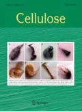
The graphical abstract represents the structural modification of bacterial nanocellulose (BNC) with the inclusion of microducts and microporosities. Furthermore, it represents the usefulness of the microducts in future applications, where, they can be used for nutrients inlet to feed the cells and to remove the wastes from the developed tissue, same as do the blood vessels.


References
Acosta Gómez AP (2006) El fibroblasto: su origen, estructura, funciones y heterogeneidad dentro del periodonto. Univ Odontol 25:26–33
Agarwal T, Maiti TK, Ghosh SK (2019) Decellularized caprine liver-derived biomimetic and pro-angiogenic scaffolds for liver tissue engineering. Mater Sci Eng C 98:939–948. https://doi.org/10.1016/j.msec.2019.01.037
Amin MC, Abadi AG, Katas H (2014) Purification, characterization and comparative studies of spray-dried bacterial cellulose microparticles. Carbohydr Polym 99:180–189. https://doi.org/10.1016/j.carbpol.2013.08.041
Aydogdu MO, Chou J, Altun E et al (2019) Production of the biomimetic small diameter blood vessels for cardiovascular tissue engineering. Int J Polym Mater Polym Biomater 68:243–255. https://doi.org/10.1080/00914037.2018.1443930
Bodin A, Bharadwaj S, Wu S, Gatenholm P, Atala A, Zhang Y (2010) Tissue-engineered conduit using urine-derived stem cells seeded bacterial cellulose polymer in urinary reconstruction and diversion. Biomaterials 31:8889–8901. https://doi.org/10.1016/j.biomaterials.2010.07.108
Castro C, Zuluaga R, Putaux JL, Caro G, Mondragon I, Gañán P (2011) Structural characterization of bacterial cellulose produced by Gluconacetobacter swingsii sp. from Colombian agroindustrial wastes. Carbohydr Polym 84:96–102. https://doi.org/10.1016/j.carbpol.2010.10.072
Castro C, Zuluaga R, Álvarez C, Putaux JL, Caro G, Rojas OJ, Mondragon I, Gañán P (2012) Bacterial cellulose produced by a new acid-resistant strain of Gluconacetobacter genus. Carbohydr Polym 89:1033–1037. https://doi.org/10.1016/j.carbpol.2012.03.045
Chiaoprakobkij N, Sanchavanakit N, Subbalekha K, Pavasant P, Phisalaphong M (2011) Characterization and biocompatibility of bacterial cellulose/alginate composite sponges with human keratinocytes and gingival fibroblasts. Carbohydr Polym 85:548–553. https://doi.org/10.1016/j.carbpol.2011.03.011
Courtenay JC, Johns MA, Galembeck F, Deneke C, Lanzoni EM, Costa CA, Scott JL, Sharma RI (2017) Surface modified cellulose scaffolds for tissue engineering. Cellulose 24:253–267. https://doi.org/10.1007/s10570-016-1111-y
Cui Q, Zheng Y, Lin Q, Song W, Qiao K, Liu S (2014) Selective oxidation of bacterial cellulose by NO2-HNO3. RSC Adv 4:1630–1639. https://doi.org/10.1039/C3RA44516J
Dang W, Kubouchi M, Yamamoto S, Sembokuya H, Tsuda K (2002) An approach to chemical recycling of epoxy resin cured with amine using nitric acid. Polymer 43:2953–2958. https://doi.org/10.1016/S0032-3861(02)00100-3
Dang W, Kubouchi M, Sembokuya H, Tsuda K (2005) Chemical recycling of glass fiber reinforced epoxy resin cured with amine using nitric acid. Polymer 46:1905–1912. https://doi.org/10.1016/j.polymer.2004.12.035
Davoodi MM, Sapuan SM, Ahmad D, Aidy A, Khalina A, Jonoobi M (2012) Effect of polybutylene terephthalate (PBT) on impact property improvement of hybrid kenaf/glass epoxy composite. Mater Lett 67:5–7. https://doi.org/10.1016/j.matlet.2011.08.101
de la Ossa MAF, Torre M, García-Ruiz C (2012) Nitrocellulose in propellants: characteristics and thermal properties. Advances in Materials Science Research. Nova Science Publishers, Hauppauge
Do AV, Khorsand B, Geary SM, Salem AK (2015) 3D printing of scaffolds for tissue regeneration applications. Adv Healthc Mater 4:1742–1762. https://doi.org/10.1002/adhm.201500168
Feldmann EM, Sundberg JF, Bobbili B, Schwarz S, Gatenholm P, Rotter N (2013) Description of a novel approach to engineer cartilage with porous bacterial nanocellulose for reconstruction of a human auricle. J Biomater Appl 28:626–640. https://doi.org/10.1177/0885328212472547
Gama M, Gatenholm P, Klemm D (2013) Bacterial nanocellulose: a sophisticated multifunctional material. CRC Press Taylor & Francis Group, Boca Raton
Gao Y (2017) Biology of vascular smooth muscle: vasoconstriction and dilatation. Springer, Singapore
George J, Ramana KV, Bawa AS (2011) Bacterial cellulose nanocrystals exhibiting high thermal stability and their polymer nanocomposites. Int J Biol Macromol 48:50–57. https://doi.org/10.1016/j.ijbiomac.2010.09.013
Gershlak JR, Hernandez S, Fontana G, Perreault LR, Hansen KJ, Larson SA, Binder BY, Dolivo DM, Yang T, Dominko T, Rolle MW, Weathers PJ, Medina-Bolivar F, Cramer CL, Murphy WL, Gaudette GR (2017) Crossing kingdoms: using decellularized plants as perfusable tissue engineering scaffolds. Biomaterials 125:13–22. https://doi.org/10.1016/j.biomaterials.2017.02.011
Giraud S, Favreau F, Chatauret N, Thuillier R, Maiga S, Hauet T (2011) Contribution of large pig for renal ischemia-reperfusion and transplantation studies: the preclinical model. J Biomed Biotechnol 2011:532127. https://doi.org/10.1155/2011/532127
Góra A, Pliszka D, Mukherjee S, Ramakrishna S (2016) Tubular tissues and organs of human body—challenges in regenerative medicine. J Nanosci Nanotechnol 16:19–39. https://doi.org/10.1166/jnn.2016.11604
Gueutin V, Deray G, Isnard-Bagnis C (2012) Renal physiology. Bull Cancer 99:237–249. https://doi.org/10.1684/bdc.2011.1482
Hagens G, Tiedemann K, Kriz W (1987) The current potential of plastination. Anat Embryol 175:411–421. https://doi.org/10.1007/bf00309677
Huling J, Min SI, Kim DS et al (2019) Kidney regeneration with biomimetic vascular scaffolds based on vascular corrosion casts. Acta Biomater 95:328–336. https://doi.org/10.1016/j.actbio.2019.04.001
Huntley CJ, Crews KD, Abdalla MA, Russell AE, Curry ML (2015) Influence of strong acid hydrolysis processing on the thermal stability and crystallinity of cellulose isolated from wheat straw. Int J Chem Eng 2015:1–11. https://doi.org/10.1155/2015/658163
Iguchi M, Yamanaka S, Budhiono A (2000) Bacterial cellulose—a masterpiece of nature’s arts. J Mater Sci 35:261–270. https://doi.org/10.1023/A:1004775229149
Imai T, Sugiyama J (1998) Nanodomains of I α and I β cellulose in algal microfibrils. Macromolecules 31:6275–6279. https://doi.org/10.1021/ma980664h
Isogai A (2013) Wood nanocelluloses: fundamentals and applications as new bio-based nanomaterials. J Wood Sci 59:449–459. https://doi.org/10.1007/s10086-013-1365-z
Kc N, Priya K, Lama S, Magar A (2007) Plastination—an unrevealed art in the medical science. Kathmandu Univ Med J 5:139–141
Kim J, Cai Z, Lee HS, Choi GS, Lee DH, Jo C (2011) Preparation and characterization of a bacterial cellulose/chitosan composite for potential biomedical application. J Polym Res 18:739–744. https://doi.org/10.1007/s10965-010-9470-9
Lei D, Yang Y, Liu Z et al (2019) 3D printing of biomimetic vasculature for tissue regeneration. Mater Horiz 6:1197–1206. https://doi.org/10.1039/c9mh00174c
Levey AS, Eckardt KU, Tsukamoto Y, Levin A, Coresh J, Rossert J, De Zeeuw D, Hostetter TH, Lameire N, Eknoyan G (2005) Definition and classification of chronic kidney disease: a position statement from kidney disease: improving global outcomes (KDIGO). Kidney Int 67:2089–2100. https://doi.org/10.1111/j.1523-1755.2005.00365.x
Li S, Cui C, Hou H, Wu Q, Zhang S (2015) The effect of hyperbranched polyester and zirconium slag nanoparticles on the impact resistance of epoxy resin thermosets. Composites B 79:342–350
McMurry J (2004) Q Organica. Thomson, Mexico
Molina-Ramirez C, Castro M, Osorio M et al (2017) Effect of different carbon sources on bacterial nanocellulose production and structure using the low pH resistant strain Komagataeibacter medellinensis. Materials 10:1–12. https://doi.org/10.3390/ma10060639
Murphy SV, Atala A (2014) 3D bioprinting of tissues and organs. Nat Biotechnol 32:773–785. https://doi.org/10.1038/nbt.2958
Osorio M, Velásquez-Cock J, Restrepo LM, Zuluaga R, Gañán P, Rojas OJ, Ortiz-Trujillo I, Castro C (2017) Bioactive 3D-shaped wound dressings synthesized from bacterial cellulose: effect on cell adhesion of polyvinyl alcohol integrated in situ. Int J Polym Sci 2017:1–10. https://doi.org/10.1155/2017/3728485
Osorio M, Fernández-Morales P, Gañán P et al (2018) Development of novel three-dimensional scaffolds based on bacterial nanocellulose for tissue engineering and regenerative medicine: effect of processing methods, pore size and surface area. J Biomed Mater Res Part A 107:348–359. https://doi.org/10.1002/jbm.a.36532
Osorio M, Cañas A, Puerta J et al (2019a) Ex vivo and in vivo biocompatibility assessment (blood and tissue) of three-dimensional bacterial nanocellulose biomaterials for soft tissue implants. Sci Rep 9:1–14. https://doi.org/10.1038/s41598-019-46918-x
Osorio M, Ortiz I, Gañán P et al (2019b) Novel surface modification of three-dimensional bacterial nanocellulose with cell-derived adhesion proteins for soft tissue engineering. Mater Sci Eng C 100:697–705. https://doi.org/10.1016/j.msec.2019.03.045
Petrochenko PE, Torgersen J, Gruber P, Hicks LA, Zheng J, Kumar G, Narayan RJ, Goering PL, Liska R, Stampfl J, Ovsianikov A (2015) Laser 3D printing with sub-microscale resolution of porous elastomeric scaffolds for supporting human bone stem cells. Adv Healthc Mater 4:739–747. https://doi.org/10.1002/adhm.201400442
Recouvreux DOS, Rambo CR, Berti FV, Carminatti CA, Antônio RV, Porto LM (2011) Novel three-dimensional cocoon-like hydrogels for soft tissue regeneration. Mater Sci Eng C 31:151–157. https://doi.org/10.1016/j.msec.2010.08.004
Rouwkema J, Rivron NC, van Blitterswijk CA (2008) Vascularization in tissue engineering. Trends Biotechnol 26:434–441. https://doi.org/10.1016/j.tibtech.2008.04.009
Rueden CT, Schindelin J, Hiner MC, DeZonia BE, Walter AE, Arena ET, Eliceiri KW (2017) Image J2: ImageJ for the next generation of scientific image data. BMC Bioinformatics 18:529. https://doi.org/10.1186/s12859-017-1934-z
Shi Z, Zang S, Jiang F, Huang L, Lu D, Ma Y, Yang G (2012) In situ nano-assembly of bacterial cellulose–polyaniline composites. RSC Adv 2:1040–1046. https://doi.org/10.1039/C1RA00719J
Shimazono Y (2007) The state of the international organ trade: a provisional picture based on integration of available information. Bull World Health Organ 85:955–962. https://doi.org/10.2471/blt.06.039370
Su X, Zhao Q, Zhang D, Dong W (2015) Synthesis and membrane performance characterization of self-emulsified waterborne nitrocellulose dispersion modified with castor oil. Appl Surf Sci 356:610–614. https://doi.org/10.1016/j.apsusc.2015.08.108
Sullivan F, Simon L, Loannidis N, Patel S, Ophir Z, Gogos C, Jaffe M, Tirmizi S, Bonnett P, Abbate P (2018) Nitration kinetics of cellulose fibers derived from wood pulp in mixed acids. Ind Eng Chem Res 57:1883–1893. https://doi.org/10.1021/acs.iecr.7b03818
UNOS (2018) National Data UNOS. https://unos.org/data/. Accessed 9 Apr 2018
von Hagens G (1979) Impregnation of soft biological specimens with thermosetting resins and elastomers. Anat Rec 194:247–255. https://doi.org/10.1002/ar.1091940206
Wang H, Liu Y, Li M, Huang H, Xu HM, Hong RJ, Shen H (2010) Multifunctional TiO2 nanowires-modified nanoparticles bilayer film for 3D dye-sensitized solar cells. Optoelectron Adv Mater Rapid Commun 4:1166–1169
Yan Z, Chen S, Wang H, Wang B, Jiang J (2008) Biosynthesis of bacterial cellulose/multi-walled carbon nanotubes in agitated culture. Carbohydr Polym 74:659–665. https://doi.org/10.1016/j.carbpol.2008.04.028
Yeong WY, Sudarmadji N, Yu HY, Chua CK, Leong KF, Venkatraman SS, Boey YC, Tan LP (2010) Porous polycaprolactone scaffold for cardiac tissue engineering fabricated by selective laser sintering. Acta Biomater 6:2028–2034. https://doi.org/10.1016/j.actbio.2009.12.033
Zhu C, Fan D, Wang Y (2014) Human-like collagen/hyaluronic acid 3D scaffolds for vascular tissue engineering. Mater Sci Eng C 34:393–401. https://doi.org/10.1016/j.msec.2013.09.044
Acknowledgments
Authors wants to acknowledgement to COLCIENCIAS (NanoBioCáncer Grand Number FP44842-211-2018) and to CIDI-UPB for the funding that allowed the realization of this work. Moreover, the authors would like to thank the Ibero-American Program of Science and Technology for Development (CYTED Networks).
Author information
Authors and Affiliations
Corresponding author
Additional information
Publisher's Note
Springer Nature remains neutral with regard to jurisdictional claims in published maps and institutional affiliations.
Electronic supplementary material
Below is the link to the electronic supplementary material.
Supplementary file1 (MP4 3131 kb)
Rights and permissions
About this article
Cite this article
Osorio, M., Martinez, E., Kooten, T.V. et al. Biomimetics of microducts in three-dimensional bacterial nanocellulose biomaterials for soft tissue regenerative medicine. Cellulose 27, 5923–5937 (2020). https://doi.org/10.1007/s10570-020-03175-w
Received:
Accepted:
Published:
Issue Date:
DOI: https://doi.org/10.1007/s10570-020-03175-w


