


The authors apologise for errors in the labelling of male and female pronuclei in Figures 1, 3, 4 and 5. The correctly labelled Figures are shown in the following pages.
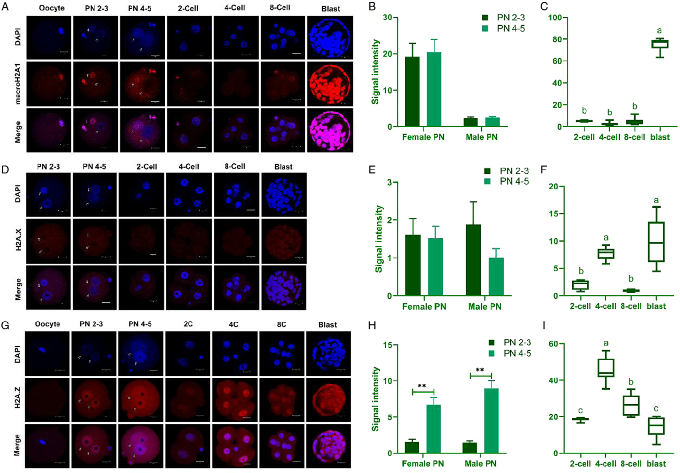
Figure 1
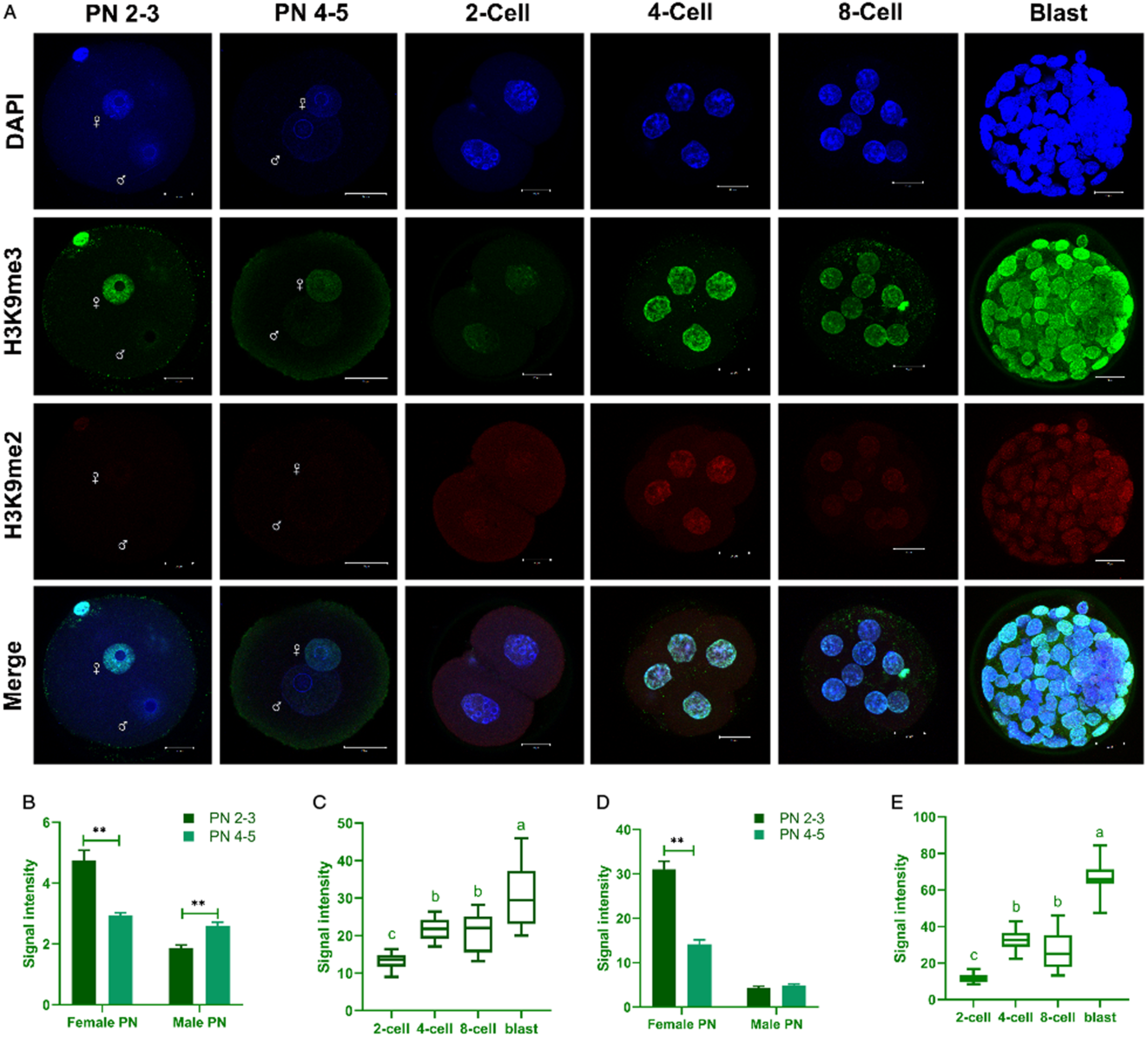
Figure 3
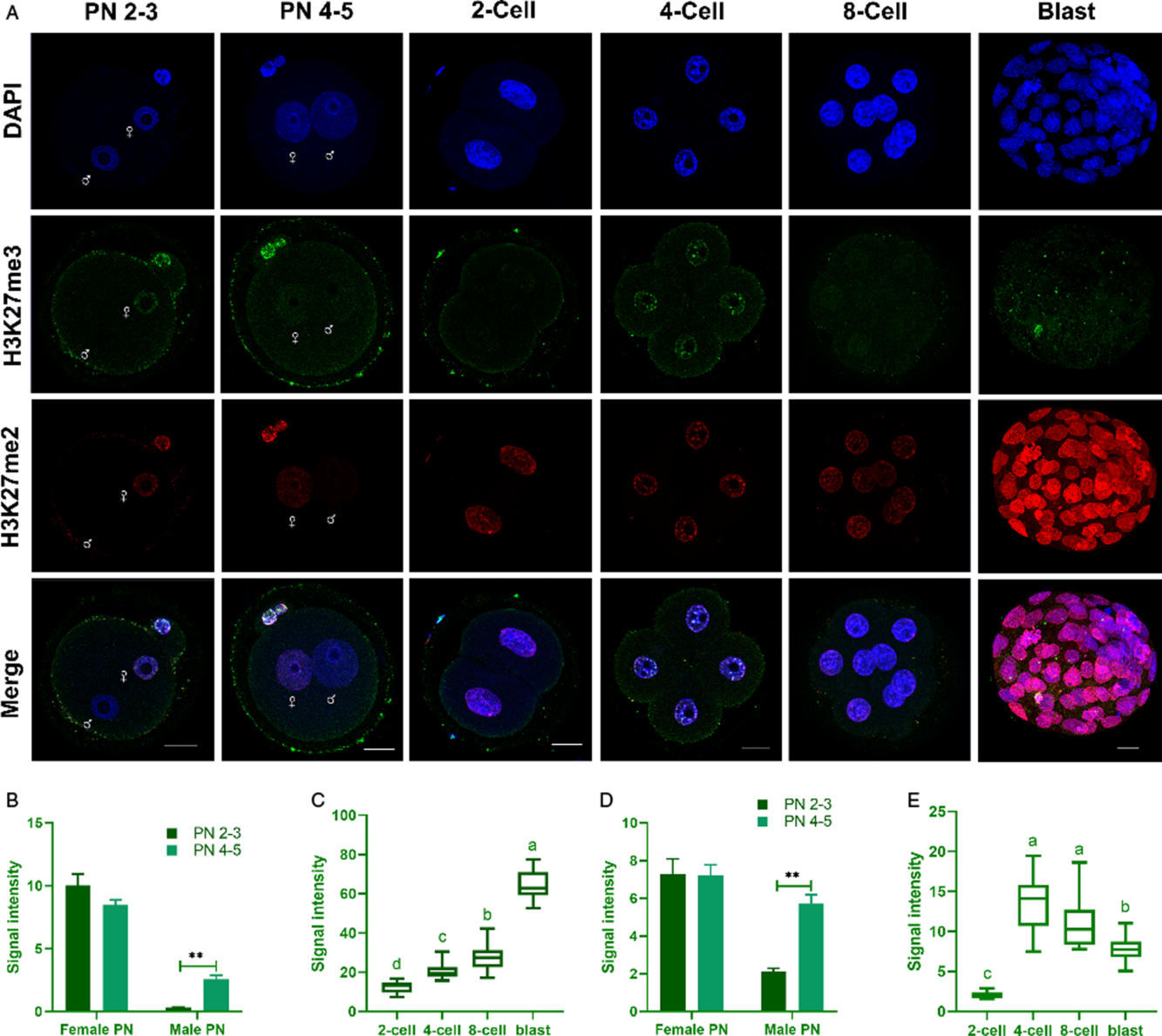
Figure 4
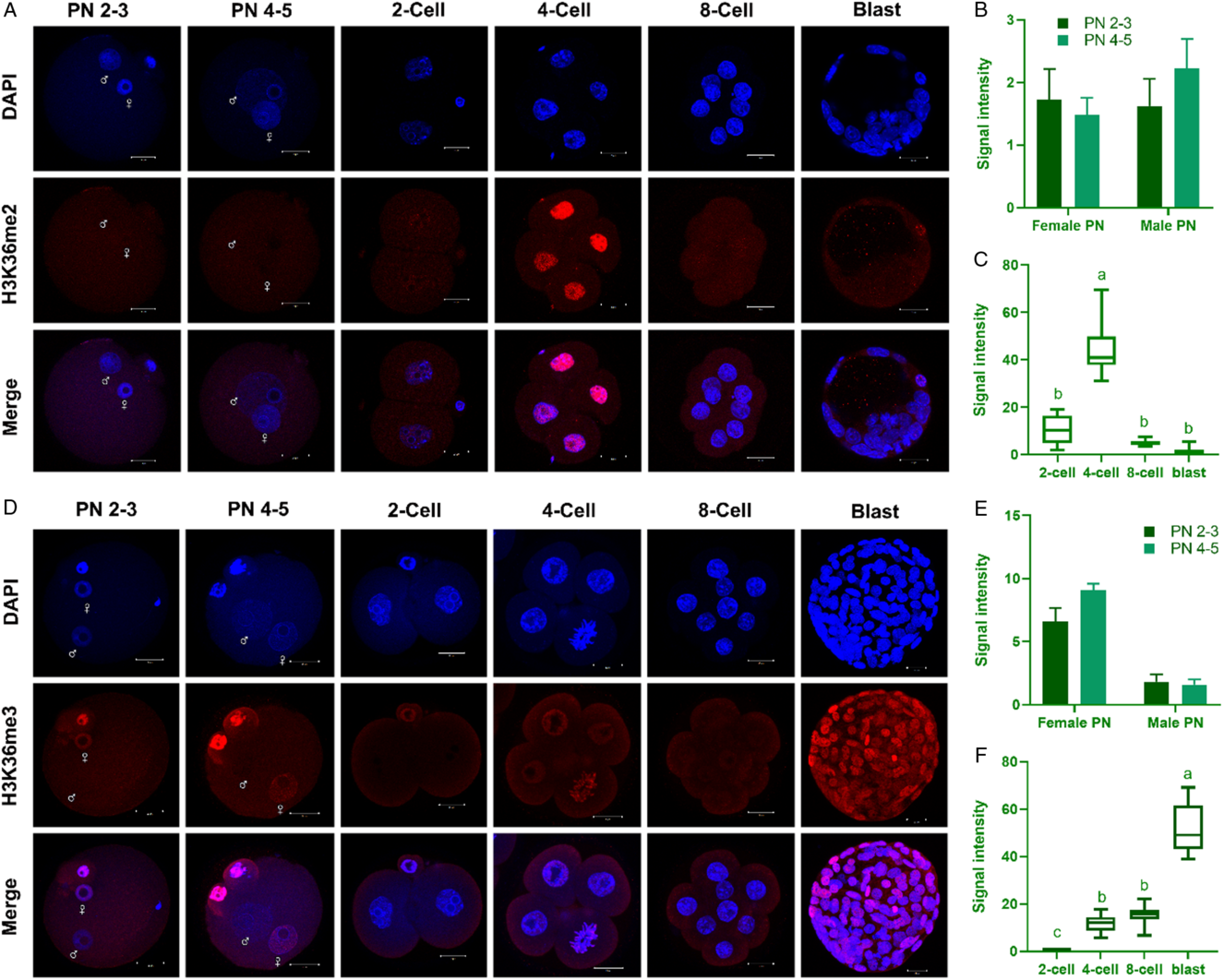
Figure 5
Published online by Cambridge University Press: 20 March 2020
The authors apologise for errors in the labelling of male and female pronuclei in Figures 1, 3, 4 and 5. The correctly labelled Figures are shown in the following pages.
Figure 1
Figure 3
Figure 4
Figure 5

Figure 1

Figure 3

Figure 4

Figure 5
 You have
Access
You have
Access
