当前位置:
X-MOL 学术
›
Prog. Nucl. Magn. Reson. Spectrosc.
›
论文详情
Our official English website, www.x-mol.net, welcomes your feedback! (Note: you will need to create a separate account there.)
Magnetic resonance thermometry and its biological applications - Physical principles and practical considerations
Progress in Nuclear Magnetic Resonance Spectroscopy ( IF 6.1 ) Pub Date : 2019-02-01 , DOI: 10.1016/j.pnmrs.2019.01.003 Henrik Odéen 1 , Dennis L Parker 1
Progress in Nuclear Magnetic Resonance Spectroscopy ( IF 6.1 ) Pub Date : 2019-02-01 , DOI: 10.1016/j.pnmrs.2019.01.003 Henrik Odéen 1 , Dennis L Parker 1
Affiliation
|
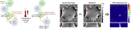
Most parameters that influence the magnetic resonance imaging (MRI) signal experience a temperature dependence. The fact that MRI can be used for non-invasive measurements of temperature and temperature change deep inside the human body has been known for over 30 years. Today, MR temperature imaging is widely used to monitor and evaluate thermal therapies such as radio frequency, microwave, laser, and focused ultrasound therapy. In this paper we cover the physical principles underlying the biological applications of MR temperature imaging and discuss practical considerations and remaining challenges. For biological tissue, the MR signal of interest comes mostly from hydrogen protons of water molecules but also from protons in, e.g., adipose tissue and various metabolites. Most of the discussed methods, such as those using the proton resonance frequency (PRF) shift, T1, T2, and diffusion only measure temperature change, but measurements of absolute temperatures are also possible using spectroscopic imaging methods (taking advantage of various metabolite signals as internal references) or various types of contrast agents. Currently, the PRF method is the most used clinically due to good sensitivity, excellent linearity with temperature, and because it is largely independent of tissue type. Because the PRF method does not work in adipose tissues, T1- and T2-based methods have recently gained interest for monitoring temperature change in areas with high fat content such as the breast and abdomen. Absolute temperature measurement methods using spectroscopic imaging and contrast agents often offer too low spatial and temporal resolution for accurate monitoring of ablative thermal procedures, but have shown great promise in monitoring the slower and usually less spatially localized temperature change observed during hyperthermia procedures. Much of the current research effort for ablative procedures is aimed at providing faster measurements, larger field-of-view coverage, simultaneous monitoring in aqueous and adipose tissues, and more motion-insensitive acquisitions for better precision measurements in organs such as the heart, liver, and kidneys. For hyperthermia applications, larger coverage, motion insensitivity, and simultaneous aqueous and adipose monitoring are also important, but great effort is also aimed at solving the problem of long-term field drift which gets interpreted as temperature change when using the PRF method.
中文翻译:

磁共振测温及其生物学应用 - 物理原理和实际考虑
大多数影响磁共振成像 (MRI) 信号的参数都具有温度依赖性。30 多年来,人们都知道 MRI 可用于对人体内部深处的温度和温度变化进行非侵入性测量。今天,MR 温度成像被广泛用于监测和评估热疗法,例如射频、微波、激光和聚焦超声疗法。在本文中,我们涵盖了 MR 温度成像生物学应用的物理原理,并讨论了实际考虑因素和剩余挑战。对于生物组织,感兴趣的 MR 信号主要来自水分子的氢质子,但也来自例如脂肪组织和各种代谢物的质子。大多数讨论的方法,例如使用质子共振频率 (PRF) 偏移、T1、T2 和扩散的那些仅测量温度变化,但也可以使用光谱成像方法(利用各种代谢物信号作为内部参考)或各种类型来测量绝对温度造影剂。目前,PRF 方法是临床上使用最多的方法,因为它具有良好的灵敏度、出色的温度线性度,并且在很大程度上与组织类型无关。由于 PRF 方法不适用于脂肪组织,因此基于 T1 和 T2 的方法最近在监测高脂肪含量区域(如乳房和腹部)的温度变化方面受到了关注。使用光谱成像和造影剂的绝对温度测量方法通常提供太低的空间和时间分辨率,无法准确监测消融热过程,但在监测热疗过程中观察到的较慢且通常在空间上较少的局部温度变化方面显示出巨大的希望。目前对消融程序的大部分研究工作旨在提供更快的测量、更大的视野覆盖、同时监测水和脂肪组织,以及更多的运动不敏感采集,以便在心脏、肝脏等器官中进行更精确的测量,和肾脏。对于热疗应用,更大的覆盖范围、运动不敏感以及同时监测水和脂肪也很重要,
更新日期:2019-02-01
中文翻译:
磁共振测温及其生物学应用 - 物理原理和实际考虑
大多数影响磁共振成像 (MRI) 信号的参数都具有温度依赖性。30 多年来,人们都知道 MRI 可用于对人体内部深处的温度和温度变化进行非侵入性测量。今天,MR 温度成像被广泛用于监测和评估热疗法,例如射频、微波、激光和聚焦超声疗法。在本文中,我们涵盖了 MR 温度成像生物学应用的物理原理,并讨论了实际考虑因素和剩余挑战。对于生物组织,感兴趣的 MR 信号主要来自水分子的氢质子,但也来自例如脂肪组织和各种代谢物的质子。大多数讨论的方法,例如使用质子共振频率 (PRF) 偏移、T1、T2 和扩散的那些仅测量温度变化,但也可以使用光谱成像方法(利用各种代谢物信号作为内部参考)或各种类型来测量绝对温度造影剂。目前,PRF 方法是临床上使用最多的方法,因为它具有良好的灵敏度、出色的温度线性度,并且在很大程度上与组织类型无关。由于 PRF 方法不适用于脂肪组织,因此基于 T1 和 T2 的方法最近在监测高脂肪含量区域(如乳房和腹部)的温度变化方面受到了关注。使用光谱成像和造影剂的绝对温度测量方法通常提供太低的空间和时间分辨率,无法准确监测消融热过程,但在监测热疗过程中观察到的较慢且通常在空间上较少的局部温度变化方面显示出巨大的希望。目前对消融程序的大部分研究工作旨在提供更快的测量、更大的视野覆盖、同时监测水和脂肪组织,以及更多的运动不敏感采集,以便在心脏、肝脏等器官中进行更精确的测量,和肾脏。对于热疗应用,更大的覆盖范围、运动不敏感以及同时监测水和脂肪也很重要,


 京公网安备 11010802027423号