当前位置:
X-MOL 学术
›
Cytom. Part A
›
论文详情
Our official English website, www.x-mol.net, welcomes your feedback! (Note: you will need to create a separate account there.)
Discriminating Bacterial Phenotypes at the Population and Single-Cell Level: A Comparison of Flow Cytometry and Raman Spectroscopy Fingerprinting.
Cytometry Part A ( IF 3.7 ) Pub Date : 2019-12-30 , DOI: 10.1002/cyto.a.23952 Cristina García-Timermans 1 , Peter Rubbens 2 , Jasmine Heyse 1 , Frederiek-Maarten Kerckhof 1 , Ruben Props 1 , Andre G Skirtach 3 , Willem Waegeman 2 , Nico Boon 1
Cytometry Part A ( IF 3.7 ) Pub Date : 2019-12-30 , DOI: 10.1002/cyto.a.23952 Cristina García-Timermans 1 , Peter Rubbens 2 , Jasmine Heyse 1 , Frederiek-Maarten Kerckhof 1 , Ruben Props 1 , Andre G Skirtach 3 , Willem Waegeman 2 , Nico Boon 1
Affiliation
|
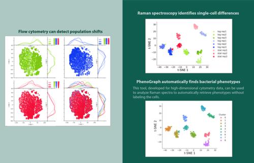
Investigating phenotypic heterogeneity can help to better understand and manage microbial communities. However, characterizing phenotypic heterogeneity remains a challenge, as there is no standardized analysis framework. Several optical tools are available, such as flow cytometry and Raman spectroscopy, which describe optical properties of the individual cell. In this work, we compare Raman spectroscopy and flow cytometry to study phenotypic heterogeneity in bacterial populations. The growth stages of three replicate Escherichia coli populations were characterized using both technologies. Our findings show that flow cytometry detects and quantifies shifts in phenotypic heterogeneity at the population level due to its high‐throughput nature. Raman spectroscopy, on the other hand, offers a much higher resolution at the single‐cell level (i.e., more biochemical information is recorded). Therefore, it can identify distinct phenotypic populations when coupled with analyses tailored toward single‐cell data. In addition, it provides information about biomolecules that are present, which can be linked to cell functionality. We propose a computational workflow to distinguish between bacterial phenotypic populations using Raman spectroscopy and validated this approach with an external data set. We recommend using flow cytometry to quantify phenotypic heterogeneity at the population level, and Raman spectroscopy to perform a more in‐depth analysis of heterogeneity at the single‐cell level. © 2019 International Society for Advancement of Cytometry
中文翻译:

在种群和单细胞水平上区分细菌表型:流式细胞术和拉曼光谱指纹的比较。
调查表型异质性有助于更好地了解和管理微生物群落。然而,表征表型异质性仍然是一个挑战,因为没有标准化的分析框架。有几种光学工具可用,例如流式细胞术和拉曼光谱,它们描述了单个细胞的光学特性。在这项工作中,我们比较了拉曼光谱和流式细胞术来研究细菌种群的表型异质性。三个重复大肠杆菌的生长阶段使用这两种技术对人群进行了表征。我们的研究结果表明,流式细胞术由于其高通量性质,可以检测和量化群体水平上表型异质性的变化。另一方面,拉曼光谱在单细胞水平上提供了更高的分辨率(即记录了更多的生化信息)。因此,当结合针对单细胞数据的分析时,它可以识别不同的表型群体。此外,它还提供有关存在的生物分子的信息,这些信息可以与细胞功能相关联。我们提出了使用拉曼光谱区分细菌表型种群的计算工作流程,并使用外部数据集验证了这种方法。我们建议使用流式细胞术在群体水平上量化表型异质性,并使用拉曼光谱在单细胞水平上对异质性进行更深入的分析。© 2019 国际细胞计量学促进会
更新日期:2019-12-30
中文翻译:
在种群和单细胞水平上区分细菌表型:流式细胞术和拉曼光谱指纹的比较。
调查表型异质性有助于更好地了解和管理微生物群落。然而,表征表型异质性仍然是一个挑战,因为没有标准化的分析框架。有几种光学工具可用,例如流式细胞术和拉曼光谱,它们描述了单个细胞的光学特性。在这项工作中,我们比较了拉曼光谱和流式细胞术来研究细菌种群的表型异质性。三个重复大肠杆菌的生长阶段使用这两种技术对人群进行了表征。我们的研究结果表明,流式细胞术由于其高通量性质,可以检测和量化群体水平上表型异质性的变化。另一方面,拉曼光谱在单细胞水平上提供了更高的分辨率(即记录了更多的生化信息)。因此,当结合针对单细胞数据的分析时,它可以识别不同的表型群体。此外,它还提供有关存在的生物分子的信息,这些信息可以与细胞功能相关联。我们提出了使用拉曼光谱区分细菌表型种群的计算工作流程,并使用外部数据集验证了这种方法。我们建议使用流式细胞术在群体水平上量化表型异质性,并使用拉曼光谱在单细胞水平上对异质性进行更深入的分析。© 2019 国际细胞计量学促进会


 京公网安备 11010802027423号