当前位置:
X-MOL 学术
›
Anal. Chem.
›
论文详情
Our official English website, www.x-mol.net, welcomes your feedback! (Note: you will need to create a separate account there.)
Quantitatively Mapping the Assembly Pattern of EpCAM on Cell Membranes with Peptide Probes.
Analytical Chemistry ( IF 7.4 ) Pub Date : 2020-01-08 , DOI: 10.1021/acs.analchem.9b03901 Yingying Jing 1, 2 , Lulu Zhou 1 , Junling Chen 1 , Haijiao Xu 1 , Jiayin Sun 1 , Mingjun Cai 1 , Junguang Jiang 1 , Jing Gao 1 , Hongda Wang 1, 2, 3
Analytical Chemistry ( IF 7.4 ) Pub Date : 2020-01-08 , DOI: 10.1021/acs.analchem.9b03901 Yingying Jing 1, 2 , Lulu Zhou 1 , Junling Chen 1 , Haijiao Xu 1 , Jiayin Sun 1 , Mingjun Cai 1 , Junguang Jiang 1 , Jing Gao 1 , Hongda Wang 1, 2, 3
Affiliation

|
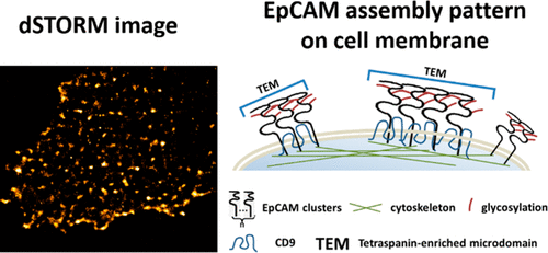
Epithelial cell adhesion molecule (EpCAM) is an important type I transmembrane protein that is overexpressed on the surfaces of most cancer cells and involved in various biological processes such as cell adhesion and cell signaling. Although it plays crucial roles in cell functions and tumorigenesis, questions concerning the detailed morphology, molecular stoichiometry, and the assembly mechanisms of EpCAM on cell membranes have not been fully elucidated. Here, we used direct stochastic optical reconstruction microscopy (dSTORM) and relied on fluorophore-conjugated peptides to quantitatively analyze the assembly pattern of EpCAM with single-molecule precision. EpCAM was found to organize heterogeneous clusters with different sizes, which contain different numbers of EpCAM molecules on MCF-7 cell membranes. Moreover, dual-color dSTORM imaging revealed a significant correlation between EpCAM and tetraspanin CD9, and part of the EpCAM clusters could be disrupted by knockdown of CD9, which indicated that EpCAM might localize in tetraspanin-enriched microdomains (TEMs) and function cooperatively with CD9 on cell membranes. In addition, the assembly of the membrane EpCAM was found to be limited by both cytoskeleton and glycosylation. Overall, our work clarified the clustered distribution of EpCAM and revealed the potential mechanisms of its clustering at the molecular level, promoting a deeper understanding of the nano-organization of membrane proteins.
中文翻译:

用肽探针定量映射EpCAM在细胞膜上的组装模式。
上皮细胞粘附分子(EpCAM)是一种重要的I型跨膜蛋白,在大多数癌细胞的表面过表达,并参与多种生物学过程,例如细胞粘附和细胞信号传导。尽管它在细胞功能和肿瘤发生中起着至关重要的作用,但有关EpCAM在细胞膜上的详细形态,分子化学计量和组装机制的问题尚未得到充分阐明。在这里,我们使用直接随机光学重建显微镜(dSTORM)并依靠荧光团偶联的肽以单分子精度定量分析EpCAM的组装模式。发现EpCAM可以组织不同大小的异质簇,这些簇在MCF-7细胞膜上包含不同数量的EpCAM分子。而且,dSTORM双色成像显示EpCAM与四跨膜CD9之间存在显着相关性,而部分敲除CD9可能会破坏EpCAM簇,这表明EpCAM可能位于富含四跨膜的微区(TEM)中,并与细胞上的CD9协同起作用膜。另外,发现膜EpCAM的组装受细胞骨架和糖基化的限制。总体而言,我们的工作阐明了EpCAM的聚集分布,并揭示了其在分子水平上聚集的潜在机制,从而促进了对膜蛋白纳米组织的更深入了解。这表明EpCAM可能位于富含四跨素的微区(TEM)中,并与CD9在细胞膜上协同发挥作用。另外,发现膜EpCAM的组装受细胞骨架和糖基化的限制。总体而言,我们的工作阐明了EpCAM的聚集分布,并揭示了其在分子水平上聚集的潜在机制,从而促进了对膜蛋白纳米组织的更深入了解。这表明EpCAM可能位于富含四跨素的微区(TEM)中,并与CD9在细胞膜上协同发挥作用。另外,发现膜EpCAM的组装受细胞骨架和糖基化的限制。总体而言,我们的工作阐明了EpCAM的聚集分布,并揭示了其在分子水平上聚集的潜在机制,从而促进了对膜蛋白纳米组织的更深入了解。
更新日期:2020-01-08
中文翻译:

用肽探针定量映射EpCAM在细胞膜上的组装模式。
上皮细胞粘附分子(EpCAM)是一种重要的I型跨膜蛋白,在大多数癌细胞的表面过表达,并参与多种生物学过程,例如细胞粘附和细胞信号传导。尽管它在细胞功能和肿瘤发生中起着至关重要的作用,但有关EpCAM在细胞膜上的详细形态,分子化学计量和组装机制的问题尚未得到充分阐明。在这里,我们使用直接随机光学重建显微镜(dSTORM)并依靠荧光团偶联的肽以单分子精度定量分析EpCAM的组装模式。发现EpCAM可以组织不同大小的异质簇,这些簇在MCF-7细胞膜上包含不同数量的EpCAM分子。而且,dSTORM双色成像显示EpCAM与四跨膜CD9之间存在显着相关性,而部分敲除CD9可能会破坏EpCAM簇,这表明EpCAM可能位于富含四跨膜的微区(TEM)中,并与细胞上的CD9协同起作用膜。另外,发现膜EpCAM的组装受细胞骨架和糖基化的限制。总体而言,我们的工作阐明了EpCAM的聚集分布,并揭示了其在分子水平上聚集的潜在机制,从而促进了对膜蛋白纳米组织的更深入了解。这表明EpCAM可能位于富含四跨素的微区(TEM)中,并与CD9在细胞膜上协同发挥作用。另外,发现膜EpCAM的组装受细胞骨架和糖基化的限制。总体而言,我们的工作阐明了EpCAM的聚集分布,并揭示了其在分子水平上聚集的潜在机制,从而促进了对膜蛋白纳米组织的更深入了解。这表明EpCAM可能位于富含四跨素的微区(TEM)中,并与CD9在细胞膜上协同发挥作用。另外,发现膜EpCAM的组装受细胞骨架和糖基化的限制。总体而言,我们的工作阐明了EpCAM的聚集分布,并揭示了其在分子水平上聚集的潜在机制,从而促进了对膜蛋白纳米组织的更深入了解。


 京公网安备 11010802027423号