当前位置:
X-MOL 学术
›
J. Hepatol.
›
论文详情
Our official English website, www.x-mol.net, welcomes your feedback! (Note: you will need to create a separate account there.)
Comparison of extracellular and hepatobiliary MR contrast agents for the diagnosis of small HCCs
Journal of Hepatology ( IF 25.7 ) Pub Date : 2020-05-01 , DOI: 10.1016/j.jhep.2019.12.011 Anita Paisant 1 , Valérie Vilgrain 2 , Jérémie Riou 3 , Frédéric Oberti 4 , Olivier Sutter 5 , Valérie Laurent 6 , Agnès Rodes 7 , Boris Guiu 8 , Christophe Cassinotto 9 , Hervé Trillaud 10 , Ivan Bricault 11 , Sophie Michalak 12 , Onorina Bruno 13 , Maxime Ronot 2 , Christophe Aubé 1
Journal of Hepatology ( IF 25.7 ) Pub Date : 2020-05-01 , DOI: 10.1016/j.jhep.2019.12.011 Anita Paisant 1 , Valérie Vilgrain 2 , Jérémie Riou 3 , Frédéric Oberti 4 , Olivier Sutter 5 , Valérie Laurent 6 , Agnès Rodes 7 , Boris Guiu 8 , Christophe Cassinotto 9 , Hervé Trillaud 10 , Ivan Bricault 11 , Sophie Michalak 12 , Onorina Bruno 13 , Maxime Ronot 2 , Christophe Aubé 1
Affiliation
|
BACKGROUND & AIMS
The aim of this study was to compare the performance of MRIs with extracellular contrast agents (ECA-MRI) to HB contrast agents (HBA-MRI) for the non-invasive diagnosis of small HCCs in a head-to-head comparison. METHODS
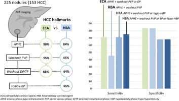
Between August 2014 and October 2017; 171 cirrhotic patients, each with 1 to 3 nodules measuring 1 to 3 cm, were included across eight centers. All patients had both an ECA-MRI and an HBA-MRI within a month. The non-invasive diagnosis of HCC was made when the nodule was hyper-enhanced at the arterial phase (HA) with wash-out at the portal phase (PP) and/or delayed phase (DP) for the ECA-MRI, or PP and/or HB phase (HBP) for the HBA-MRI. The gold standard was defined by a composite algorithm previously published. RESULTS
225 nodules, of which 153 were HCCs and 72 were not HCCs, were included. Both MRI sensitivities were similar (71.2% [63.4-78.3]). Specificity was 83.3% [72.7-91.1] for the ECA-MRI and 68.1% [56.0-78.6] for the HBA-MRI. With regard to HCCs, on ECA-MRI, 138 were HA, 84 had wash-out at PP and 104 at DP; on HBA-MRI, 128 were HA, 71 had wash-out at PP and 99 at HBP. For nodules from 2 to 3 cm, sensitivity and specificity were similar with 70.9% [57.1-82.4] and 75.0% [47.6-92.7] respectively. For nodules from 1 to 2 cm, specificity dropped to 66.1% [52.2-78.2] for the HBA-MRI vs. 85.7% [73.8-93.6] for the ECA-MRI. CONCLUSIONS
HBA-MRI specificity is lower than ECA-MRI for diagnosing small HCCs on cirrhotic patients. These results raise the question of the proper use of HBA-MRI in algorithms for the non-invasive diagnosis of small HCCs.
中文翻译:

细胞外和肝胆 MR 造影剂对小 HCC 诊断的比较
背景和目的 本研究的目的是比较 MRI 与细胞外造影剂 (ECA-MRI) 和 HB 造影剂 (HBA-MRI) 在头对头比较中无创诊断小 HCC 的性能. 方法 2014 年 8 月至 2017 年 10 月;8 个中心共纳入 171 名肝硬化患者,每名患者有 1 至 3 个直径为 1 至 3 厘米的结节。所有患者均在一个月内进行了 ECA-MRI 和 HBA-MRI。当结节在动脉期 (HA) 超强化,门脉期 (PP) 和/或延迟期 (DP) 消失时,ECA-MRI 或 PP和/或 HBA-MRI 的 HB 期 (HBP)。黄金标准是由先前发布的复合算法定义的。结果 包括 225 个结节,其中 153 个是 HCC,72 个不是 HCC。两种 MRI 敏感性相似(71.2% [63.4-78.3])。ECA-MRI 的特异性为 83.3% [72.7-91.1],HBA-MRI 的特异性为 68.1% [56.0-78.6]。关于 HCC,ECA-MRI 显示 138 例为 HA,84 例为 PP 洗脱,104 例为 DP;在 HBA-MRI 上,128 例为 HA,71 例为 PP 洗脱,99 例为 HBP。对于 2 至 3 cm 的结节,敏感性和特异性相似,分别为 70.9% [57.1-82.4] 和 75.0% [47.6-92.7]。对于 1 至 2 cm 的结节,HBA-MRI 的特异性降至 66.1% [52.2-78.2],而 ECA-MRI 的特异性降至 85.7% [73.8-93.6]。结论 HBA-MRI 诊断肝硬化患者小 HCC 的特异性低于 ECA-MRI。这些结果提出了在小 HCC 非侵入性诊断算法中正确使用 HBA-MRI 的问题。1] 对于 ECA-MRI 和 68.1% [56.0-78.6] 对于 HBA-MRI。关于 HCC,ECA-MRI 显示 138 例为 HA,84 例为 PP 洗脱,104 例为 DP;在 HBA-MRI 上,128 例为 HA,71 例为 PP 洗脱,99 例为 HBP。对于 2 至 3 cm 的结节,敏感性和特异性相似,分别为 70.9% [57.1-82.4] 和 75.0% [47.6-92.7]。对于 1 至 2 cm 的结节,HBA-MRI 的特异性降至 66.1% [52.2-78.2],而 ECA-MRI 的特异性降至 85.7% [73.8-93.6]。结论 HBA-MRI 诊断肝硬化患者小 HCC 的特异性低于 ECA-MRI。这些结果提出了在小 HCC 非侵入性诊断算法中正确使用 HBA-MRI 的问题。1] 对于 ECA-MRI 和 68.1% [56.0-78.6] 对于 HBA-MRI。关于 HCC,ECA-MRI 显示 138 例为 HA,84 例为 PP 洗脱,104 例为 DP;在 HBA-MRI 上,128 例为 HA,71 例为 PP 洗脱,99 例为 HBP。对于 2 至 3 cm 的结节,敏感性和特异性相似,分别为 70.9% [57.1-82.4] 和 75.0% [47.6-92.7]。对于 1 至 2 cm 的结节,HBA-MRI 的特异性降至 66.1% [52.2-78.2],而 ECA-MRI 的特异性降至 85.7% [73.8-93.6]。结论 HBA-MRI 诊断肝硬化患者小 HCC 的特异性低于 ECA-MRI。这些结果提出了在小 HCC 非侵入性诊断算法中正确使用 HBA-MRI 的问题。对于 2 至 3 cm 的结节,敏感性和特异性相似,分别为 70.9% [57.1-82.4] 和 75.0% [47.6-92.7]。对于 1 至 2 cm 的结节,HBA-MRI 的特异性降至 66.1% [52.2-78.2],而 ECA-MRI 的特异性降至 85.7% [73.8-93.6]。结论 HBA-MRI 诊断肝硬化患者小 HCC 的特异性低于 ECA-MRI。这些结果提出了在小 HCC 非侵入性诊断算法中正确使用 HBA-MRI 的问题。对于 2 至 3 cm 的结节,敏感性和特异性相似,分别为 70.9% [57.1-82.4] 和 75.0% [47.6-92.7]。对于 1 至 2 cm 的结节,HBA-MRI 的特异性降至 66.1% [52.2-78.2],而 ECA-MRI 的特异性降至 85.7% [73.8-93.6]。结论 HBA-MRI 诊断肝硬化患者小 HCC 的特异性低于 ECA-MRI。这些结果提出了在小 HCC 非侵入性诊断算法中正确使用 HBA-MRI 的问题。
更新日期:2020-05-01
中文翻译:
细胞外和肝胆 MR 造影剂对小 HCC 诊断的比较
背景和目的 本研究的目的是比较 MRI 与细胞外造影剂 (ECA-MRI) 和 HB 造影剂 (HBA-MRI) 在头对头比较中无创诊断小 HCC 的性能. 方法 2014 年 8 月至 2017 年 10 月;8 个中心共纳入 171 名肝硬化患者,每名患者有 1 至 3 个直径为 1 至 3 厘米的结节。所有患者均在一个月内进行了 ECA-MRI 和 HBA-MRI。当结节在动脉期 (HA) 超强化,门脉期 (PP) 和/或延迟期 (DP) 消失时,ECA-MRI 或 PP和/或 HBA-MRI 的 HB 期 (HBP)。黄金标准是由先前发布的复合算法定义的。结果 包括 225 个结节,其中 153 个是 HCC,72 个不是 HCC。两种 MRI 敏感性相似(71.2% [63.4-78.3])。ECA-MRI 的特异性为 83.3% [72.7-91.1],HBA-MRI 的特异性为 68.1% [56.0-78.6]。关于 HCC,ECA-MRI 显示 138 例为 HA,84 例为 PP 洗脱,104 例为 DP;在 HBA-MRI 上,128 例为 HA,71 例为 PP 洗脱,99 例为 HBP。对于 2 至 3 cm 的结节,敏感性和特异性相似,分别为 70.9% [57.1-82.4] 和 75.0% [47.6-92.7]。对于 1 至 2 cm 的结节,HBA-MRI 的特异性降至 66.1% [52.2-78.2],而 ECA-MRI 的特异性降至 85.7% [73.8-93.6]。结论 HBA-MRI 诊断肝硬化患者小 HCC 的特异性低于 ECA-MRI。这些结果提出了在小 HCC 非侵入性诊断算法中正确使用 HBA-MRI 的问题。1] 对于 ECA-MRI 和 68.1% [56.0-78.6] 对于 HBA-MRI。关于 HCC,ECA-MRI 显示 138 例为 HA,84 例为 PP 洗脱,104 例为 DP;在 HBA-MRI 上,128 例为 HA,71 例为 PP 洗脱,99 例为 HBP。对于 2 至 3 cm 的结节,敏感性和特异性相似,分别为 70.9% [57.1-82.4] 和 75.0% [47.6-92.7]。对于 1 至 2 cm 的结节,HBA-MRI 的特异性降至 66.1% [52.2-78.2],而 ECA-MRI 的特异性降至 85.7% [73.8-93.6]。结论 HBA-MRI 诊断肝硬化患者小 HCC 的特异性低于 ECA-MRI。这些结果提出了在小 HCC 非侵入性诊断算法中正确使用 HBA-MRI 的问题。1] 对于 ECA-MRI 和 68.1% [56.0-78.6] 对于 HBA-MRI。关于 HCC,ECA-MRI 显示 138 例为 HA,84 例为 PP 洗脱,104 例为 DP;在 HBA-MRI 上,128 例为 HA,71 例为 PP 洗脱,99 例为 HBP。对于 2 至 3 cm 的结节,敏感性和特异性相似,分别为 70.9% [57.1-82.4] 和 75.0% [47.6-92.7]。对于 1 至 2 cm 的结节,HBA-MRI 的特异性降至 66.1% [52.2-78.2],而 ECA-MRI 的特异性降至 85.7% [73.8-93.6]。结论 HBA-MRI 诊断肝硬化患者小 HCC 的特异性低于 ECA-MRI。这些结果提出了在小 HCC 非侵入性诊断算法中正确使用 HBA-MRI 的问题。对于 2 至 3 cm 的结节,敏感性和特异性相似,分别为 70.9% [57.1-82.4] 和 75.0% [47.6-92.7]。对于 1 至 2 cm 的结节,HBA-MRI 的特异性降至 66.1% [52.2-78.2],而 ECA-MRI 的特异性降至 85.7% [73.8-93.6]。结论 HBA-MRI 诊断肝硬化患者小 HCC 的特异性低于 ECA-MRI。这些结果提出了在小 HCC 非侵入性诊断算法中正确使用 HBA-MRI 的问题。对于 2 至 3 cm 的结节,敏感性和特异性相似,分别为 70.9% [57.1-82.4] 和 75.0% [47.6-92.7]。对于 1 至 2 cm 的结节,HBA-MRI 的特异性降至 66.1% [52.2-78.2],而 ECA-MRI 的特异性降至 85.7% [73.8-93.6]。结论 HBA-MRI 诊断肝硬化患者小 HCC 的特异性低于 ECA-MRI。这些结果提出了在小 HCC 非侵入性诊断算法中正确使用 HBA-MRI 的问题。


 京公网安备 11010802027423号