当前位置:
X-MOL 学术
›
Light Sci. Appl.
›
论文详情
Our official English website, www.x-mol.net, welcomes your feedback! (Note: you will need to create a separate account there.)
Full noncontact laser ultrasound: first human data.
Light: Science & Applications ( IF 19.4 ) Pub Date : 2019-12-20 , DOI: 10.1038/s41377-019-0229-8 Xiang Zhang 1, 2 , Jonathan R Fincke 1, 2 , Charles M Wynn 3 , Matt R Johnson 3 , Robert W Haupt 3 , Brian W Anthony 1, 2
Light: Science & Applications ( IF 19.4 ) Pub Date : 2019-12-20 , DOI: 10.1038/s41377-019-0229-8 Xiang Zhang 1, 2 , Jonathan R Fincke 1, 2 , Charles M Wynn 3 , Matt R Johnson 3 , Robert W Haupt 3 , Brian W Anthony 1, 2
Affiliation
|
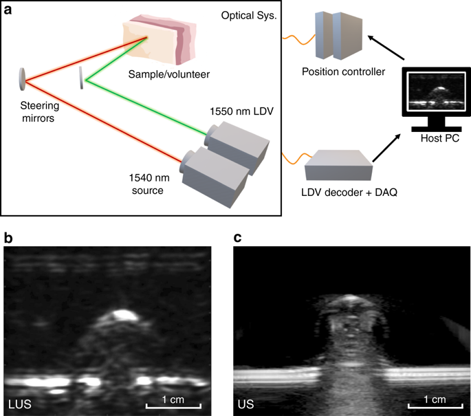
Full noncontact laser ultrasound (LUS) imaging has several distinct advantages over current medical ultrasound (US) technologies: elimination of the coupling mediums (gel/water), operator-independent image quality, improved repeatability, and volumetric imaging. Current light-based ultrasound utilizing tissue-penetrating photoacoustics (PA) generally uses traditional piezoelectric transducers in contact with the imaged tissue or carries an optical fiber detector close to the imaging site. Unlike PA, the LUS design presented here minimizes the optical penetration and specifically restricts optical-to-acoustic energy transduction at the tissue surface, maximizing the generated acoustic source amplitude. With an appropriate optical design and interferometry, any exposed tissue surfaces can become viable acoustic sources and detectors. LUS operates analogously to conventional ultrasound but uses light instead of piezoelectric elements. Here, we present full noncontact LUS results, imaging targets at ~5 cm depths and at a meter-scale standoff from the target surface. Experimental results demonstrating volumetric imaging and the first LUS images on humans are presented, all at eye- and skin-safe optical exposure levels. The progression of LUS imaging from tissue-mimicking phantoms, to excised animal tissue, to humans in vivo is shown, with validation from conventional ultrasound images. The LUS system design insights and results presented here inspire further LUS development and are a significant step toward the clinical implementation of LUS.
中文翻译:

完全非接触式激光超声:人类的第一手资料。
完整的非接触式激光超声(LUS)成像相对于当前的医学超声(US)技术具有几个明显的优势:消除了耦合介质(凝胶/水),独立于操作员的图像质量,改进的可重复性和体积成像。当前利用组织穿透光声(PA)的基于光的超声通常使用传统的压电换能器与被成像的组织接触,或者携带靠近成像部位的光纤检测器。与PA不同,此处介绍的LUS设计可最大程度地减少光学穿透力,并特别限制组织表面的光声能量传导,从而最大程度地提高产生的声源振幅。通过适当的光学设计和干涉测量,任何暴露的组织表面都可以成为可行的声源和检测器。LUS的操作类似于常规超声,但是使用光代替压电元件。在这里,我们展示了完整的非接触LUS结果,可以在距目标表面约5厘米深度和一米尺度的距离内对目标成像。实验结果证明了人体的体积成像和第一批LUS图像均在对眼睛和皮肤安全的光学曝光水平下进行。显示了LUS成像在体内的模拟组织幻像,到切除的动物组织,再到人体的过程,并得到了常规超声图像的验证。本文介绍的LUS系统设计见解和结果激发了LUS的进一步发展,并且是向LUS临床实施迈出的重要一步。可以在距目标表面约5厘米深度和一米尺度的距离内对目标成像。实验结果证明了人体的体积成像和第一批LUS图像均在对眼睛和皮肤安全的光学曝光水平下进行。显示了LUS成像在体内的模拟组织幻像,到切除的动物组织,再到人体的过程,并得到了常规超声图像的验证。本文介绍的LUS系统设计见解和结果激发了LUS的进一步发展,并且是向LUS临床实施迈出的重要一步。可以在距目标表面约5厘米深度和一米尺度的距离内对目标成像。实验结果证明了人体的体积成像和第一批LUS图像均在对眼睛和皮肤安全的光学曝光水平下进行。显示了LUS成像在体内的模拟组织幻像,到切除的动物组织,再到人体的过程,并得到了常规超声图像的验证。本文介绍的LUS系统设计见解和结果激发了LUS的进一步发展,并且是向LUS临床实施迈出的重要一步。显示了LUS成像在体内的模拟组织幻像,到切除的动物组织,再到人体的过程,并得到了常规超声图像的验证。本文介绍的LUS系统设计见解和结果激发了LUS的进一步发展,并且是向LUS临床实施迈出的重要一步。显示了LUS成像在体内的模拟组织幻像,到切除的动物组织,再到人体的过程,并得到了常规超声图像的验证。本文介绍的LUS系统设计见解和结果激发了LUS的进一步发展,并且是向LUS临床实施迈出的重要一步。
更新日期:2019-12-20
中文翻译:
完全非接触式激光超声:人类的第一手资料。
完整的非接触式激光超声(LUS)成像相对于当前的医学超声(US)技术具有几个明显的优势:消除了耦合介质(凝胶/水),独立于操作员的图像质量,改进的可重复性和体积成像。当前利用组织穿透光声(PA)的基于光的超声通常使用传统的压电换能器与被成像的组织接触,或者携带靠近成像部位的光纤检测器。与PA不同,此处介绍的LUS设计可最大程度地减少光学穿透力,并特别限制组织表面的光声能量传导,从而最大程度地提高产生的声源振幅。通过适当的光学设计和干涉测量,任何暴露的组织表面都可以成为可行的声源和检测器。LUS的操作类似于常规超声,但是使用光代替压电元件。在这里,我们展示了完整的非接触LUS结果,可以在距目标表面约5厘米深度和一米尺度的距离内对目标成像。实验结果证明了人体的体积成像和第一批LUS图像均在对眼睛和皮肤安全的光学曝光水平下进行。显示了LUS成像在体内的模拟组织幻像,到切除的动物组织,再到人体的过程,并得到了常规超声图像的验证。本文介绍的LUS系统设计见解和结果激发了LUS的进一步发展,并且是向LUS临床实施迈出的重要一步。可以在距目标表面约5厘米深度和一米尺度的距离内对目标成像。实验结果证明了人体的体积成像和第一批LUS图像均在对眼睛和皮肤安全的光学曝光水平下进行。显示了LUS成像在体内的模拟组织幻像,到切除的动物组织,再到人体的过程,并得到了常规超声图像的验证。本文介绍的LUS系统设计见解和结果激发了LUS的进一步发展,并且是向LUS临床实施迈出的重要一步。可以在距目标表面约5厘米深度和一米尺度的距离内对目标成像。实验结果证明了人体的体积成像和第一批LUS图像均在对眼睛和皮肤安全的光学曝光水平下进行。显示了LUS成像在体内的模拟组织幻像,到切除的动物组织,再到人体的过程,并得到了常规超声图像的验证。本文介绍的LUS系统设计见解和结果激发了LUS的进一步发展,并且是向LUS临床实施迈出的重要一步。显示了LUS成像在体内的模拟组织幻像,到切除的动物组织,再到人体的过程,并得到了常规超声图像的验证。本文介绍的LUS系统设计见解和结果激发了LUS的进一步发展,并且是向LUS临床实施迈出的重要一步。显示了LUS成像在体内的模拟组织幻像,到切除的动物组织,再到人体的过程,并得到了常规超声图像的验证。本文介绍的LUS系统设计见解和结果激发了LUS的进一步发展,并且是向LUS临床实施迈出的重要一步。


 京公网安备 11010802027423号