当前位置:
X-MOL 学术
›
Hypertens. Res.
›
论文详情
Our official English website, www.x-mol.net, welcomes your feedback! (Note: you will need to create a separate account there.)
Brain angiotensin type-1 and type-2 receptors: cellular locations under normal and hypertensive conditions
Hypertension Research ( IF 5.4 ) Pub Date : 2019-12-18 , DOI: 10.1038/s41440-019-0374-8 Colin Sumners 1 , Amy Alleyne 2 , Vermalí Rodríguez 1 , David J Pioquinto 2 , Jacob A Ludin 2 , Shormista Kar 1 , Zachary Winder 1, 2 , Yuma Ortiz 2 , Meng Liu 1 , Eric G Krause 2 , Annette D de Kloet 1
Hypertension Research ( IF 5.4 ) Pub Date : 2019-12-18 , DOI: 10.1038/s41440-019-0374-8 Colin Sumners 1 , Amy Alleyne 2 , Vermalí Rodríguez 1 , David J Pioquinto 2 , Jacob A Ludin 2 , Shormista Kar 1 , Zachary Winder 1, 2 , Yuma Ortiz 2 , Meng Liu 1 , Eric G Krause 2 , Annette D de Kloet 1
Affiliation
|
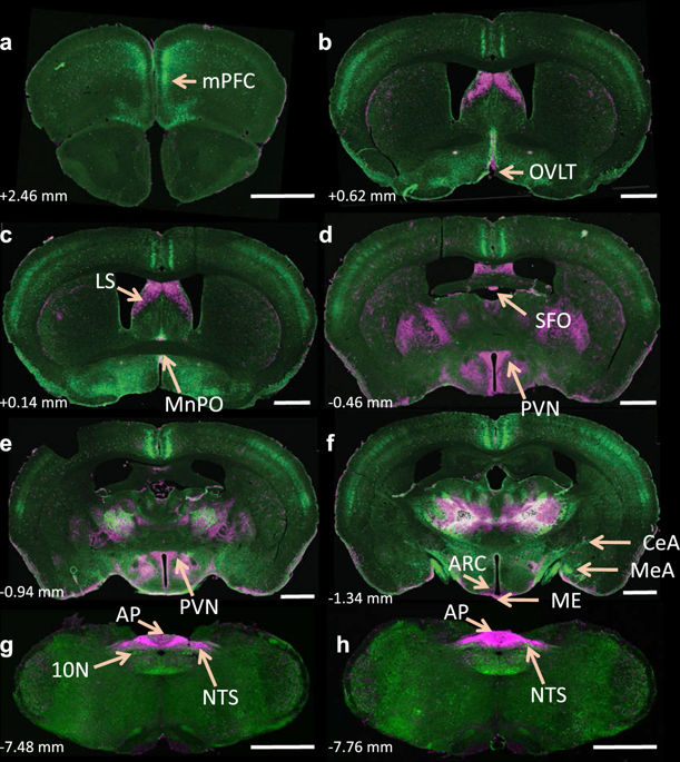
Brain angiotensin-II (Ang-II) type-1 receptors (AT1Rs), which exert profound effects on normal cardiovascular, fluid, and metabolic homeostasis, are overactivated in and contribute to chronic sympathoexcitation and hypertension. Accumulating evidence indicates that the activation of Ang-II type-2 receptors (AT2Rs) in the brain exerts effects that are opposite to those of AT1Rs, lowering blood pressure, and reducing hypertension. Thus, it would be interesting to understand the relative cellular localization of AT1R and AT2R in the brain under normal conditions and whether this localization changes during hypertension. Here, we developed a novel AT1aR-tdTomato reporter mouse strain in which the location of brain AT1aR was largely consistent with that determined in the previous studies. This AT1aR-tdTomato reporter mouse strain was crossed with our previously described AT2R-eGFP reporter mouse strain to yield a novel dual AT1aR/AT2R reporter mouse strain, which allowed us to determine that AT1aR and AT2R are primarily localized to different populations of neurons in brain regions controlling cardiovascular, fluid, and metabolic homeostasis. Using the individual AT1aR-tdTomato reporter mice, we also demonstrated that during hypertension induced by the administration of deoxycorticosterone acetate-salt, there was no shift in the expression of AT1aR from neurons to microglia or astrocytes in the paraventricular nucleus, a brain area important for sympathetic regulation. Using AT2R-eGFP reporter mice under similar hypertensive conditions, we demonstrated that the same was true of AT2R expression in the nucleus of the solitary tract (NTS), an area critical for baroreflex control. Collectively, these findings provided a novel means to assess the colocalization of AT1R and AT2R in the brain and a novel view of their cellular localization in hypertension.
中文翻译:

脑血管紧张素 1 型和 2 型受体:正常和高血压条件下的细胞位置
脑血管紧张素-II (Ang-II) 1 型受体 (AT1Rs) 对正常心血管、体液和代谢稳态产生深远影响,在慢性交感神经兴奋和高血压中过度激活并导致它们。越来越多的证据表明,大脑中Ang-II 2型受体(AT2Rs)的激活产生与AT1Rs相反的作用,降低血压,降低高血压。因此,了解正常情况下大脑中 AT1R 和 AT2R 的相对细胞定位以及这种定位在高血压期间是否会发生变化将是很有趣的。在这里,我们开发了一种新的 AT1aR-tdTomato 报告小鼠品系,其中大脑 AT1aR 的位置与先前研究中确定的位置基本一致。这种 AT1aR-tdTomato 报告小鼠品系与我们之前描述的 AT2R-eGFP 报告小鼠品系杂交,产生一种新的双 AT1aR/AT2R 报告小鼠品系,这使我们能够确定 AT1aR 和 AT2R 主要定位于大脑中不同的神经元群体控制心血管、体液和代谢稳态的区域。使用单独的 AT1aR-tdTomato 报告小鼠,我们还证明,在由醋酸脱氧皮质酮盐诱导的高血压期间,AT1aR 的表达没有从神经元向室旁核中的小胶质细胞或星形胶质细胞转变,室旁核是一个重要的大脑区域。同情调节。在类似的高血压条件下使用 AT2R-eGFP 报告小鼠,我们证明了孤束核 (NTS) 中的 AT2R 表达也是如此,这是一个对压力反射控制至关重要的区域。总的来说,这些发现提供了一种新的方法来评估 AT1R 和 AT2R 在大脑中的共定位以及它们在高血压中的细胞定位的新观点。
更新日期:2019-12-18
中文翻译:
脑血管紧张素 1 型和 2 型受体:正常和高血压条件下的细胞位置
脑血管紧张素-II (Ang-II) 1 型受体 (AT1Rs) 对正常心血管、体液和代谢稳态产生深远影响,在慢性交感神经兴奋和高血压中过度激活并导致它们。越来越多的证据表明,大脑中Ang-II 2型受体(AT2Rs)的激活产生与AT1Rs相反的作用,降低血压,降低高血压。因此,了解正常情况下大脑中 AT1R 和 AT2R 的相对细胞定位以及这种定位在高血压期间是否会发生变化将是很有趣的。在这里,我们开发了一种新的 AT1aR-tdTomato 报告小鼠品系,其中大脑 AT1aR 的位置与先前研究中确定的位置基本一致。这种 AT1aR-tdTomato 报告小鼠品系与我们之前描述的 AT2R-eGFP 报告小鼠品系杂交,产生一种新的双 AT1aR/AT2R 报告小鼠品系,这使我们能够确定 AT1aR 和 AT2R 主要定位于大脑中不同的神经元群体控制心血管、体液和代谢稳态的区域。使用单独的 AT1aR-tdTomato 报告小鼠,我们还证明,在由醋酸脱氧皮质酮盐诱导的高血压期间,AT1aR 的表达没有从神经元向室旁核中的小胶质细胞或星形胶质细胞转变,室旁核是一个重要的大脑区域。同情调节。在类似的高血压条件下使用 AT2R-eGFP 报告小鼠,我们证明了孤束核 (NTS) 中的 AT2R 表达也是如此,这是一个对压力反射控制至关重要的区域。总的来说,这些发现提供了一种新的方法来评估 AT1R 和 AT2R 在大脑中的共定位以及它们在高血压中的细胞定位的新观点。


 京公网安备 11010802027423号