当前位置:
X-MOL 学术
›
Cell Calcium
›
论文详情
Our official English website, www.x-mol.net, welcomes your feedback! (Note: you will need to create a separate account there.)
Noise analysis of cytosolic calcium image data.
Cell Calcium ( IF 4 ) Pub Date : 2019-12-18 , DOI: 10.1016/j.ceca.2019.102152 Divya Swaminathan 1 , George D Dickinson 1 , Angelo Demuro 1 , Ian Parker 2
Cell Calcium ( IF 4 ) Pub Date : 2019-12-18 , DOI: 10.1016/j.ceca.2019.102152 Divya Swaminathan 1 , George D Dickinson 1 , Angelo Demuro 1 , Ian Parker 2
Affiliation
|
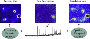
Cellular Ca2+ signals are often constrained to cytosolic micro- or nano-domains where stochastic openings of Ca2+ channels cause large fluctuations in local Ca2+ concentration (Ca2+ 'noise'). With the advent of TIRF microscopy to image the fluorescence of Ca2+-sensitive probes from attoliter volumes it has become possible to directly monitor these signals, which closely track the gating of plasmalemmal and ER Ca2+-permeable channels. Nevertheless, it is likely that many physiologically important Ca2+ signals are too small to resolve as discrete events in fluorescence recordings. By analogy with noise analysis of electrophysiological data, we explore here the use of statistical approaches to detect and analyze such Ca2+ noise in images obtained using Ca2+-sensitive indicator dyes. We describe two techniques - power spectrum analysis and spatio-temporal correlation - and demonstrate that both effectively identify discrete, spatially localized calcium release events (Ca2+ puffs). Moreover, we show they are able to detect localized noise fluctuations in a case where discrete events cannot directly be resolved.
中文翻译:

胞质钙图像数据的噪声分析。
细胞中的Ca2 +信号通常被限制在胞质的微域或纳米域,其中Ca2 +通道的随机开放会引起局部Ca2 +浓度的大幅波动(Ca2 +“噪声”)。随着TIRF显微镜技术的出现,从attoliter体积对Ca2 +敏感探针的荧光成像,可以直接监视这些信号,从而密切跟踪血浆和ER Ca2 +渗透通道的门控。然而,很可能许多生理上重要的Ca2 +信号太小而无法解析为荧光记录中的离散事件。通过类似于电生理数据的噪声分析,我们在这里探索使用统计方法来检测和分析使用Ca2 +敏感指示剂染料获得的图像中的此类Ca2 +噪声。我们描述了两种技术-功率谱分析和时空相关性-并证明这两种技术都能有效地识别离散的,空间局部的钙释放事件(Ca2 +泡芙)。此外,我们表明在离散事件无法直接解决的情况下,它们能够检测局部噪声波动。
更新日期:2019-12-19
中文翻译:
胞质钙图像数据的噪声分析。
细胞中的Ca2 +信号通常被限制在胞质的微域或纳米域,其中Ca2 +通道的随机开放会引起局部Ca2 +浓度的大幅波动(Ca2 +“噪声”)。随着TIRF显微镜技术的出现,从attoliter体积对Ca2 +敏感探针的荧光成像,可以直接监视这些信号,从而密切跟踪血浆和ER Ca2 +渗透通道的门控。然而,很可能许多生理上重要的Ca2 +信号太小而无法解析为荧光记录中的离散事件。通过类似于电生理数据的噪声分析,我们在这里探索使用统计方法来检测和分析使用Ca2 +敏感指示剂染料获得的图像中的此类Ca2 +噪声。我们描述了两种技术-功率谱分析和时空相关性-并证明这两种技术都能有效地识别离散的,空间局部的钙释放事件(Ca2 +泡芙)。此外,我们表明在离散事件无法直接解决的情况下,它们能够检测局部噪声波动。


 京公网安备 11010802027423号