当前位置:
X-MOL 学术
›
Spinal Cord Ser. Cases
›
论文详情
Our official English website, www.x-mol.net, welcomes your feedback! (Note: you will need to create a separate account there.)
Quantitative electrodiagnostic patterns of damage and recovery after spinal cord injury: a pilot study.
Spinal Cord Series and Cases Pub Date : 2019-12-12 , DOI: 10.1038/s41394-019-0246-0 Elissa C Zakrasek 1 , Jeffrey P Jaramillo 1 , Zoia C Lateva 1 , Vandana Punj 1 , B Jenny Kiratli 1 , Kevin C McGill 1
Spinal Cord Series and Cases Pub Date : 2019-12-12 , DOI: 10.1038/s41394-019-0246-0 Elissa C Zakrasek 1 , Jeffrey P Jaramillo 1 , Zoia C Lateva 1 , Vandana Punj 1 , B Jenny Kiratli 1 , Kevin C McGill 1
Affiliation
|
Study design
Prospective observational pilot study.
Objectives
To compare quantitative electromyographic (EMG), imaging and strength data at two time points in individuals with cervical spinal cord injury (SCI).
Setting
SCI center, Veterans Affairs Health Care System, Palo Alto, California, USA.
Methods
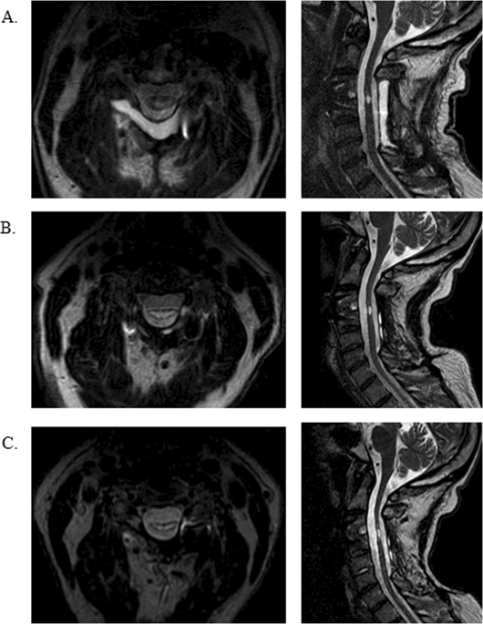
Subjects without suspected peripheral nerve injury were recruited within 3 months of injury. Needle EMG examination was performed in myotomes above, at, and below the SCI level around 11- and 12-months post injury. EMG data were decomposed using custom software into constituent motor unit trains and each distinct motor unit was analyzed for firing rate and amplitude. Strength measurements were made with dynamometry and according to the International Standard of Neurologic Classification of SCI (ISNCSCI). Cervical magnetic resonance images (MRI) were evaluated by two neuroradiologists for gray and white matter damage around the SCI. Here, we compare the EMG, strength, and imaging findings of the one of the four participants who completed both 3- and 12-month EMG evaluations.
Results
There was an increase in force generation in all muscles tested at 1 year. Localized findings of very fast firing motor units helped localize spinal cord damage and revealed gray matter damage in spinal segments where MRI was normal. Meanwhile, improvement in strength over time corresponded with different electrophysiologic patterns.
Conclusions
Electromyographic decomposition at two time points provides valuable information about localization of spinal cord damage, integrity of motor neuron pools and may provide a unique understanding of neural recovery mechanisms.
中文翻译:

脊髓损伤后损伤和恢复的定量电诊断模式:一项试点研究。
研究设计 前瞻性观察性试点研究。目的 比较颈脊髓损伤 (SCI) 患者两个时间点的定量肌电图 (EMG)、成像和力量数据。设置 SCI 中心,退伍军人事务医疗保健系统,帕洛阿尔托,加利福尼亚州,美国。方法 招募受伤后 3 个月内没有疑似周围神经损伤的受试者。受伤后 11 个月和 12 个月左右,在 SCI 水平上方、上方和下方的肌节中进行针肌电图检查。使用定制软件将肌电图数据分解为组成的运动单元序列,并分析每个不同运动单元的放电频率和幅度。力量测量采用测力法并根据 SCI 神经学分类国际标准 (ISNCSCI) 进行。两名神经放射科医生对颈椎磁共振图像 (MRI) 进行了评估,以了解 SCI 周围的灰质和白质损伤情况。在这里,我们比较了完成 3 个月和 12 个月肌电图评估的四名参与者之一的肌电图、肌力和影像学结果。结果一年后,所有测试的肌肉产生的力量都有所增加。快速放电运动单位的局部发现有助于定位脊髓损伤,并揭示 MRI 正常的脊髓节段的灰质损伤。同时,随着时间的推移,力量的提高与不同的电生理模式相对应。结论 两个时间点的肌电图分解提供了有关脊髓损伤定位、运动神经元池完整性的有价值的信息,并可能提供对神经恢复机制的独特理解。
更新日期:2019-12-12
中文翻译:
脊髓损伤后损伤和恢复的定量电诊断模式:一项试点研究。
研究设计 前瞻性观察性试点研究。目的 比较颈脊髓损伤 (SCI) 患者两个时间点的定量肌电图 (EMG)、成像和力量数据。设置 SCI 中心,退伍军人事务医疗保健系统,帕洛阿尔托,加利福尼亚州,美国。方法 招募受伤后 3 个月内没有疑似周围神经损伤的受试者。受伤后 11 个月和 12 个月左右,在 SCI 水平上方、上方和下方的肌节中进行针肌电图检查。使用定制软件将肌电图数据分解为组成的运动单元序列,并分析每个不同运动单元的放电频率和幅度。力量测量采用测力法并根据 SCI 神经学分类国际标准 (ISNCSCI) 进行。两名神经放射科医生对颈椎磁共振图像 (MRI) 进行了评估,以了解 SCI 周围的灰质和白质损伤情况。在这里,我们比较了完成 3 个月和 12 个月肌电图评估的四名参与者之一的肌电图、肌力和影像学结果。结果一年后,所有测试的肌肉产生的力量都有所增加。快速放电运动单位的局部发现有助于定位脊髓损伤,并揭示 MRI 正常的脊髓节段的灰质损伤。同时,随着时间的推移,力量的提高与不同的电生理模式相对应。结论 两个时间点的肌电图分解提供了有关脊髓损伤定位、运动神经元池完整性的有价值的信息,并可能提供对神经恢复机制的独特理解。


 京公网安备 11010802027423号