当前位置:
X-MOL 学术
›
J. Hepatol.
›
论文详情
Our official English website, www.x-mol.net, welcomes your feedback! (Note: you will need to create a separate account there.)
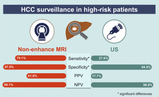
Non-enhanced Magnetic Resonance Imaging as a Surveillance Tool for Hepatocellular Carcinoma: Comparison with Ultrasound
Journal of Hepatology ( IF 25.7 ) Pub Date : 2020-04-01 , DOI: 10.1016/j.jhep.2019.12.001 Hyo Jung Park 1 , Hye Young Jang 2 , So Yeon Kim 1 , So Jung Lee 1 , Hyung Jin Won 1 , Jae Ho Byun 1 , Sang Hyun Choi 1 , Seung Soo Lee 1 , Jihyun An 3 , Young-Suk Lim 4
Journal of Hepatology ( IF 25.7 ) Pub Date : 2020-04-01 , DOI: 10.1016/j.jhep.2019.12.001 Hyo Jung Park 1 , Hye Young Jang 2 , So Yeon Kim 1 , So Jung Lee 1 , Hyung Jin Won 1 , Jae Ho Byun 1 , Sang Hyun Choi 1 , Seung Soo Lee 1 , Jihyun An 3 , Young-Suk Lim 4
Affiliation
|
BACKGROUND & AIMS
Recently revised international guidelines for hepatocellular carcinoma (HCC) suggest that patients with inadequate ultrasonography be assessed by alternative imaging modalities. Given short scan time and the absence of contrast agent-associated risks, non-enhanced magnetic resonance imaging (MRI) has potential as a surveillance tool. This study compared the performance of non-enhanced MRI and ultrasonography for HCC surveillance in high-risk patients. METHODS
We included 382 high-risk patients in a prospective cohort who underwent 1 to 3 rounds of paired gadoxetic acid-enhanced MRI and ultrasonography. Non-enhanced MRI, consisting of diffusion-weighted imaging (DWI) and T2-weighted imaging, was simulated and retrospectively analyzed, with results considered positive when lesion(s) ≥ 1cm showed diffusion restriction or mild-moderate T2 hyperintensity. Ultrasonography results were retrieved from patient records. HCC was diagnosed histologically and/or radiologically. Sensitivity, positive predictive value (PPV), specificity, and negative predictive value (NPV) were evaluated using generalized estimating equations. RESULTS
Forty-eight HCCs were diagnosed in 43 patients. Per-lesion and per-exam sensitivities of non-enhanced MRI were 77.1% and 79.1%, respectively, higher than ultrasonography (25.0% and 27.9%, respectively, P <0.001). Per-lesion and per-exam PPVs were higher for non-enhanced MRI (56.9% and 61.8%, respectively) than ultrasonography (16.7% and 17.7%, respectively). Specificities of non-enhanced MRI (97.9%) and ultrasonography (94.5%) differ significantly (P <0.001). NPV was higher for non-enhanced MRI (99.1%) than ultrasonography (96.9%). Estimated scan time of non-enhanced MRI was <6 minutes. CONCLUSION
Given the high performance, short scan time, and the lack of contrast agent-associated risks, non-enhanced MRI is a promising option for HCC surveillance in high-risk patients.
中文翻译:

非增强磁共振成像作为肝细胞癌的监测工具:与超声的比较
背景和目的 最近修订的肝细胞癌 (HCC) 国际指南建议超声检查不充分的患者通过替代成像方式进行评估。鉴于扫描时间短且不存在造影剂相关风险,非增强磁共振成像 (MRI) 具有作为监测工具的潜力。本研究比较了非增强 MRI 和超声检查对高危患者 HCC 监测的性能。方法 我们在一个前瞻性队列中纳入了 382 名高危患者,他们接受了 1 到 3 轮配对钆塞酸增强 MRI 和超声检查。非增强 MRI,包括弥散加权成像 (DWI) 和 T2 加权成像,被模拟和回顾性分析,当病变≥ 1cm 显示弥散受限或轻中度 T2 高信号时,结果被认为是阳性的。从患者记录中检索超声检查结果。HCC 通过组织学和/或放射学诊断。使用广义估计方程评估敏感性、阳性预测值 (PPV)、特异性和阴性预测值 (NPV)。结果 43 名患者中诊断出 48 例 HCC。非增强 MRI 的按病灶和每次检查的敏感性分别为 77.1% 和 79.1%,高于超声检查(分别为 25.0% 和 27.9%,P <0.001)。非增强 MRI 的每病灶和每检查 PPV(分别为 56.9% 和 61.8%)高于超声检查(分别为 16.7% 和 17.7%)。非增强 MRI (97.9%) 和超声检查 (94.5%) 的特异性差异显着(P < 0. 001)。非增强 MRI (99.1%) 的 NPV 高于超声检查 (96.9%)。非增强 MRI 的估计扫描时间 <6 分钟。结论鉴于高性能、短扫描时间和缺乏造影剂相关风险,非增强 MRI 是高危患者 HCC 监测的有希望的选择。
更新日期:2020-04-01
中文翻译:
非增强磁共振成像作为肝细胞癌的监测工具:与超声的比较
背景和目的 最近修订的肝细胞癌 (HCC) 国际指南建议超声检查不充分的患者通过替代成像方式进行评估。鉴于扫描时间短且不存在造影剂相关风险,非增强磁共振成像 (MRI) 具有作为监测工具的潜力。本研究比较了非增强 MRI 和超声检查对高危患者 HCC 监测的性能。方法 我们在一个前瞻性队列中纳入了 382 名高危患者,他们接受了 1 到 3 轮配对钆塞酸增强 MRI 和超声检查。非增强 MRI,包括弥散加权成像 (DWI) 和 T2 加权成像,被模拟和回顾性分析,当病变≥ 1cm 显示弥散受限或轻中度 T2 高信号时,结果被认为是阳性的。从患者记录中检索超声检查结果。HCC 通过组织学和/或放射学诊断。使用广义估计方程评估敏感性、阳性预测值 (PPV)、特异性和阴性预测值 (NPV)。结果 43 名患者中诊断出 48 例 HCC。非增强 MRI 的按病灶和每次检查的敏感性分别为 77.1% 和 79.1%,高于超声检查(分别为 25.0% 和 27.9%,P <0.001)。非增强 MRI 的每病灶和每检查 PPV(分别为 56.9% 和 61.8%)高于超声检查(分别为 16.7% 和 17.7%)。非增强 MRI (97.9%) 和超声检查 (94.5%) 的特异性差异显着(P < 0. 001)。非增强 MRI (99.1%) 的 NPV 高于超声检查 (96.9%)。非增强 MRI 的估计扫描时间 <6 分钟。结论鉴于高性能、短扫描时间和缺乏造影剂相关风险,非增强 MRI 是高危患者 HCC 监测的有希望的选择。


 京公网安备 11010802027423号