Cell Stem Cell ( IF 23.9 ) Pub Date : 2019-12-05 , DOI: 10.1016/j.stem.2019.10.003 Leo Kunz 1 , Timm Schroeder 1
|
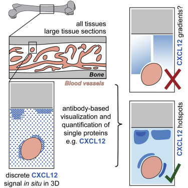
Technological limitations have hampered understanding of how individual molecules, including putative stem cell regulators, are distributed throughout tissues and stem cell niches. Here, we report adaptation of the proximity ligation assay (PLA) for large-volume, in situ imaging of individual proteins with multiple additional fluorescent channels with integrated 3D quantification strategies and software. Using this platform, we quantified the bone marrow (BM) distribution of individual CXCL12 chemokine proteins, both before and after their depletion by granulocyte-colony stimulating factor (G-CSF) treatment. We found ubiquitous CXCL12 distributions with local enrichments but no long-range gradients, in contrast to current assumptions about how CXCL12 controls migration of hematopoietic stem and progenitor cells (HSPCs) within BM. This pipeline for discrete digital quantitative, large-volume, multicolor imaging, with up to single-molecule sensitivity, may be broadly applied to any antibody epitope and tissue, enabling further insights into molecular organization of tissues and cellular interactions.
中文翻译:
用于定量分泌分子的3D组织全数字成像管道显示骨髓中不存在CXCL12梯度。
技术局限性阻碍了人们对包括推定的干细胞调节剂在内的各个分子如何分布在整个组织和干细胞壁ni中的理解。在这里,我们报告了针对大批量原位检测的邻近结扎分析(PLA)的适应性具有集成的3D定量策略和软件的多个附加荧光通道可对单个蛋白质进行成像。使用此平台,我们定量了单个CXCL12趋化因子蛋白在粒细胞集落刺激因子(G-CSF)处理消耗之前和之后的骨髓(BM)分布。我们发现普遍存在的CXCL12分布具有局部富集,但没有长程梯度,这与当前关于CXCL12如何控制BM中造血干细胞和祖细胞(HSPC)迁移的假设相反。这种用于离散数字定量,大容量,多色成像的管线,具有高达单分子的灵敏度,可广泛应用于任何抗体表位和组织,从而能够进一步洞察组织的分子组织和细胞相互作用。


 京公网安备 11010802027423号