当前位置:
X-MOL 学术
›
J. Biophotonics
›
论文详情
Our official English website, www.x-mol.net, welcomes your feedback! (Note: you will need to create a separate account there.)
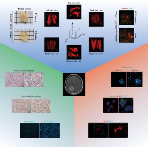
The combination of two-dimensional and three-dimensional analysis methods contributes to the understanding of glioblastoma spatial heterogeneity.
Journal of Biophotonics ( IF 2.8 ) Pub Date : 2019-12-04 , DOI: 10.1002/jbio.201900196 Runwei Yang 1, 2 , Jinglin Guo 1, 2 , Zhiying Lin 1, 2 , Haimin Song 1, 2 , Zhanpeng Feng 1, 2 , Yichao Ou 1, 2 , Mingfeng Zhou 1, 2 , Yaomin Li 1, 2 , Guozhong Yi 1, 2 , Ke Li 2 , Kaishu Li 1, 2 , Manlan Guo 2 , Xiran Wang 1, 2 , Guanglong Huang 1, 2, 3 , Zhifeng Liu 4 , Songtao Qi 1, 2, 3 , Yawei Liu 1, 2
Journal of Biophotonics ( IF 2.8 ) Pub Date : 2019-12-04 , DOI: 10.1002/jbio.201900196 Runwei Yang 1, 2 , Jinglin Guo 1, 2 , Zhiying Lin 1, 2 , Haimin Song 1, 2 , Zhanpeng Feng 1, 2 , Yichao Ou 1, 2 , Mingfeng Zhou 1, 2 , Yaomin Li 1, 2 , Guozhong Yi 1, 2 , Ke Li 2 , Kaishu Li 1, 2 , Manlan Guo 2 , Xiran Wang 1, 2 , Guanglong Huang 1, 2, 3 , Zhifeng Liu 4 , Songtao Qi 1, 2, 3 , Yawei Liu 1, 2
Affiliation
|
Heterogeneity is regarded as the major factor leading to the poor outcomes of glioblastoma (GBM) patients. However, conventional two‐dimensional (2D) analysis methods, such as immunohistochemistry and immunofluorescence, have limited capacity to reveal GBM spatial heterogeneity. Thus, we sought to develop an effective analysis strategy to increase the understanding of GBM spatial heterogeneity. Here, 2D and three‐dimensional (3D) analysis methods were compared for the examination of cell morphology, cell distribution and large intact structures, and both types of methods were employed to dissect GBM spatial heterogeneity. The results showed that 2D assays showed only cross‐sections of specimens but provided a full view. To visualize intact GBM specimens in 3D without sectioning, the optical tissue clearing methods CUBIC and iDISCO+ were used to clear opaque specimens so that they would become more transparent, after which the specimens were imaged with a two‐photon microscope. The 3D analysis methods showed specimens at a large spatial scale at cell‐level resolution and had overwhelming advantages in comparison to the 2D methods. Furthermore, in 3D, heterogeneity in terms of cell stemness, the microvasculature, and immune cell infiltration within GBM was comprehensively observed and analysed. Overall, we propose that 2D and 3D analysis methods should be combined to provide much greater detail to increase the understanding of GBM spatial heterogeneity.
中文翻译:

二维和三维分析方法的结合有助于了解胶质母细胞瘤的空间异质性。
异质性被认为是导致胶质母细胞瘤(GBM)患者预后不良的主要因素。但是,常规的二维(2D)分析方法(如免疫组织化学和免疫荧光法)显示GBM空间异质性的能力有限。因此,我们寻求开发一种有效的分析策略,以增加对GBM空间异质性的了解。在这里,比较了2D和3D(3D)分析方法以检查细胞形态,细胞分布和大的完整结构,并且两种方法都用于剖析GBM空间异质性。结果表明,二维检测仅显示了标本的横截面,但提供了完整的视图。要在不进行切片的情况下以3D形式显示完整的GBM标本,使用光学组织清除方法CUBIC和iDISCO +清除不透明的标本,使其变得更加透明,然后用双光子显微镜对标本成像。3D分析方法显示的标本在细胞级别的分辨率下具有较大的空间比例,并且与2D方法相比具有压倒性的优势。此外,在3D中,全面观察和分析了GBM中在细胞干性,微脉管系统和免疫细胞浸润方面的异质性。总体而言,我们建议将2D和3D分析方法结合起来,以提供更多详细信息,以增加对GBM空间异质性的理解。3D分析方法显示的标本在细胞级别的分辨率下具有较大的空间比例,并且与2D方法相比具有压倒性的优势。此外,在3D中,全面观察和分析了GBM内在细胞干性,微脉管系统和免疫细胞浸润方面的异质性。总体而言,我们建议将2D和3D分析方法结合起来,以提供更多详细信息,以增加对GBM空间异质性的理解。3D分析方法显示的标本在细胞级别的分辨率下具有较大的空间比例,并且与2D方法相比具有压倒性的优势。此外,在3D中,全面观察和分析了GBM中在细胞干性,微脉管系统和免疫细胞浸润方面的异质性。总体而言,我们建议将2D和3D分析方法结合起来,以提供更多详细信息,以增加对GBM空间异质性的理解。
更新日期:2019-12-04
中文翻译:
二维和三维分析方法的结合有助于了解胶质母细胞瘤的空间异质性。
异质性被认为是导致胶质母细胞瘤(GBM)患者预后不良的主要因素。但是,常规的二维(2D)分析方法(如免疫组织化学和免疫荧光法)显示GBM空间异质性的能力有限。因此,我们寻求开发一种有效的分析策略,以增加对GBM空间异质性的了解。在这里,比较了2D和3D(3D)分析方法以检查细胞形态,细胞分布和大的完整结构,并且两种方法都用于剖析GBM空间异质性。结果表明,二维检测仅显示了标本的横截面,但提供了完整的视图。要在不进行切片的情况下以3D形式显示完整的GBM标本,使用光学组织清除方法CUBIC和iDISCO +清除不透明的标本,使其变得更加透明,然后用双光子显微镜对标本成像。3D分析方法显示的标本在细胞级别的分辨率下具有较大的空间比例,并且与2D方法相比具有压倒性的优势。此外,在3D中,全面观察和分析了GBM中在细胞干性,微脉管系统和免疫细胞浸润方面的异质性。总体而言,我们建议将2D和3D分析方法结合起来,以提供更多详细信息,以增加对GBM空间异质性的理解。3D分析方法显示的标本在细胞级别的分辨率下具有较大的空间比例,并且与2D方法相比具有压倒性的优势。此外,在3D中,全面观察和分析了GBM内在细胞干性,微脉管系统和免疫细胞浸润方面的异质性。总体而言,我们建议将2D和3D分析方法结合起来,以提供更多详细信息,以增加对GBM空间异质性的理解。3D分析方法显示的标本在细胞级别的分辨率下具有较大的空间比例,并且与2D方法相比具有压倒性的优势。此外,在3D中,全面观察和分析了GBM中在细胞干性,微脉管系统和免疫细胞浸润方面的异质性。总体而言,我们建议将2D和3D分析方法结合起来,以提供更多详细信息,以增加对GBM空间异质性的理解。


 京公网安备 11010802027423号