当前位置:
X-MOL 学术
›
Faraday Discuss.
›
论文详情
Our official English website, www.x-mol.net, welcomes your feedback! (Note: you will need to create a separate account there.)
The effects of drying technique and surface pre-treatment on the cytotoxicity and dissolution rate of luminescent porous silicon quantum dots in model fluids and living cells.
Faraday Discussions ( IF 3.4 ) Pub Date : 2019-11-01 , DOI: 10.1039/c9fd00107g Maxim B Gongalsky 1 , Uliana A Tsurikova , Catherine J Storey , Yana V Evstratova , Andrew A Kudryavtsev , Leigh T Canham , Liubov A Osminkina
Faraday Discussions ( IF 3.4 ) Pub Date : 2019-11-01 , DOI: 10.1039/c9fd00107g Maxim B Gongalsky 1 , Uliana A Tsurikova , Catherine J Storey , Yana V Evstratova , Andrew A Kudryavtsev , Leigh T Canham , Liubov A Osminkina
Affiliation
|
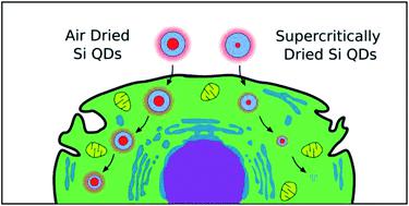
Tailoring of the biodegradation of photoluminescent silicon quantum dots (Si QDs) is important for their future applications in diagnostics and therapy. Here, the effect of drying and surface pretreatment on the dissolution rate of Si QDs in model liquids and living cells was studied in vitro using a combination of photoluminescence and Raman micro-spectroscopy. Porous silicon particles were obtained by mechanical milling of electrochemically etched mesoporous silicon films, and consist of interlinked silicon nanocrystals (QDs) and pores. The samples were subjected to super-critical drying with CO2 solvent (SCD) or air drying (AD) and then annealed at 600 °C for 16 hours in 1% oxygen to obtain nano-sized Si QDs. The obtained samples were characterized by a core–shell structure with a crystalline silicon core and a SiO2 layer on the surface. The sizes of the crystalline silicon cores, calculated from Raman scattering spectra, were about 4.5 nm for the initial AD-SiQDs, and about 2 nm for the initial SCD-SiQDs. Both the AD-Si QDs and the SCD-Si QDs exhibited visible photoluminescence (PL) properties due to quantum confinement effects. The dissolution of the nanocrystals was evaluated through their PL quenching, as well as by the presence of a low-frequency shift, broadening, and a decrease in the intensity of the Raman signal. The stability of the AD-Si QDs and the complete dissolution of the SCD-Si QDs during 24 hours of incubation with cells have been demonstrated. This might explain the apparent lower cytotoxicity observed for SCD-Si QDs.
中文翻译:

干燥技术和表面预处理对模型流体和活细胞中发光多孔硅量子点的细胞毒性和溶解速率的影响。
量身定制光致发光硅量子点(Si QDs)的生物降解对其在诊断和治疗中的未来应用非常重要。在此,干燥和表面预处理的在Si量子点在模型液体和活细胞的溶解率的影响进行了研究在体外使用的光致发光和拉曼显微光谱的组合。多孔硅颗粒是通过对电化学蚀刻的中孔硅膜进行机械研磨而获得的,并且由相互连接的硅纳米晶体(QD)和孔组成。样品用CO 2进行超临界干燥溶剂(SCD)或空气干燥(AD),然后在1%的氧气中于600°C退火16小时,以获得纳米级的Si QD。获得的样品的特征是具有结晶硅核和SiO 2的核-壳结构表面上的一层。根据拉曼散射光谱计算的结晶硅核的尺寸,对于初始AD-SiQD约为4.5 nm,对于初始SCD-SiQD约为2 nm。由于量子限制效应,AD-Si QD和SCD-Si QD均显示可见光致发光(PL)特性。纳米晶体的溶解通过其PL猝灭以及低频移动,拉宽和拉曼信号强度降低的存在来评估。已经证明了在与细胞温育24小时期间AD-Si QD的稳定性和SCD-Si QD的完全溶解。这可能解释了SCD-Si QDs明显降低的细胞毒性。
更新日期:2019-11-01
中文翻译:
干燥技术和表面预处理对模型流体和活细胞中发光多孔硅量子点的细胞毒性和溶解速率的影响。
量身定制光致发光硅量子点(Si QDs)的生物降解对其在诊断和治疗中的未来应用非常重要。在此,干燥和表面预处理的在Si量子点在模型液体和活细胞的溶解率的影响进行了研究在体外使用的光致发光和拉曼显微光谱的组合。多孔硅颗粒是通过对电化学蚀刻的中孔硅膜进行机械研磨而获得的,并且由相互连接的硅纳米晶体(QD)和孔组成。样品用CO 2进行超临界干燥溶剂(SCD)或空气干燥(AD),然后在1%的氧气中于600°C退火16小时,以获得纳米级的Si QD。获得的样品的特征是具有结晶硅核和SiO 2的核-壳结构表面上的一层。根据拉曼散射光谱计算的结晶硅核的尺寸,对于初始AD-SiQD约为4.5 nm,对于初始SCD-SiQD约为2 nm。由于量子限制效应,AD-Si QD和SCD-Si QD均显示可见光致发光(PL)特性。纳米晶体的溶解通过其PL猝灭以及低频移动,拉宽和拉曼信号强度降低的存在来评估。已经证明了在与细胞温育24小时期间AD-Si QD的稳定性和SCD-Si QD的完全溶解。这可能解释了SCD-Si QDs明显降低的细胞毒性。


 京公网安备 11010802027423号