npj Parkinson's Disease ( IF 9.304 ) Pub Date : 2019-07-04 , DOI: 10.1038/s41531-019-0085-5 M. Isabel Vanegas , Annabelle Blangero , James E. Galvin , Alessandro Di Rocco , Angelo Quartarone , M. Felice Ghilardi , Simon P. Kelly
|
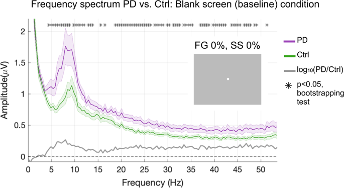
Over the last decades, psychophysical and electrophysiological studies in patients and animal models of Parkinson’s disease (PD), have consistently revealed a number of visual abnormalities. In particular, specific alterations of contrast sensitivity curves, electroretinogram (ERG), and visual-evoked potentials (VEP), have been attributed to dopaminergic retinal depletion. However, fundamental mechanisms of cortical visual processing, such as normalization or “gain control” computations, have not yet been examined in PD patients. Here, we measured electrophysiological indices of gain control in both space (surround suppression) and time (sensory adaptation) in PD patients based on steady-state VEP (ssVEP). Compared with controls, patients exhibited a significantly higher initial ssVEP amplitude that quickly decayed over time, and greater relative suppression of ssVEP amplitude as a function of surrounding stimulus contrast. Meanwhile, EEG frequency spectra were broadly elevated in patients relative to controls. Thus, contrary to what might be expected given the reduced contrast sensitivity often reported in PD, visual neural responses are not weaker; rather, they are initially larger but undergo an exaggerated degree of spatial and temporal gain control and are embedded within a greater background noise level. These differences may reflect cortical mechanisms that compensate for dysfunctional center-surround interactions at the retinal level.
中文翻译:
帕金森氏病中视觉环境相互作用的动态变化
在过去的几十年中,对帕金森氏病(PD)的患者和动物模型进行的心理物理和电生理研究一直显示出许多视觉异常。特别是,对比敏感度曲线,视网膜电图(ERG)和视觉诱发电位(VEP)的特定改变已归因于多巴胺能视网膜的耗竭。但是,尚未在PD患者中检查皮质视觉处理的基本机制,例如归一化或“增益控制”计算。在这里,我们基于稳态VEP(ssVEP)测量了PD患者在空间(周围抑制)和时间(感觉适应)方面的增益控制的电生理指标。与对照组相比,患者表现出明显更高的初始ssVEP振幅,并随时间迅速衰减,ssVEP振幅的更大相对抑制,其与周围刺激对比度的函数关系。同时,相对于对照,患者的脑电图频谱显着升高。因此,与PD中经常报告的对比敏感度降低的情况相反,视觉神经反应并不弱。相反,它们最初较大,但经过了夸大的空间和时间增益控制,并被嵌入更大的背景噪声水平内。这些差异可能反映了皮质机制,以补偿视网膜水平上功能失调的中心-周围相互作用。鉴于PD中经常报告的对比敏感度降低,这与预期相反,视觉神经反应并不弱。相反,它们最初较大,但经过了夸大的空间和时间增益控制,并被嵌入更大的背景噪声水平内。这些差异可能反映了皮质机制,以补偿视网膜水平上功能失调的中心-周围相互作用。鉴于PD中经常报告的对比敏感度降低,这与预期相反,视觉神经反应并不弱。相反,它们最初较大,但经过了夸大的空间和时间增益控制,并被嵌入更大的背景噪声水平内。这些差异可能反映了皮质机制,以补偿视网膜水平上功能失调的中心-周围相互作用。


 京公网安备 11010802027423号