当前位置:
X-MOL 学术
›
Theranostics
›
论文详情
Our official English website, www.x-mol.net, welcomes your feedback! (Note: you will need to create a separate account there.)
Dual-Channel Fluorescence Imaging of Hydrogel Degradation and Tissue Regeneration in the Brain.
Theranostics ( IF 12.4 ) Pub Date : 2019-01-01 , DOI: 10.7150/thno.35606 G Kate Park 1, 2 , Su-Hwan Kim 3 , Kyungmin Kim 3 , Priyanka Das 2 , Byung-Gee Kim 1, 3, 4 , Satoshi Kashiwagi 2 , Hak Soo Choi 2 , Nathaniel S Hwang 1, 3, 4
Theranostics ( IF 12.4 ) Pub Date : 2019-01-01 , DOI: 10.7150/thno.35606 G Kate Park 1, 2 , Su-Hwan Kim 3 , Kyungmin Kim 3 , Priyanka Das 2 , Byung-Gee Kim 1, 3, 4 , Satoshi Kashiwagi 2 , Hak Soo Choi 2 , Nathaniel S Hwang 1, 3, 4
Affiliation
|
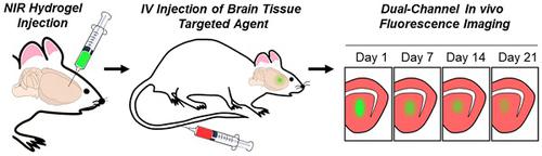
The ability of brain tissue to regenerate is limited; therefore, brain diseases (i.e., trauma, stroke, tumors) often lead to irreversible motor and cognitive impairments. Therapeutic interventions using various types of injectable biomaterials have been investigated to promote endogenous neural differentiation. Despite promising results in pre-clinical studies, the translation of regenerative medicine to the clinic has many challenges due to the lack of reliable imaging systems to achieve accurate evaluation of the treatment efficacy. Methods: In this study, we developed a dual-channel fluorescence imaging technique to simultaneously monitor tissue ingrowth and scaffold disintegration. Enzymatically crosslinked gelatin-hyaluronic acid hydrogel was labeled with 800 nm fluorophore, ZW800-3a, while the regenerated tissue was highlighted with 700 nm brain-specific contrast agent, Ox1. Results: Using the multichannel fluorescence imaging system, tissue growth and degradation of the NIR hydrogel were simultaneously imaged in the brain of mice. Images were further analyzed and reconstructed to show both visual and quantitative information of each stage of a therapeutic period. Conclusion: Dual-channel in vivo imaging systems can provide highly accurate visual and quantitative information of the brain tissue ingrowth for the evaluation of the therapeutic effect of NIR hydrogel through a simple and fast operating procedure.
中文翻译:

脑中水凝胶降解和组织再生的双通道荧光成像。
脑组织的再生能力有限;因此,脑部疾病(即外伤,中风,肿瘤)通常会导致不可逆的运动和认知障碍。已经研究了使用各种类型的可注射生物材料的治疗性干预措施,以促进内源性神经分化。尽管在临床前研究中取得了令人鼓舞的结果,但由于缺乏可靠的成像系统来实现对治疗功效的准确评估,再生医学到临床的翻译仍然面临许多挑战。方法:在这项研究中,我们开发了一种双通道荧光成像技术,以同时监测组织向内生长和支架崩解。酶促交联的明胶-透明质酸水凝胶用800 nm荧光团ZW800-3a标记,而再生组织则用700 nm脑特异性造影剂Ox1突出显示。结果:使用多通道荧光成像系统,可同时在小鼠的大脑中对NIR水凝胶的组织生长和降解进行成像。进一步分析和重建图像,以显示治疗期各个阶段的视觉和定量信息。结论:双通道体内成像系统可通过简单,快速的操作程序,为脑组织向内生长提供高度准确的视觉和定量信息,以评估NIR水凝胶的治疗效果。进一步分析和重建图像,以显示治疗期各个阶段的视觉和定量信息。结论:双通道体内成像系统可通过简单,快速的操作程序,为脑组织向内生长提供高度准确的视觉和定量信息,以评估NIR水凝胶的治疗效果。进一步分析和重建图像,以显示治疗期各个阶段的视觉和定量信息。结论:双通道体内成像系统可通过简单,快速的操作程序,为脑组织向内生长提供高度准确的视觉和定量信息,以评估NIR水凝胶的治疗效果。
更新日期:2019-01-01
中文翻译:
脑中水凝胶降解和组织再生的双通道荧光成像。
脑组织的再生能力有限;因此,脑部疾病(即外伤,中风,肿瘤)通常会导致不可逆的运动和认知障碍。已经研究了使用各种类型的可注射生物材料的治疗性干预措施,以促进内源性神经分化。尽管在临床前研究中取得了令人鼓舞的结果,但由于缺乏可靠的成像系统来实现对治疗功效的准确评估,再生医学到临床的翻译仍然面临许多挑战。方法:在这项研究中,我们开发了一种双通道荧光成像技术,以同时监测组织向内生长和支架崩解。酶促交联的明胶-透明质酸水凝胶用800 nm荧光团ZW800-3a标记,而再生组织则用700 nm脑特异性造影剂Ox1突出显示。结果:使用多通道荧光成像系统,可同时在小鼠的大脑中对NIR水凝胶的组织生长和降解进行成像。进一步分析和重建图像,以显示治疗期各个阶段的视觉和定量信息。结论:双通道体内成像系统可通过简单,快速的操作程序,为脑组织向内生长提供高度准确的视觉和定量信息,以评估NIR水凝胶的治疗效果。进一步分析和重建图像,以显示治疗期各个阶段的视觉和定量信息。结论:双通道体内成像系统可通过简单,快速的操作程序,为脑组织向内生长提供高度准确的视觉和定量信息,以评估NIR水凝胶的治疗效果。进一步分析和重建图像,以显示治疗期各个阶段的视觉和定量信息。结论:双通道体内成像系统可通过简单,快速的操作程序,为脑组织向内生长提供高度准确的视觉和定量信息,以评估NIR水凝胶的治疗效果。


 京公网安备 11010802027423号