Our official English website, www.x-mol.net, welcomes your feedback! (Note: you will need to create a separate account there.)
In vivo STED microscopy: a roadmap to nanoscale imaging in the living mouse
Methods ( IF 4.8 ) Pub Date : 2020-03-01 , DOI: 10.1016/j.ymeth.2019.05.020 Heinz Steffens , Waja Wegner , Katrin I. Willig
Methods ( IF 4.8 ) Pub Date : 2020-03-01 , DOI: 10.1016/j.ymeth.2019.05.020 Heinz Steffens , Waja Wegner , Katrin I. Willig
|
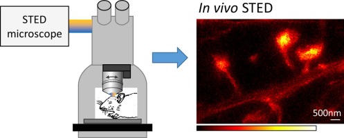
Superresolution microscopy techniques are now widely used, but their application in living animals remains a challenging task. The first superresolution imaging in a live vertebrate was demonstrated with STED microscopy in the visual cortex of an anaesthetized mouse. Here, we explain the requirements for a simple but robust in vivo STED microscope as well as the surgical preparation of the cranial window and the mounting of the mouse in detail. We have developed a mounting stage with a heating plate to keep the mouse body temperature stable and that can be adjusted to the optical axis of the microscope. We have optimised the design to avoid inducing thermal drift, which is critical for nanoscale imaging. STED microscopy with a resolution of 60 nm requires special cranial window preparation to avoid motion artefacts. We have implemented a drain tube to reduce the fluid between the glass window and the surface of the brain, which has been identified as the main cause for the motion artefacts. Together, these advances in the preparation allow the use of a simple intraperitoneal anaesthesia and make the previously used venous infusion and artificial respiration obsolete.
中文翻译:

体内 STED 显微镜:活体小鼠纳米级成像的路线图
超分辨率显微镜技术现已被广泛使用,但它们在活体动物中的应用仍然是一项具有挑战性的任务。在麻醉小鼠的视觉皮层中使用 STED 显微镜证明了活脊椎动物的第一次超分辨率成像。在这里,我们详细解释了对简单但坚固的体内 STED 显微镜的要求,以及颅窗的手术准备和鼠标的安装。我们开发了一个带有加热板的安装台,以保持鼠标体温稳定,并且可以根据显微镜的光轴进行调整。我们优化了设计以避免引起热漂移,这对于纳米级成像至关重要。分辨率为 60 nm 的 STED 显微镜需要特殊的颅窗准备以避免运动伪影。我们已经安装了一个引流管来减少玻璃窗和大脑表面之间的液体,这已被确定为运动伪影的主要原因。总之,这些在制备方面的进步允许使用简单的腹膜内麻醉,并使以前使用的静脉输液和人工呼吸过时。
更新日期:2020-03-01
中文翻译:
体内 STED 显微镜:活体小鼠纳米级成像的路线图
超分辨率显微镜技术现已被广泛使用,但它们在活体动物中的应用仍然是一项具有挑战性的任务。在麻醉小鼠的视觉皮层中使用 STED 显微镜证明了活脊椎动物的第一次超分辨率成像。在这里,我们详细解释了对简单但坚固的体内 STED 显微镜的要求,以及颅窗的手术准备和鼠标的安装。我们开发了一个带有加热板的安装台,以保持鼠标体温稳定,并且可以根据显微镜的光轴进行调整。我们优化了设计以避免引起热漂移,这对于纳米级成像至关重要。分辨率为 60 nm 的 STED 显微镜需要特殊的颅窗准备以避免运动伪影。我们已经安装了一个引流管来减少玻璃窗和大脑表面之间的液体,这已被确定为运动伪影的主要原因。总之,这些在制备方面的进步允许使用简单的腹膜内麻醉,并使以前使用的静脉输液和人工呼吸过时。


 京公网安备 11010802027423号