当前位置:
X-MOL 学术
›
Bioconjugate Chem.
›
论文详情
Our official English website, www.x-mol.net, welcomes your feedback! (Note: you will need to create a separate account there.)
Multimodal Positron Emission Tomography Imaging to Quantify Uptake of 89Zr-Labeled Liposomes in the Atherosclerotic Vessel Wall.
Bioconjugate Chemistry ( IF 4.7 ) Pub Date : 2019-06-07 , DOI: 10.1021/acs.bioconjchem.9b00256 Mark E Lobatto 1, 2 , Tina Binderup 1, 3 , Philip M Robson 1 , Luuk F P Giesen 1 , Claudia Calcagno 1 , Julia Witjes 1 , Francois Fay 1, 4 , Samantha Baxter 1 , Chang Ho Wessel 1 , Mootaz Eldib 1 , Jason Bini 1 , Sean D Carlin 5 , Erik S G Stroes 6 , Gert Storm 7, 8 , Andreas Kjaer 3 , Jason S Lewis 5, 9 , Thomas Reiner 5, 10 , Zahi A Fayad 1 , Willem J M Mulder 1, 11, 12 , Carlos Pérez-Medina 1, 13
Bioconjugate Chemistry ( IF 4.7 ) Pub Date : 2019-06-07 , DOI: 10.1021/acs.bioconjchem.9b00256 Mark E Lobatto 1, 2 , Tina Binderup 1, 3 , Philip M Robson 1 , Luuk F P Giesen 1 , Claudia Calcagno 1 , Julia Witjes 1 , Francois Fay 1, 4 , Samantha Baxter 1 , Chang Ho Wessel 1 , Mootaz Eldib 1 , Jason Bini 1 , Sean D Carlin 5 , Erik S G Stroes 6 , Gert Storm 7, 8 , Andreas Kjaer 3 , Jason S Lewis 5, 9 , Thomas Reiner 5, 10 , Zahi A Fayad 1 , Willem J M Mulder 1, 11, 12 , Carlos Pérez-Medina 1, 13
Affiliation

|
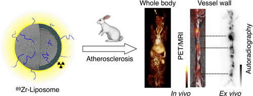
Nanotherapy has recently emerged as an experimental treatment option for atherosclerosis. To fulfill its promise, robust noninvasive imaging approaches for subject selection and treatment evaluation are warranted. To that end, we present here a positron emission tomography (PET)-based method for quantification of liposomal nanoparticle uptake in the atherosclerotic vessel wall. We evaluated a modular procedure to label liposomal nanoparticles with the radioisotope zirconium-89 (89Zr). Their biodistribution and vessel wall targeting in a rabbit atherosclerosis model was evaluated up to 15 days after intravenous injection by PET/computed tomography (CT) and PET/magnetic resonance imaging (PET/MRI). Vascular permeability was assessed in vivo using three-dimensional dynamic contrast-enhanced MRI (3D DCE-MRI) and ex vivo using near-infrared fluorescence (NIRF) imaging. The 89Zr-radiolabeled liposomes displayed a biodistribution pattern typical of long-circulating nanoparticles. Importantly, they markedly accumulated in atherosclerotic lesions in the abdominal aorta, as evident on PET/MRI and confirmed by autoradiography, and this uptake moderately correlated with vascular permeability. The method presented herein facilitates the development of nanotherapy for atherosclerotic disease as it provides a tool to screen for nanoparticle targeting in individual subjects' plaques.
中文翻译:

多峰正电子发射断层显像成像以量化动脉粥样硬化血管壁中89Zr标签的脂质体的摄取。
纳米疗法最近已成为动脉粥样硬化的实验性治疗选择。为了兑现其诺言,必须为对象选择和治疗评估提供可靠的无创成像方法。为此,我们在此介绍一种基于正电子发射断层扫描(PET)的方法,用于量化动脉粥样硬化血管壁中脂质体纳米颗粒的摄取。我们评估了用放射性同位素锆89(89Zr)标记脂质体纳米颗粒的模块化程序。在兔动脉粥样硬化模型中,通过PET /计算机断层扫描(CT)和PET /磁共振成像(PET / MRI)静脉注射后长达15天,评估了它们的生物分布和血管壁靶向性。在体内使用三维动态对比增强MRI(3D DCE-MRI)评估血管通透性,在体外使用近红外荧光(NIRF)成像评估血管通透性。89Zr放射性标记的脂质体表现出长循环纳米颗粒特有的生物分布模式。重要的是,它们显着积聚在腹主动脉的动脉粥样硬化病变中,这在PET / MRI上很明显,并通过放射自显影得以证实,并且这种摄取与血管通透性呈中度相关。本文提出的方法促进了针对动脉粥样硬化疾病的纳米疗法的发展,因为它提供了筛选针对个体受试者斑块中的纳米粒子的工具。它们在PET / MRI上很明显地聚集在腹主动脉的动脉粥样硬化病变中,并经放射自显影证实,并且这种摄取与血管通透性呈中等相关性。本文提出的方法促进了针对动脉粥样硬化疾病的纳米疗法的发展,因为它提供了筛选针对个体受试者斑块中的纳米粒子的工具。它们在PET / MRI上很明显地聚集在腹主动脉的动脉粥样硬化病变中,并经放射自显影证实,并且这种摄取与血管通透性相关。本文提出的方法促进了针对动脉粥样硬化疾病的纳米疗法的发展,因为它提供了筛选针对个体受试者斑块中的纳米粒子的工具。
更新日期:2019-11-18
中文翻译:

多峰正电子发射断层显像成像以量化动脉粥样硬化血管壁中89Zr标签的脂质体的摄取。
纳米疗法最近已成为动脉粥样硬化的实验性治疗选择。为了兑现其诺言,必须为对象选择和治疗评估提供可靠的无创成像方法。为此,我们在此介绍一种基于正电子发射断层扫描(PET)的方法,用于量化动脉粥样硬化血管壁中脂质体纳米颗粒的摄取。我们评估了用放射性同位素锆89(89Zr)标记脂质体纳米颗粒的模块化程序。在兔动脉粥样硬化模型中,通过PET /计算机断层扫描(CT)和PET /磁共振成像(PET / MRI)静脉注射后长达15天,评估了它们的生物分布和血管壁靶向性。在体内使用三维动态对比增强MRI(3D DCE-MRI)评估血管通透性,在体外使用近红外荧光(NIRF)成像评估血管通透性。89Zr放射性标记的脂质体表现出长循环纳米颗粒特有的生物分布模式。重要的是,它们显着积聚在腹主动脉的动脉粥样硬化病变中,这在PET / MRI上很明显,并通过放射自显影得以证实,并且这种摄取与血管通透性呈中度相关。本文提出的方法促进了针对动脉粥样硬化疾病的纳米疗法的发展,因为它提供了筛选针对个体受试者斑块中的纳米粒子的工具。它们在PET / MRI上很明显地聚集在腹主动脉的动脉粥样硬化病变中,并经放射自显影证实,并且这种摄取与血管通透性呈中等相关性。本文提出的方法促进了针对动脉粥样硬化疾病的纳米疗法的发展,因为它提供了筛选针对个体受试者斑块中的纳米粒子的工具。它们在PET / MRI上很明显地聚集在腹主动脉的动脉粥样硬化病变中,并经放射自显影证实,并且这种摄取与血管通透性相关。本文提出的方法促进了针对动脉粥样硬化疾病的纳米疗法的发展,因为它提供了筛选针对个体受试者斑块中的纳米粒子的工具。


 京公网安备 11010802027423号