npj Regenerative Medicine ( IF 7.2 ) Pub Date : 2019-04-16 , DOI: 10.1038/s41536-019-0070-y Debora Kehl , Melanie Generali , Anna Mallone , Manfred Heller , Anne-Christine Uldry , Phil Cheng , Benjamin Gantenbein , Simon P. Hoerstrup , Benedikt Weber
|
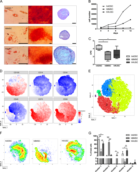
Human mesenchymal stromal cell (hMSC) secretomes have shown to influence the microenvironment upon injury, promoting cytoprotection, angiogenesis, and tissue repair. The angiogenic potential is of particular interest for the treatment of ischemic diseases. Interestingly, hMSC secretomes isolated from different tissue sources have shown dissimilarities with respect to their angiogenic profile. This study compares angiogenesis of hMSC secretomes from adipose tissue (hADSCs), bone marrow (hBMSCs), and umbilical cord Wharton’s jelly (hWJSCs). hMSC secretomes were obtained under xenofree conditions and analyzed by liquid chromatography tandem mass spectrometry (LC/MS-MS). Biological processes related to angiogenesis were found to be enriched in the proteomic profile of hMSC secretomes. hWJSC secretomes revealed a more complete angiogenic network with higher concentrations of angiogenesis related proteins, followed by hBMSC secretomes. hADSC secretomes lacked central angiogenic proteins and expressed most detected proteins to a significantly lower level. In vivo all secretomes induced vascularization of subcutaneously implanted Matrigel plugs in mice. Differences in secretome composition were functionally analyzed with monocyte and endothelial cell (EC) in vitro co-culture experiments using vi-SNE based multidimensional flow cytometry data analysis. Functional responses between hBMSC and hWJSC secretomes were comparable, with significantly higher migration of CD14++ CD16− monocytes and enhanced macrophage differentiation compared with hADSC secretomes. Both secretomes also induced a more profound pro-angiogenic phenotype of ECs. These results suggest hWJSCs secretome as the most potent hMSC source for inflammation-mediated angiogenesis induction, while the potency of hADSC secretomes was lowest. This systematic analysis may have implication on the selection of hMSCs for future clinical studies.
中文翻译:
人间质基质细胞分泌蛋白组学的蛋白质组学分析:血管生成潜力的系统比较
人间质基质细胞(hMSC)分泌组已显示在损伤时影响微环境,促进细胞保护,血管生成和组织修复。血管生成潜力对于缺血性疾病的治疗尤为重要。有趣的是,从不同组织来源分离出的hMSC分泌组在其血管生成方面显示出差异。这项研究比较了来自脂肪组织(hADSCs),骨髓(hBMSCs)和脐带沃顿氏胶体(hWJSCs)的hMSC分泌组的血管生成。在无异种条件下获得hMSC分泌组,并通过液相色谱串联质谱法(LC / MS-MS)进行分析。发现与血管生成有关的生物学过程在hMSC分泌组的蛋白质组学特征中得到丰富。hWJSC分泌组显示了一个更完整的血管生成网络,其中血管生成相关蛋白的浓度更高,其次是hBMSC分泌组。hADSC分泌蛋白缺乏中央血管生成蛋白,大多数检测到的蛋白表达水平明显降低。在体内,所有的分泌组都诱导小鼠皮下植入的基质胶塞的血管形成。使用基于vi-SNE的多维流式细胞术数据分析,通过单核细胞和内皮细胞(EC)在体外共培养实验中对分泌组组成的差异进行功能分析。hBMSC和hWJSC分泌组之间的功能反应相当,CD14的迁移明显更高 hADSC分泌组缺乏中央的血管生成蛋白,并且将大多数检测到的蛋白表达到明显较低的水平。在体内,所有的分泌组都诱导小鼠皮下植入的基质胶塞的血管形成。使用基于vi-SNE的多维流式细胞术数据分析,通过单核细胞和内皮细胞(EC)在体外共培养实验中对分泌组组成的差异进行功能分析。hBMSC和hWJSC分泌组之间的功能反应相当,CD14的迁移明显更高 hADSC分泌蛋白缺乏中央血管生成蛋白,大多数检测到的蛋白表达水平明显降低。在体内,所有的分泌组都诱导小鼠皮下植入的基质胶塞的血管形成。使用基于vi-SNE的多维流式细胞术数据分析,通过单核细胞和内皮细胞(EC)在体外共培养实验中对分泌组组成的差异进行功能分析。hBMSC和hWJSC分泌组之间的功能反应相当,CD14的迁移明显更高 使用基于vi-SNE的多维流式细胞术数据分析,通过单核细胞和内皮细胞(EC)在体外共培养实验中对分泌组组成的差异进行功能分析。hBMSC和hWJSC分泌组之间的功能反应相当,CD14的迁移明显更高 使用基于vi-SNE的多维流式细胞术数据分析,通过单核细胞和内皮细胞(EC)在体外共培养实验中对分泌组组成的差异进行功能分析。hBMSC和hWJSC分泌组之间的功能反应相当,CD14的迁移明显更高++ CD16 -单核细胞和巨噬细胞增强分化与hADSC secretomes比较。两种分泌组还诱导了更深的促血管生成前表型EC。这些结果表明,hWJSCs分泌组是炎症介导的血管生成诱导的最有效的hMSC来源,而hADSC分泌组的效力最低。这种系统的分析可能对hMSCs的选择有一定的指导意义,以供将来的临床研究之用。


 京公网安备 11010802027423号