Our official English website, www.x-mol.net, welcomes your feedback! (Note: you will need to create a separate account there.)
Post-operative perfusion and diffusion MR imaging and tumor progression in high-grade gliomas.
PLOS ONE ( IF 3.7 ) Pub Date : 2019-03-18 , DOI: 10.1371/journal.pone.0213905 Matthew L White 1 , Yan Zhang 1 , Fang Yu 2 , Nicole Shonka 3 , Michele R Aizenberg 4 , Pavani Adapa 5 , Syed A Jaffar Kazmi 6
PLOS ONE ( IF 3.7 ) Pub Date : 2019-03-18 , DOI: 10.1371/journal.pone.0213905 Matthew L White 1 , Yan Zhang 1 , Fang Yu 2 , Nicole Shonka 3 , Michele R Aizenberg 4 , Pavani Adapa 5 , Syed A Jaffar Kazmi 6
Affiliation
|
PURPOSE
Perfusion and diffusion magnetic resonance imaging (MRI) provide important biomarkers for brain tumor analysis. Our aim was to investigate if regions of increased perfusion or tumor with restricted diffusion on the immediate post-operative MRI examination would be predictive of time to tumor progression in patients with high-grade gliomas.
MATERIALS AND METHODS
Twenty-three patients with high-grade gliomas were retrospectively analyzed. We measured the perfusion at the resection area and evaluated the presence or absence of the restricted diffusion in residual tumor masses. The associations of the perfusion, diffusion and contrast enhancement (delayed static enhancement (DSE)) characteristics with time to tumor progression were statistically calculated. We also evaluated if the location of the tumor progression was concordant to the areas of the elevated perfusion, tumor type restricted diffusion and enhancement.
RESULTS
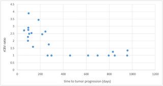
Patients with >200 days to progression are more likely to have no elevated relative cerebral blood volume (rCBV) ratio (p = 0.0004), no tumor restriction (p = 0.024), and no DSE (p = 0.052). The elevated mean rCBV ratio (p<0.001) and tumor type restricted diffusion (p = 0.002) were significantly associated with a higher risk of progression. All cases with rCBV ratio of >1.5 progressed in 275 days or earlier. Tumors tended to progress at the area where patients with post-operative MRIs showed elevated perfusion (p = 0.006), tumor-type restricted diffusion (p = 0.005) and DSE (p = 0.008).
CONCLUSIONS
Post-operative analysis of rCBV, tumor type restricted diffusion and enhancement characteristics are predictive of time to progression, risk of progression and where tumor progression is likely to occur.
中文翻译:

高度脑胶质瘤的术后灌注和扩散MR成像及肿瘤进展。
目的灌注和扩散磁共振成像(MRI)为脑肿瘤分析提供了重要的生物标记。我们的目的是调查在术后MRI即时检查中灌注增加或肿瘤扩散受限的区域是否可预测高级别神经胶质瘤患者的肿瘤进展时间。材料与方法回顾性分析23例高级神经胶质瘤患者。我们测量了切除区域的灌注,并评估了残留肿瘤块中是否存在限制扩散。统计地计算出灌注,扩散和造影剂增强(延迟静态增强(DSE))特征与肿瘤进展时间之间的关系。我们还评估了肿瘤进展的位置是否与高灌注区域一致,肿瘤类型限制了扩散和增强。结果进展> 200天的患者更有可能没有相对脑血容量(rCBV)比升高(p = 0.0004),无肿瘤限制(p = 0.024)和无DSE(p = 0.052)。平均rCBV比值升高(p <0.001)和肿瘤类型限制扩散(p = 0.002)与进展风险较高显着相关。rCBV比率> 1.5的所有病例均在275天或更早时间内进展。术后MRI表现为灌注升高(p = 0.006),肿瘤类型限制扩散(p = 0.005)和DSE(p = 0.008)的区域,肿瘤倾向于发展。结论rCBV的术后分析
更新日期:2019-03-19
中文翻译:
高度脑胶质瘤的术后灌注和扩散MR成像及肿瘤进展。
目的灌注和扩散磁共振成像(MRI)为脑肿瘤分析提供了重要的生物标记。我们的目的是调查在术后MRI即时检查中灌注增加或肿瘤扩散受限的区域是否可预测高级别神经胶质瘤患者的肿瘤进展时间。材料与方法回顾性分析23例高级神经胶质瘤患者。我们测量了切除区域的灌注,并评估了残留肿瘤块中是否存在限制扩散。统计地计算出灌注,扩散和造影剂增强(延迟静态增强(DSE))特征与肿瘤进展时间之间的关系。我们还评估了肿瘤进展的位置是否与高灌注区域一致,肿瘤类型限制了扩散和增强。结果进展> 200天的患者更有可能没有相对脑血容量(rCBV)比升高(p = 0.0004),无肿瘤限制(p = 0.024)和无DSE(p = 0.052)。平均rCBV比值升高(p <0.001)和肿瘤类型限制扩散(p = 0.002)与进展风险较高显着相关。rCBV比率> 1.5的所有病例均在275天或更早时间内进展。术后MRI表现为灌注升高(p = 0.006),肿瘤类型限制扩散(p = 0.005)和DSE(p = 0.008)的区域,肿瘤倾向于发展。结论rCBV的术后分析


 京公网安备 11010802027423号