Our official English website, www.x-mol.net, welcomes your feedback! (Note: you will need to create a separate account there.)
Posterior scleral deformations around optic disc are associated with visual field damage in open-angle glaucoma patients with myopia.
PLOS ONE ( IF 3.7 ) Pub Date : 2019-03-15 , DOI: 10.1371/journal.pone.0213714 Eun Kyoung Kim 1, 2 , Hae-Young Lopilly Park 1, 2 , Chan Kee Park 1, 2
PLOS ONE ( IF 3.7 ) Pub Date : 2019-03-15 , DOI: 10.1371/journal.pone.0213714 Eun Kyoung Kim 1, 2 , Hae-Young Lopilly Park 1, 2 , Chan Kee Park 1, 2
Affiliation
|
PURPOSE
To identify important variables associated with visual field (VF) defects in open-angle glaucoma (OAG) with myopia.
MATERIALS AND METHODS
A total of 105 OAG with myopia were enrolled in this cross-sectional study. The disc tilt ratio, disc torsion degree, disc-foveal angle, and area of peripapillary atrophy (PPA) were measured from red-free fundus photographs. Patients underwent Swept-source optical coherence tomography to measure peripapillary retinal nerve fiber layer (RNFL), subfoveal choroidal, and sufoveal scleral thicknesses. Functional evaluation was performed using 24-2 standard automated perimetry. For statistical analyses, logistic regression, artificial neural networks (ANN), and hierarchical cluster analysis were performed.
RESULTS
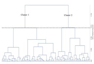
Logistic regression demonstrated peripapillary RNFL thickness as a significant variable for the presence of VF defects, otherwise ANN identified PPA area, peripapillary RNFL thickness, disc-foveal angle, and disc torsion degree as significant clinical variables in OAG with myopia. Two clusters were made after a hierarchical cluster analysis. Cluster 2 showed significantly worse VF damage than cluster 1 (MD = -5.20±5.25 dB for cluster 2 and -1.84±3.02 dB for cluster 1, P < .001). Cluster 2 had significantly greater disc tilt ratio, disc-foveal angle, and PPA area compared with cluster 1 (P < .001, 0.005, and < .001, respectively).
CONCLUSIONS
Generally peripapillary RNFL thickness is considered as an important variable associated with visual field defects in glaucoma patients. ANN identified parameters associated with posterior scleral deformations around optic disc induced by myopic change including PPA area, disc torsion degree, and disc-foveal angle as significant clinical variables for visual field damage in OAG with myopia.
中文翻译:

在开角型青光眼近视患者中,视盘周围的巩膜后部变形与视野损害有关。
目的确定与近视性开角型青光眼(OAG)视野(VF)缺陷相关的重要变量。材料与方法这项横断面研究共入选了105例OAG近视眼。从无红眼底照片中测量椎间盘倾斜率,椎间盘扭转度,椎间盘凹角和乳头周围萎缩(PPA)面积。患者接受扫频光学相干断层扫描,以测量乳头周围视网膜神经纤维层(RNFL),小凹脉络膜和小凹巩膜厚度。使用24-2标准自动视野检查法进行功能评估。对于统计分析,执行了逻辑回归,人工神经网络(ANN)和层次聚类分析。结果Logistic回归表明,对于存在VF缺损的患者,乳头周围RNFL厚度是重要变量,否则ANN将PPA面积,乳头周围RNFL厚度,椎间盘凹角和椎间盘扭转程度确定为OAG伴有近视的重要临床变量。在进行层次聚类分析后,创建了两个聚类。群集2显示的VF损坏比群集1严重得多(群集2的MD = -5.20±5.25 dB,群集1的MD = -1.84±3.02 dB,P <.001)。与群集1相比,群集2具有明显更大的椎间盘倾斜率,椎间盘凹角和PPA面积(分别为P <.001、0.005和<.001)。结论通常,青光眼患者的视乳头周围RNFL厚度被认为是与视野缺损相关的重要变量。
更新日期:2019-03-17
中文翻译:
在开角型青光眼近视患者中,视盘周围的巩膜后部变形与视野损害有关。
目的确定与近视性开角型青光眼(OAG)视野(VF)缺陷相关的重要变量。材料与方法这项横断面研究共入选了105例OAG近视眼。从无红眼底照片中测量椎间盘倾斜率,椎间盘扭转度,椎间盘凹角和乳头周围萎缩(PPA)面积。患者接受扫频光学相干断层扫描,以测量乳头周围视网膜神经纤维层(RNFL),小凹脉络膜和小凹巩膜厚度。使用24-2标准自动视野检查法进行功能评估。对于统计分析,执行了逻辑回归,人工神经网络(ANN)和层次聚类分析。结果Logistic回归表明,对于存在VF缺损的患者,乳头周围RNFL厚度是重要变量,否则ANN将PPA面积,乳头周围RNFL厚度,椎间盘凹角和椎间盘扭转程度确定为OAG伴有近视的重要临床变量。在进行层次聚类分析后,创建了两个聚类。群集2显示的VF损坏比群集1严重得多(群集2的MD = -5.20±5.25 dB,群集1的MD = -1.84±3.02 dB,P <.001)。与群集1相比,群集2具有明显更大的椎间盘倾斜率,椎间盘凹角和PPA面积(分别为P <.001、0.005和<.001)。结论通常,青光眼患者的视乳头周围RNFL厚度被认为是与视野缺损相关的重要变量。


 京公网安备 11010802027423号