当前位置:
X-MOL 学术
›
Contrast Media Mol. Imaging
›
论文详情
Our official English website, www.x-mol.net, welcomes your feedback! (Note: you will need to create a separate account there.)
64CuS‐labeled nanoparticles: a new sentinel‐lymph‐node‐mapping agent for PET–CT and photoacoustic tomography
Contrast Media & Molecular Imaging ( IF 3.009 ) Pub Date : 2016-08-15 , DOI: 10.1002/cmmi.1709 Qiufang Liu 1, 2, 3 , Min Zhou 4 , Panli Li 1, 2, 3 , Geng Ku 4 , Gang Huang 1, 2, 3 , Chun Li 4 , Shaoli Song 1, 2, 3
Contrast Media & Molecular Imaging ( IF 3.009 ) Pub Date : 2016-08-15 , DOI: 10.1002/cmmi.1709 Qiufang Liu 1, 2, 3 , Min Zhou 4 , Panli Li 1, 2, 3 , Geng Ku 4 , Gang Huang 1, 2, 3 , Chun Li 4 , Shaoli Song 1, 2, 3
Affiliation
|
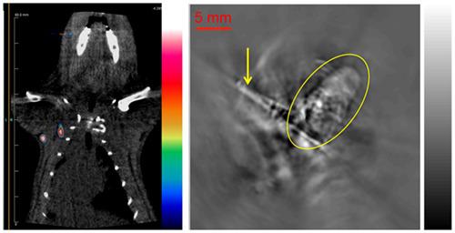
Determining sentinel lymph node (SLN) status is critical to cancer staging and treatment decisions. Currently, in clinical practice, 99mTc‐radiocolloid‐mediated planar scintigraphy and single‐photon emission computed tomography (SPECT) are used to guide the biopsy and resection of SLNs. Recently, an emerging technique that combines positron emission tomography (PET) and photoacoustic tomography (PAT; PET–PAT) may offer accurate information in detecting SLNs. Herein, we report a kind of 64CuS‐labeled nanoparticle (64CuS‐NP) for the detection of SLNs with PET–PAT. We subcutaneously injected 64CuS‐NPs into the rats’ forepaw pads. After 24 h, the rats’ first draining axillary lymph nodes (i.e. the SLNs) could be clearly visualized with micro‐PET (μPET)–CT. Rats were sacrificed after μPET–CT imaging, their axillary lymph nodes were surgically identified, and then PAT was employed to discover 64CuS‐NP‐avid SLNs, which were embedded inside tissues. Biodistribution, autoradiography, and copper staining analyses confirmed the SLNs’ high uptake of 64CuS‐NPs. Our study indicates that 64CuS‐NPs are a promising dual‐function agent for both PET–CT and PAT and could be used with multi‐modal imaging strategies such as PET–PAT to identify SLNs in a clinical setting. Copyright © 2016 John Wiley & Sons, Ltd.
中文翻译:

64CuS标记的纳米颗粒:一种用于PET-CT和光声层析成像的新型前哨淋巴结映射剂
确定前哨淋巴结(SLN)状态对于癌症分期和治疗决策至关重要。目前,在临床实践中,使用99m Tc放射性胶体介导的平面闪烁显像和单光子发射计算机断层扫描(SPECT)来指导SLN的活检和切除。最近,结合正电子发射断层扫描(PET)和光声断层扫描(PAT; PET–PAT)的新兴技术可能会提供检测SLN的准确信息。在本文中,我们报告了一种用PET-PAT检测SLN的64 CuS标记纳米颗粒(64 CuS-NP)。我们皮下注射64CuS-NPs进入大鼠的前爪垫。24小时后,可使用microPET(μPET)–CT清晰地观察大鼠的第一个引流腋窝淋巴结(即SLN)。在μPET-CT成像后处死大鼠,通过外科手术鉴定其腋窝淋巴结,然后使用PAT来发现64枚CuS-NP-avid SLN,它们嵌入组织内部。生物分布,放射自显影和铜染色分析证实了SLNs对64 CuS-NPs的高摄取。我们的研究表明,64种CuS-NPs是用于PET-CT和PAT的有前途的双重功能药物,可与PET-PAT等多模态成像策略一起用于临床诊断SLN。版权所有©2016 John Wiley&Sons,Ltd.
更新日期:2016-08-15
中文翻译:
64CuS标记的纳米颗粒:一种用于PET-CT和光声层析成像的新型前哨淋巴结映射剂
确定前哨淋巴结(SLN)状态对于癌症分期和治疗决策至关重要。目前,在临床实践中,使用99m Tc放射性胶体介导的平面闪烁显像和单光子发射计算机断层扫描(SPECT)来指导SLN的活检和切除。最近,结合正电子发射断层扫描(PET)和光声断层扫描(PAT; PET–PAT)的新兴技术可能会提供检测SLN的准确信息。在本文中,我们报告了一种用PET-PAT检测SLN的64 CuS标记纳米颗粒(64 CuS-NP)。我们皮下注射64CuS-NPs进入大鼠的前爪垫。24小时后,可使用microPET(μPET)–CT清晰地观察大鼠的第一个引流腋窝淋巴结(即SLN)。在μPET-CT成像后处死大鼠,通过外科手术鉴定其腋窝淋巴结,然后使用PAT来发现64枚CuS-NP-avid SLN,它们嵌入组织内部。生物分布,放射自显影和铜染色分析证实了SLNs对64 CuS-NPs的高摄取。我们的研究表明,64种CuS-NPs是用于PET-CT和PAT的有前途的双重功能药物,可与PET-PAT等多模态成像策略一起用于临床诊断SLN。版权所有©2016 John Wiley&Sons,Ltd.


 京公网安备 11010802027423号