当前位置:
X-MOL 学术
›
Contrast Media Mol. Imaging
›
论文详情
Our official English website, www.x-mol.net, welcomes your feedback! (Note: you will need to create a separate account there.)
In vivo quantification of magnetically labelled cells by MRI relaxometry
Contrast Media & Molecular Imaging ( IF 3.009 ) Pub Date : 2016-10-21 , DOI: 10.1002/cmmi.1715 Ulysse Gimenez 1 , Hélène Lajous 1 , Michèle El Atifi 1 , Marie Bidart 1 , Vincent Auboiroux 2 , Pascal Henry Fries 3 , François Berger 1 , Hana Lahrech 1
Contrast Media & Molecular Imaging ( IF 3.009 ) Pub Date : 2016-10-21 , DOI: 10.1002/cmmi.1715 Ulysse Gimenez 1 , Hélène Lajous 1 , Michèle El Atifi 1 , Marie Bidart 1 , Vincent Auboiroux 2 , Pascal Henry Fries 3 , François Berger 1 , Hana Lahrech 1
Affiliation
|
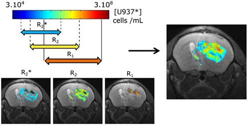
Cellular MRI, which visualizes magnetically labelled cells (cells*), is an active research field for in vivo cell therapy and tracking. The simultaneous relaxation rate measurements (R2*, R2, R1) are the basis of a quantitative cellular MRI method proposed here. U937 cells were labelled with Molday ION Rhodamine B, a bi‐functional superparamagnetic and fluorescent nanoparticle (U937*). U937* viability and proliferation were not affected in vitro. In vitro relaxometry was performed in a cell concentration range of [2.5 × 10⁴–108] cells/mL. These measurements show the existence of complementary cell concentration intervals where these rates vary linearly. The juxtaposition of these intervals delineates a wide cell concentration range over which one of the relaxation rates in a voxel of an in vivo image can be converted into an absolute cell concentration. The linear regime was found at high concentrations for R1 in the range of [106– 2 × 10⁸] cells/mL, at intermediate concentrations for R2 in [2.5 × 10⁵– 5 × 10⁷] cells/mL and at low concentrations for R2* in [8 × 10⁴– 5 × 10⁶] cells/mL. In vivo relaxometry was performed in a longitudinal study, with labelled U937 cells injected into a U87 glioma mouse model. Using in vitro data, maps of in vivo U937* concentrations were obtained by converting one of the in vivo relaxation rates to cell concentration maps. MRI results were compared with the corresponding optical images of the same brains, showing the usefulness of our method to accurately follow therapeutic cell biodistribution in a longitudinal study. Results also demonstrate that the method quantifies a large range of magnetically labelled cells*. Copyright © 2016 John Wiley & Sons, Ltd.
中文翻译:

通过MRI弛豫法对磁性标记细胞进行体内定量
可视化磁性标记细胞(cells *)的细胞MRI是体内细胞治疗和追踪的活跃研究领域。同时松弛率测量(R 2 *,R 2,R 1)是此处提出的定量细胞MRI方法的基础。U937细胞用Molday ION罗丹明B(双功能超顺磁性和荧光纳米颗粒(U937 *))标记。U937 *的活力和增殖在体外不受影响。体外弛豫法在细胞浓度范围[2.5×10 4 –10 8]细胞/ mL。这些测量表明存在互补的细胞浓度间隔,其中这些速率线性变化。这些间隔的并置描绘了宽的细胞浓度范围,在该范围内可以将体内图像体素中的弛豫率之一转换为绝对细胞浓度。在高浓度下,R 1的线性范围为[10 6 – 2×10 8 ]个细胞/ mL,在中等浓度下,R 2的线性范围为[2.5×10 5 – 5×10 7 ]个细胞/ mL,在低浓度下对- [R 2 * [8×10 4 - 5×10 6]细胞/ mL。在纵向研究中进行体内弛豫测定,将标记的U937细胞注射到U87胶质瘤小鼠模型中。使用体外数据,通过将体内弛豫率之一转换为细胞浓度图,可获得体内U937 *浓度的图。MRI结果与相同大脑的相应光学图像进行了比较,显示了我们的方法在纵向研究中准确跟踪治疗性细胞生物分布的有用性。结果还表明,该方法可量化大范围的磁性标记细胞*。版权所有©2016 John Wiley&Sons,Ltd.
更新日期:2016-10-21
中文翻译:
通过MRI弛豫法对磁性标记细胞进行体内定量
可视化磁性标记细胞(cells *)的细胞MRI是体内细胞治疗和追踪的活跃研究领域。同时松弛率测量(R 2 *,R 2,R 1)是此处提出的定量细胞MRI方法的基础。U937细胞用Molday ION罗丹明B(双功能超顺磁性和荧光纳米颗粒(U937 *))标记。U937 *的活力和增殖在体外不受影响。体外弛豫法在细胞浓度范围[2.5×10 4 –10 8]细胞/ mL。这些测量表明存在互补的细胞浓度间隔,其中这些速率线性变化。这些间隔的并置描绘了宽的细胞浓度范围,在该范围内可以将体内图像体素中的弛豫率之一转换为绝对细胞浓度。在高浓度下,R 1的线性范围为[10 6 – 2×10 8 ]个细胞/ mL,在中等浓度下,R 2的线性范围为[2.5×10 5 – 5×10 7 ]个细胞/ mL,在低浓度下对- [R 2 * [8×10 4 - 5×10 6]细胞/ mL。在纵向研究中进行体内弛豫测定,将标记的U937细胞注射到U87胶质瘤小鼠模型中。使用体外数据,通过将体内弛豫率之一转换为细胞浓度图,可获得体内U937 *浓度的图。MRI结果与相同大脑的相应光学图像进行了比较,显示了我们的方法在纵向研究中准确跟踪治疗性细胞生物分布的有用性。结果还表明,该方法可量化大范围的磁性标记细胞*。版权所有©2016 John Wiley&Sons,Ltd.


 京公网安备 11010802027423号