当前位置:
X-MOL 学术
›
Contrast Media Mol. Imaging
›
论文详情
Our official English website, www.x-mol.net, welcomes your feedback! (Note: you will need to create a separate account there.)
USPIO enhanced lymph node MRI using 3D multi‐echo GRE in a rabbit model
Contrast Media & Molecular Imaging ( IF 3.009 ) Pub Date : 2016-12-15 , DOI: 10.1002/cmmi.1716 Sung Hun Kim 1 , Soon Nam Oh 1 , Hyun Seok Choi 1 , Hyun Sil Lee 1 , Jaeseop Jun 1 , Yoonho Nam 1 , Sung Hak Lee 2 , Jin-Kwon Lee 3 , Hae Giu Lee 1
Contrast Media & Molecular Imaging ( IF 3.009 ) Pub Date : 2016-12-15 , DOI: 10.1002/cmmi.1716 Sung Hun Kim 1 , Soon Nam Oh 1 , Hyun Seok Choi 1 , Hyun Sil Lee 1 , Jaeseop Jun 1 , Yoonho Nam 1 , Sung Hak Lee 2 , Jin-Kwon Lee 3 , Hae Giu Lee 1
Affiliation
|
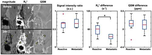
Ultrasmallsuperparamagnetic iron oxide (USPIO) has been suggested to be a negative MR contrast agent to detect metastatic lymph nodes. Previously reported studies have evaluated the diagnostic performance of USPIO‐enhanced MR lymph node imaging based on signal intensity. In this study, we investigate the specific performance of three different parametric approaches (normalized signal intensity, R2* and susceptibility) using 3D multi‐echo gradient echo to quantify the USPIO particles in lymph nodes. Nine rabbits with VX2 tumor implants were scanned before and after USPIO injection. From 3D multi‐echo GRE magnitude and phase data, we generated multi‐echo combined T2*‐weighted images, an R2* map, and a quantitative susceptibility map. Eighteen lymph nodes (nine reactive and nine metastatic) were evaluated and showed remarkable signal drops in the area of USPIO accumulation. On parametric analysis, the R2* difference before and after USPIO injection was significantly different (p < 0.05) between reactive and metastatic lymph nodes; in contrast, the normalized signal intensity and susceptibility were not significantly different between the nodes. Our study showed the potential utility of USPIO‐enhanced MRI using R2* mapping from 3D multi‐echo GRE for the detection of lymph node metastasis and parametric analysis of lymph node status in a rabbit model. Copyright © 2016 John Wiley & Sons, Ltd.
中文翻译:

在兔模型中使用3D多回波GRE的USPIO增强的淋巴结MRI
有人建议将超小型超顺磁性氧化铁(USPIO)用作检测转移性淋巴结的阴性MR造影剂。先前报道的研究已经基于信号强度评估了USPIO增强的MR淋巴结成像的诊断性能。在这项研究中,我们研究了使用3D多回波梯度回波来量化淋巴结中USPIO颗粒的三种不同参数方法(标准化信号强度,R 2 *和磁化率)的具体性能。在USPIO注射前后,对9具VX2肿瘤植入物的兔子进行了扫描。根据3D多回波GRE幅度和相位数据,我们生成了多回波组合的T 2 *加权图像,即R 2*地图,以及定量磁化率地图。评估了18个淋巴结(9个反应性和9个转移性),并在USPIO积聚区域显示出明显的信号下降。在参数分析中,USPIO注射前后的R 2 *差异在 反应性和转移性淋巴结之间存在显着差异(p <0.05);相反,节点之间的标准化信号强度和磁化率没有显着差异。我们的研究表明,使用来自3D多回波GRE的R2 *映射进行USPIO增强的MRI在检测兔模型中的淋巴结转移和参数化淋巴结状态方面具有潜在的实用性。版权所有©2016 John Wiley&Sons,Ltd.
更新日期:2016-12-15
中文翻译:
在兔模型中使用3D多回波GRE的USPIO增强的淋巴结MRI
有人建议将超小型超顺磁性氧化铁(USPIO)用作检测转移性淋巴结的阴性MR造影剂。先前报道的研究已经基于信号强度评估了USPIO增强的MR淋巴结成像的诊断性能。在这项研究中,我们研究了使用3D多回波梯度回波来量化淋巴结中USPIO颗粒的三种不同参数方法(标准化信号强度,R 2 *和磁化率)的具体性能。在USPIO注射前后,对9具VX2肿瘤植入物的兔子进行了扫描。根据3D多回波GRE幅度和相位数据,我们生成了多回波组合的T 2 *加权图像,即R 2*地图,以及定量磁化率地图。评估了18个淋巴结(9个反应性和9个转移性),并在USPIO积聚区域显示出明显的信号下降。在参数分析中,USPIO注射前后的R 2 *差异在 反应性和转移性淋巴结之间存在显着差异(p <0.05);相反,节点之间的标准化信号强度和磁化率没有显着差异。我们的研究表明,使用来自3D多回波GRE的R2 *映射进行USPIO增强的MRI在检测兔模型中的淋巴结转移和参数化淋巴结状态方面具有潜在的实用性。版权所有©2016 John Wiley&Sons,Ltd.


 京公网安备 11010802027423号