The International Journal of Cardiovascular Imaging ( IF 2.1 ) Pub Date : 2023-05-15 , DOI: 10.1007/s10554-023-02867-1 Xin Jiang 1 , Yan-Xiang Zhou 1 , Qing Zhou 1 , Sheng Cao 1
|
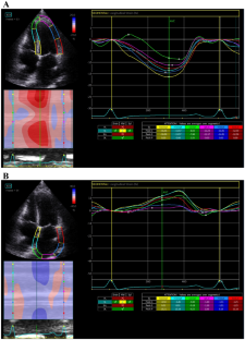
This study analyzed the differences and explored the donor/recipient factors between marginal and standard donor heart recipients after heart transplantation (HT) by speckle tracking echocardiography (STE). Seventy-two HT patients were enrolled: 25 standard and 47 marginal donor heart recipients. Thirty HT patients completed 2-year continuous follow-up (1, 6, 12, 24 months). Thirty healthy volunteers were controls. STE was used to track the strain characteristics of the left ventricle and atrium for detecting early changes in marginal donor heart recipients, including left ventricular global longitudinal, circumferential and radial strain (LVGLS, LVGCS, LVGRS) and left atrial strain in systole (LAS-S) and late diastole (LAS-A). The perioperative parameters were similar between the standard and marginal groups. No significant differences were found in left heart size, systolic and diastolic function parameters. Left ventricular systolic strain (LVGLS, LVGCS, LVGRS) and systolic and late diastolic left atrial strain (LAS-S, LAS-A) were lower in the HT recipients than the control group (P < 0.05), but there was no difference between the marginal and standard groups (P > 0.05). LVGLS, LVGCS, and LAS-S were low in the marginal group 1 month after surgery but recovered gradually at 6 months. The patients with donor/recipient body weight ratio < 0.8 group had lower LVGLS and LAS-S. STE showed no significant difference between marginal and standard donor recipients. The LVGLS and LAS-S were lower in those with a smaller donor/recipient body weight ratio. The cardiac function of HT patients was lower in the early postoperative period but gradually recovered over time.
中文翻译:
斑点追踪超声心动图评估边缘供心术后2年左心功能
本研究通过斑点追踪超声心动图(STE)分析了心脏移植(HT)后边缘供体心脏受体和标准供体心脏受体之间的差异并探讨了供体/受体因素。招募了 72 名 HT 患者:25 名标准供体心脏接受者和 47 名边缘供体心脏接受者。30 名 HT 患者完成了 2 年连续随访(1、6、12、24 个月)。三十名健康志愿者作为对照。STE用于追踪左心室和心房的应变特征,以检测边缘供心受者的早期变化,包括左心室整体纵向、周向和径向应变(LVGLS、LVGCS、LVGRS)和收缩期左心房应变(LAS- S) 和舒张晚期 (LAS-A)。标准组和边缘组的围手术期参数相似。左心大小、收缩和舒张功能参数没有发现显着差异。HT 组左心室收缩期应变(LVGLS、LVGCS、LVGRS)以及收缩期和舒张末期左心房应变(LAS-S、LAS-A)均低于对照组( P < 0.05),但两组间差异无统计学意义(P < 0.05 ) 。边缘组和标准组(P > 0.05)。术后 1 个月边缘组的 LVGLS、LVGCS 和 LAS-S 较低,但在 6 个月时逐渐恢复。供受者体重比<0.8组患者的LVGLS和LAS-S较低。STE 在边缘捐赠者和标准捐赠者之间没有显着差异。供体/受体体重比较小的患者的 LVGLS 和 LAS-S 较低。HT患者的心功能在术后早期较低,但随着时间的推移逐渐恢复。


 京公网安备 11010802027423号