Neuroradiology ( IF 2.8 ) Pub Date : 2022-10-26 , DOI: 10.1007/s00234-022-03077-7 Mengxing Li 1 , Jian Liu 1 , Feng Chen 2 , Chengzhe Fan 3 , Xinjian Yang 1 , Xuan Sun 4
|
Purpose
Contrast-induced encephalopathy (CIE) was defined as new onset of neurological deficits after exposure to contrast media, which could be observed after the endovascular treatment for intracranial aneurysms.
Methods
We enrolled a consecutive cohort of patients who underwent endovascular treatment for unruptured intracranial aneurysms. CIE was defined as a reversible neuropathic syndrome that occurred after interventional therapy, accompanied by imaging abnormalities and excluding other diseases. Multivariable Poisson regression analysis was performed to show risk factors by incidence rate ratio (IRR) and a clinical strategy was proposed.
Results
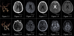
Among the 579 patients who underwent interventional therapy for intracranial aneurysms, the crude incidence rate of CIE was 2.4% (95% CI, 1.2–3.6%) at our center. Headache, hemiplegia, and disorientation could be initial symptoms, and cortical blindness was the most common localized deficit. Cerebral edema and sulci effacement on CT were observed, and re-revaluation after treatments on CT/MRI showed absent lesions. The risk factors were history of stroke (IRR, 7.752; P = 0.007), history of hypertension (IRR, 1.064; P = 0.042), posterior circulation aneurysms (IRR, 9.412; P = 0.004) and higher dosage of contrast agents (IRR, 1.018; P = 0.007). After the strategy of accelerating excretion of contrast agents, reduction of intracranial pressure and anti-inflammation/vasospasm therapy, the prognosis was favorable with most patients fully recovered within 72 h.
Conclusion
History of stroke and posterior circulation aneurysms were main risk factors for CIE. A higher dosage of contrast agents might induce CIE, and the history of hypertension should be considered as well.
中文翻译:
颅内动脉瘤血管内治疗后造影剂脑病——危险因素分析及临床对策
目的
造影剂脑病(CIE)被定义为暴露于造影剂后新发的神经功能缺损,可在颅内动脉瘤血管内治疗后观察到。
方法
我们连续招募了一组因未破裂颅内动脉瘤接受血管内治疗的患者。CIE定义为介入治疗后出现的可逆性神经病变综合征,伴有影像学异常,排除其他疾病。进行多变量泊松回归分析以按发病率比 (IRR) 显示危险因素,并提出了临床策略。
结果
在接受颅内动脉瘤介入治疗的 579 名患者中,我们中心的 CIE 粗发生率为 2.4%(95% CI,1.2-3.6%)。头痛、偏瘫和定向障碍可能是最初的症状,皮质盲是最常见的局部缺陷。在 CT 上观察到脑水肿和脑沟消失,并且在 CT/MRI 上治疗后重新评估显示没有病灶。危险因素为中风史(IRR,7.752;P = 0.007)、高血压病史(IRR,1.064;P = 0.042)、后循环动脉瘤(IRR,9.412;P = 0.004)和造影剂剂量较高(IRR , 1.018 ; = 0.007)。经加速造影剂排泄、降低颅内压和抗炎/血管痉挛治疗后,预后良好,大部分患者在72小时内完全康复。
结论
中风史和后循环动脉瘤是 CIE 的主要危险因素。较高剂量的造影剂可能诱发 CIE,还应考虑高血压病史。


 京公网安备 11010802027423号