Minerals Engineering ( IF 4.8 ) Pub Date : 2022-09-18 , DOI: 10.1016/j.mineng.2022.107748 A. Roslin , M. Marsh , N. Piché , B. Provencher , T.R. Mitchell , I.A. Onederra , C.R. Leonardi
|
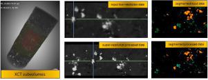
X-ray micro-computed tomography (micro-CT) is widely used for three-dimensional analysis of many rock types. However, the practical implementation of this method for micro-porous samples requires a compromise between the resolution of the images and the obtainable field of view (FOV). Generally, resolution enhancement results in a reduction of the FOV. The generation of high-quality micro-CT images is an expensive and time consuming task due to the competing requirements of a large FOV and fine resolution. To alleviate this, super-resolution processing, based on deep learning, is proposed to improve the quality of low-resolution images that can obtain a large FOV. In this research, a super-resolution technique employing the three-dimensional U-Net convolutional neural network (CNN) architecture was applied to enhance the resolution of granodiorite rock sample images. This was undertaken using two sets of micro-CT image triplexes, where the first triplex contained 3-, 6-, and 12-micron resolution sets, and the second triplex contained 1-, 2-, and 4-micron resolution sets. For each triplex, 80% of the images were used for training the neural network with the remaining 20% used for validation. Further validation was performed by comparing the processed results to images obtained from scanning electron microscopy (SEM). It was observed that super-resolution processing can significantly improve the low-resolution micro-CT image quality without physically reducing the sample size typically required for high-resolution scanning. It is expected that this technique could assist practitioners reveal features absent in small samples (e.g. large fractures and or rock textures). Furthermore, images restored through super-resolution processing maintain the FOV of the lower resolution scan, a task that would be comparatively expensive and time consuming to acquire in a high-resolution scan. The workflow proposed in this study has a significant impact on a range of fields including the numerical prediction of rock permeability, and segmentation for advanced mineral analysis.
中文翻译:
使用卷积神经网络 (CNN) 处理花岗闪长岩样品的 micro-CT 图像,第一部分:使用 3D CNN 的超分辨率增强
X 射线微型计算机断层扫描(micro-CT) 广泛用于对多种岩石类型进行三维分析。然而,该方法对微孔样品的实际实施需要在图像分辨率和可获得的视场 (FOV) 之间进行折衷。通常,分辨率增强会导致 FOV 减小。由于大视场和高分辨率的竞争要求,生成高质量的微 CT 图像是一项昂贵且耗时的任务。为了缓解这种情况,提出了基于深度学习的超分辨率处理,以提高可以获得大视场的低分辨率图像的质量。在这项研究中,采用三维 U-Net 卷积神经网络的超分辨率技术(CNN)架构被应用于提高花岗闪长岩的分辨率岩石样本图像。这是使用两组微型 CT 图像三联体进行的,其中第一个三联体包含 3、6 和 12 微米分辨率组,第二个三联体包含 1、2 和 4 微米分辨率组。对于每个三联体,80% 的图像用于训练神经网络,其余 20% 用于验证。通过将处理结果与从扫描电子显微镜 (SEM) 获得的图像进行比较来进行进一步验证。据观察,超分辨率处理可以显着提高低分辨率显微 CT 图像质量,而无需物理减少高分辨率扫描通常所需的样本大小。预计这种技术可以帮助从业者揭示小样本中不存在的特征(例如大裂缝和/或岩石纹理)。此外,通过超分辨率处理恢复的图像保持较低分辨率扫描的 FOV,在高分辨率扫描中获取该任务相对昂贵且耗时。本研究中提出的工作流程对一系列领域产生了重大影响,包括岩石渗透率的数值预测和高级矿物分析的分割。


 京公网安备 11010802027423号