Skeletal Radiology ( IF 2.1 ) Pub Date : 2022-09-07 , DOI: 10.1007/s00256-022-04171-w Henry Kwok 1 , Meera Hameed 2 , Sinchun Hwang 1
|
Objective
To evaluate MR features and clinical course of malignant melanotic nerve sheath tumor (MMNST), previously known as melanotic schwannoma and considered indolent and rarely metastasizing.
Materials and methods
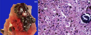
This IRB-approved retrospective study searched 31 patients (20 male: 11 female, mean age 48; range 15–76) with histologically confirmed MMNST in a single tertiary cancer center over 22 years. Pre-treatment MR was available in 12 patients and evaluated by two radiologists in consensus regarding lesion location, size, morphology, signal characteristics, contrast enhancement, local invasion, and presence of classic signs of peripheral nerve sheath tumors. Clinical outcomes, including local recurrence, metastasis, and survival, were examined in 12 patients for whom follow-up was available.
Results
The spine was the most frequent site (13/31) among all identified cases. In 12 cases with MR, lesions were well-circumscribed in 11/12 cases, with a mean size of 4.5 cm (2.3–13.0 cm). Ten of 12 cases showed T1 hyperintensity. In 5/9 spinal MRI, tumor involved multiple levels. All lesions showed contrast enhancement, and local bone invasion in > 50%. A dumb-bell shape was common to all spinal lesions. Classical signs of nerve sheath tumors were uncommon. Among 12 patients with a mean follow-up of 4.8 years (range 1.3–10.2 years), six were disease-free, while two had recurrence or metastases, and four had died of metastases.
Conclusion
MMNST usually presents as a T1 hyperintense enhancing dumb-bell shaped mass in the spine. Multi-level involvement and bone invasion are common. MMNST is clinically aggressive with high rates of metastases and death.
中文翻译:
恶性黑色素神经鞘瘤的磁共振成像特征和临床过程:二十年的单机构经验
客观的
评估恶性黑色素神经鞘瘤 (MMNST) 的 MR 特征和临床过程,以前称为黑色素神经鞘瘤,被认为是惰性和很少转移的。
材料和方法
这项 IRB 批准的回顾性研究在 22 年的时间里在一个三级癌症中心搜索了 31 名经组织学证实为 MMNST 的患者(20 名男性:11 名女性,平均年龄 48 岁;范围 15-76 岁)。治疗前 MR 可用于 12 名患者,并由两名放射科医师就病灶位置、大小、形态、信号特征、对比增强、局部浸润和周围神经鞘瘤的典型体征的存在进行共识评估。临床结果,包括局部复发、转移和生存,在 12 名可进行随访的患者中进行了检查。
结果
在所有确定的病例中,脊柱是最常见的部位 (13/31)。在 12 例 MR 病例中,11/12 例病灶边界清楚,平均大小为 4.5 厘米(2.3-13.0 厘米)。12例中有10例表现为T1高信号。在 5/9 脊柱 MRI 中,肿瘤涉及多个水平。所有病灶均呈对比增强,局部骨质浸润 > 50%。所有脊柱病变均呈哑铃状。神经鞘瘤的典型体征并不常见。在平均随访 4.8 年(范围 1.3-10.2 年)的 12 名患者中,6 名无病,2 名出现复发或转移,4 名死于转移。
结论
MMNST 通常表现为脊柱中的 T1 高信号增强哑铃形肿块。多节段受累和骨侵犯很常见。MMNST 具有高转移率和高死亡率的临床侵袭性。


 京公网安备 11010802027423号