Skeletal Radiology ( IF 2.1 ) Pub Date : 2022-09-07 , DOI: 10.1007/s00256-022-04176-5 Huda S Alsahlawi 1 , Ali Hassan 1 , Sayed Mohamed Hasan 1 , Safa A Alshaikh 2 , Hasan A Al-Aradi 3
|
Background
Most of the foot and ankle soft tissue tumors are benign. Although lipomas are the most common soft tissue tumors, intra-articular lipomas are an extremely rare entity. The majority of described cases of intra-articular lipomas occur in the knee joint. In particular, an intra-articular true lipoma of the ankle has not been reported.
Case presentation
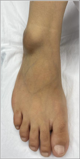
We report the case of a 45-year-old woman who presented with a progressively growing ankle mass over 5 years, which started to restrict the range of motion. It was not preceded by any trauma or sports activity. On examination, a non-tender firm mass was evident on the lateral aspect of the right ankle joint with no signs of inflammation. It was non-compressible, non-mobile, and did not transilluminate. The clinical diagnosis was probable for a soft tissue ganglion. A radiograph revealed a soft tissue opacity over the anterolateral aspect of the tibiotalar joint. Magnetic resonance imaging (MRI) demonstrated a well-defined, multilobulated, encapsulated lesion located at the lateral aspect of the tibiotalar joint; with intra- and extra-articular components and an analogous signal intensity to fat. The patient underwent surgical excision of the tumor, and the histopathological examination showed mature adipose tissue representing an intra-articular lipoma. At the follow-up visit, the patient had complete resolution of symptoms and no active complaints.
Conclusion
An intra-articular true lipoma of the ankle is an extremely rare tumor. MRI is an invaluable diagnostic tool to make a reliable diagnosis of intra-articular masses.
中文翻译:
一例罕见的关节内真性踝关节脂肪瘤病例
背景
大多数足部和脚踝软组织肿瘤是良性的。尽管脂肪瘤是最常见的软组织肿瘤,但关节内脂肪瘤极为罕见。大多数描述的关节内脂肪瘤病例发生在膝关节。特别是,尚未见踝关节内真性脂肪瘤的报道。
案例展示
我们报告了一名 45 岁女性的病例,她的脚踝肿块在 5 年内逐渐增大,开始限制活动范围。它之前没有任何创伤或体育活动。经检查,右踝关节外侧明显有无压痛的硬质肿块,无炎症迹象。它是不可压缩的,不可移动的,并且不透光。临床诊断很可能是软组织神经节。X 光片显示胫距关节前外侧软组织不透明。磁共振成像 (MRI) 显示位于胫距关节外侧的边界清楚、多分叶、包裹性病变;具有关节内和关节外成分以及与脂肪类似的信号强度。患者接受了肿瘤切除手术,组织病理学检查显示成熟脂肪组织代表关节内脂肪瘤。随访时,患者症状完全消退,无主诉。
结论
踝关节内真性脂肪瘤是一种极为罕见的肿瘤。MRI 是对关节内肿块进行可靠诊断的宝贵诊断工具。


 京公网安备 11010802027423号