Skeletal Radiology ( IF 2.1 ) Pub Date : 2022-09-05 , DOI: 10.1007/s00256-022-04170-x Hyang Sook Jeong 1 , Seul Ki Lee 2 , Jee-Young Kim 2 , Changyoung Yoo 1 , Min Wook Joo 3 , Jun-Ho Kim 4
|
Objective
To compare the MRI findings between the localized- and diffuse-type tenosynovial giant cell tumors (TSGCTs) of digits with pathology correlation.
Methods
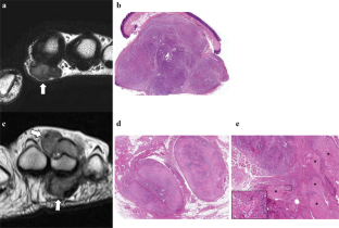
Twenty-eight patients with newly diagnosed TSGCTs of digits (22 localized and 6 diffuse types) who underwent preoperative MRI and surgical excision were included from Jan. 2015 to September 2021. MRI findings regarding nodularity, margins, morphology of hypointensity with pathology correlation, and disease extent (bone erosion, articular involvement, muscle involvement, tendon destruction, and neurovascular encasement) were assessed.
Results
Diffuse type was significantly larger (P = 0.006), more multinodular on both MRI and pathology (P = 0.038, both) with significant agreement, and infiltrative on both MRI and pathology (P < 0.001, both) with substantial agreement, and showed central granular on MRI and strong hemosiderin deposition on pathology (P = 0.022 and P = 0.021) with moderate agreement than localized type. Localized type showed significantly more frequent peripheral capsules on both MRI and pathology (P < 0.001, both) with moderate agreement than diffuse type. However, the septum on both MRI and pathology showed no statistically significant difference between the two groups (P = 0.529 and P = 0.372) without significant agreement. The disease extent was more severe in the diffuse type than the localized type regarding articular involvement (P < 0.001), muscle involvement (P < 0.001), and tendon destruction (P = 0.010). No statistically significant differences were found between the two groups regarding bone erosion (P = 0.196) or neurovascular bundle encasement (P = 0.165).
Conclusions
Diffuse-type TSGCTs of digits presented as locally aggressive lesions with larger, multinodular, infiltrative masses exhibiting stronger hemosiderin deposition and more severe disease extents of articular, muscle, and tendon involvement than the localized type.
中文翻译:
手指腱鞘巨细胞瘤:局限型和弥漫型的 MRI 鉴别与病理相关性
客观的
比较手指局部型和弥漫型腱鞘巨细胞瘤 (TSGCT) 的 MRI 表现与病理学相关性。
方法
从 2015 年 1 月到 2021 年 9 月,纳入了 28 名新诊断的数字 TSGCT 患者(22 名局部型和 6 名弥漫型),他们接受了术前 MRI 和手术切除。关于结节、边缘、低信号形态与病理相关性的 MRI 结果,以及评估疾病程度(骨侵蚀、关节受累、肌肉受累、肌腱破坏和神经血管包绕)。
结果
弥漫型显着较大(P = 0.006),MRI 和病理学上更多结节性(P = 0.038,两者)具有显着一致性,浸润型在 MRI 和病理学上(P < 0.001,两者)具有显着一致性,并显示中央MRI 上为颗粒状,病理学上为强含铁血黄素沉积(P = 0.022 和P = 0.021),与局限型相比具有中等一致性。局部型在 MRI 和病理学上显示出明显更多的外周包膜(P < 0.001,两者),与弥散型相比具有中等一致性。然而,MRI 和病理学上的隔膜在两组之间无统计学差异(P = 0.529 和P = 0.372)没有显着一致。在关节受累 ( P < 0.001)、肌肉受累 ( P < 0.001) 和肌腱破坏 ( P = 0.010)方面,弥漫型的疾病程度比局限型更严重。两组之间在骨质侵蚀( P = 0.196)或神经血管束包裹(P = 0.165)方面没有统计学差异。
结论
数字的弥漫型 TSGCT 表现为局部侵袭性病变,具有较大的多结节浸润性肿块,与局部型相比表现出更强的含铁血黄素沉积和更严重的关节、肌肉和肌腱受累疾病程度。


 京公网安备 11010802027423号