Skeletal Radiology ( IF 2.1 ) Pub Date : 2022-09-02 , DOI: 10.1007/s00256-022-04174-7 Flávio Duarte Silva 1 , Fernando Zorzenoni 1 , Lucas Nakasone Matos da Silva 1 , Afranio Dos Reis Teixeira Neto 1 , Marco Tulio Gonzalez 1 , Alípio Gomes Ormond Filho 1 , Júlio Brandão Guimarães 1
|
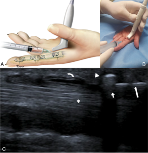
Imaging-guided tendon procedures aim to reduce pain and increase function by controlling inflammation and stimulating healing. Ultrasound is the preferable guiding modality due to its high resolution and real-time demonstration of the tendinous anatomy and needle positioning. The technique includes appropriate patient positioning, which varies depending on the targeted tendon, as well as sterile and proper draping. For most procedures, we prefer the “in-plane” approach, which demonstrates the entire needle as it advances through different tissue layers. Upper limb injections commonly use corticosteroids and anesthetics with different reported short- and long-term results depending on the tendon treated; better results are obtained in the treatment of tenosynovitis (sliding tendons such as trigger finger and De Quervain’s tenosynovitis). Shoulder and elbow tendinopathies (anchor tendons) may also benefit from injections containing irritants or healing stimulants such as dextrose (prolotherapy) and platelet-rich plasma or by the stimulation of healing via tendon perforations (fenestration). The hyaluronic acid injection has also been used in the treatment of both tenosynovitis and tendinopathies. For tendons passing through osteofibrous tunnels, an additional release may be performed, and the techniques are discussed in this review. Therefore, this article provides practicing musculoskeletal radiologists and trainees with a comprehensive review of tendon injection musculoskeletal image-guided procedures.
中文翻译:
肌腱注射——上肢
成像引导的肌腱手术旨在通过控制炎症和刺激愈合来减轻疼痛和增强功能。超声波是首选的引导方式,因为它具有高分辨率和肌腱解剖结构和针定位的实时演示。该技术包括适当的患者定位,这取决于目标肌腱,以及无菌和适当的悬垂。对于大多数程序,我们更喜欢“平面内”方法,它展示了整个针头穿过不同组织层的过程。上肢注射通常使用皮质类固醇和麻醉剂,根据所治疗的肌腱,报告的短期和长期结果不同;治疗腱鞘炎(扳机指、De Quervain氏腱鞘炎等滑肌腱炎)取得较好效果。肩部和肘部肌腱病(锚定肌腱)也可能受益于含有刺激物或愈合兴奋剂的注射,例如葡萄糖(增生疗法)和富含血小板的血浆,或通过肌腱穿孔(开窗)刺激愈合。透明质酸注射液也已用于治疗腱鞘炎和肌腱病。对于穿过骨纤维隧道的肌腱,可以进行额外的松解,本综述将讨论这些技术。因此,本文为执业肌肉骨骼放射科医师和受训人员提供了肌腱注射肌肉骨骼图像引导程序的全面回顾。


 京公网安备 11010802027423号