npj Parkinson's Disease ( IF 9.304 ) Pub Date : 2022-08-29 , DOI: 10.1038/s41531-022-00372-1 Matthew Amandola 1 , Agniva Sinha 1 , Mark J Amandola 2 , Hoi-Chung Leung 1
|
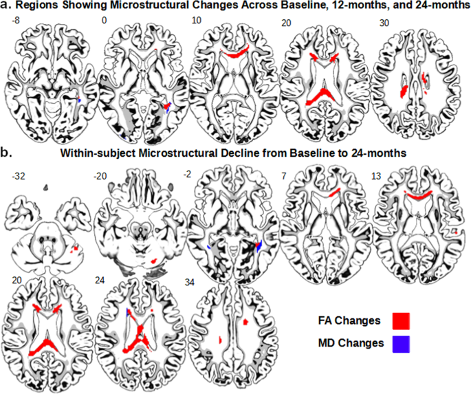
Previous diffusion tensor imaging (DTI) studies of Parkinson’s disease (PD) show reduced microstructural integrity of the corpus callosum (CC) relative to controls, although the characteristics of such callosal degradation remain poorly understood. Here, we utilized a longitudinal approach to identify microstructural decline in the entire volume of the CC and its functional subdivisions over 2 years and related the callosal changes to motor symptoms in early-stage PD. The study sample included 61 PD subjects (N = 61, aged 45–82, 38 M & 23 F, H&Y ≤ 2) from the Parkinson’s Progressive Markers Initiative database (PPMI). Whole-brain voxel-wise results revealed significant fractional anisotropy (FA) and mean diffusivity (MD) changes in the CC, especially in the genu and splenium. Using individually drawn CC regions of interest (ROI), our analysis further revealed that almost all subdivisions of the CC show significant decline in FA to certain extents over the two-year timeframe. Additionally, FA seemed lower in the right hemisphere of the CC at both time-points, and callosal FA decline was associated with FA and MD decline in widespread cortical and subcortical areas. Notably, multiple regression analysis revealed that across-subject akinetic-rigid severity was negatively associated with callosal FA at baseline and 24 months follow-up, and the effect was strongest in the anterior portion of the CC. These results suggest that callosal microstructure alterations in the anterior CC may serve as a viable biomarker for akinetic-rigid symptomology and disease progression, even in early PD.
中文翻译:
早期帕金森病纵向胼胝体微观结构下降与运动僵硬症状严重程度相关
先前对帕金森氏病 (PD) 的弥散张量成像 (DTI) 研究表明,与对照组相比,胼胝体 (CC) 的微观结构完整性有所降低,尽管对这种胼胝体退化的特征仍知之甚少。在这里,我们利用纵向方法来确定 CC 的整个体积及其功能细分的微观结构下降超过 2 年,并将胼胝体变化与早期 PD 的运动症状相关联。研究样本包括 61 名 PD 受试者 ( N = 61,年龄 45–82,38 M 和 23 F,H&Y ≤ 2) 来自帕金森进步标记倡议数据库 (PPMI)。全脑体素结果显示 CC 中的分数各向异性 (FA) 和平均扩散系数 (MD) 发生显着变化,尤其是膝部和压部。使用单独绘制的 CC 感兴趣区域 (ROI),我们的分析进一步表明,几乎所有 CC 的细分在两年的时间范围内都显示出一定程度的 FA 显着下降。此外,在两个时间点,CC 右半球的 FA 似乎较低,胼胝体 FA 下降与广泛皮质和皮质下区域的 FA 和 MD 下降有关。值得注意的是,多元回归分析显示,在基线和 24 个月的随访中,跨受试者运动僵硬的严重程度与胼胝体 FA 呈负相关,CC 的前部效果最强。这些结果表明,即使在早期 PD 中,前 CC 中的胼胝体微结构改变也可以作为运动僵硬症状和疾病进展的可行生物标志物。


 京公网安备 11010802027423号