Skeletal Radiology ( IF 2.1 ) Pub Date : 2022-08-25 , DOI: 10.1007/s00256-022-04161-y Danoob Dalili 1 , Amanda Isaac 2, 3 , Jan Fritz 4
|
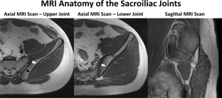
Common etiologies of low back pain include degenerative arthrosis and inflammatory arthropathy of the sacroiliac joints. The diagnostic workup revolves around identifying and confirming the sacroiliac joints as a pain generator. Diagnostic sacroiliac joint injections often serve as functional additions to the diagnostic workup through eliciting a pain response that tests the hypothesis that the sacroiliac joints do or do not contribute to the patient’s pain syndrome. Therapeutic sacroiliac joint injections aim to provide medium- to long-term relief of symptoms and reduce inflammatory activity and, ultimately, irreversible structural damage. Ultrasonography, fluoroscopy, computed tomography, and magnetic resonance imaging (MRI) may be used to guide sacroiliac joint injections. The populations that may benefit most from MRI-guided sacroiliac joint procedures include children, adolescents, adults of childbearing age, and patients receiving serial injections due to the ability of interventional MRI to avoid radiation exposure. Most clinical wide-bore MRI systems can be used for MRI-guided sacroiliac joint injections. Turbo spin echo pulse sequences optimized for interventional needle display visualize the needle tip with an error margin of < 1 mm or less. Published success rates of intra-articular sacroiliac joint drug delivery with MRI guidance range between 87 and 100%. The time required for MR-guided sacroiliac joint injections in adults range between 23–35 min and 40 min in children. In this article, we describe techniques for MRI-guided sacroiliac joint injections, share our practice of incorporating interventional MRI in the care of patients with sacroiliac joint mediated pain, discuss the rationales, benefits, and limitations of interventional MRI, and conclude with future developments.
中文翻译:
MRI 引导的儿童和成人骶髂关节注射:当前实践和未来发展
腰痛的常见病因包括退行性关节病和骶髂关节的炎性关节病。诊断检查围绕识别和确认骶髂关节作为疼痛发生器。诊断性骶髂关节注射通常作为诊断检查的功能补充,通过引发疼痛反应来检验骶髂关节是否会导致患者疼痛综合征的假设。治疗性骶髂关节注射旨在中长期缓解症状并减少炎症活动,并最终减少不可逆的结构损伤。超声检查、荧光检查、计算机断层扫描和磁共振成像 (MRI) 可用于指导骶髂关节注射。可能从 MRI 引导的骶髂关节手术中获益最多的人群包括儿童、青少年、育龄成人以及由于介入 MRI 避免辐射暴露的能力而接受连续注射的患者。大多数临床大口径 MRI 系统可用于 MRI 引导的骶髂关节注射。为介入针显示优化的 Turbo 自旋回波脉冲序列可视化针尖,误差小于 1 毫米或更小。MRI 引导下关节内骶髂关节给药的已发表成功率在 87% 到 100% 之间。成人 MR 引导的骶髂关节注射所需时间在 23-35 分钟和儿童 40 分钟之间。在这篇文章中,我们描述了 MRI 引导的骶髂关节注射技术,


 京公网安备 11010802027423号